This website uses cookies to ensure you get the best experience on our website.


- Table of Contents
Plan EIF3B chromogenic IHC on paraffin sections using the general cytoplasmic pattern and highly stained colon glandular cells as a reference (HPA tissue IHC). This guide covers fixation consistency, controls, antibody titration and cytoplasmic scoring.

Expected localisation, validated protocols, controls and antibodies — the at-a-glance facts below, then the full design guide.
| Expected localisation | Cytoplasmic staining (HPA tissue IHC) | |
| Staining pattern | General cytoplasmic staining in glandular cells (HPA tissue IHC) | |
| Antigen retrieval | EDTA pH 8.0 HIER, heat-mediated (datasheet A04318-1) | |
| Positive control | Adrenal gland+4 more · see all | |
| Negative control | None in HPA (detected in all 45 tissues); use no-primary + isotype controls |
| Fixation | Keep fixation consistent across sections. (standard IHC practice; not target-specific) | |
| Caveat | Staining and RNA have medium consistency (HPA tissue IHC) | |
| Regulation | Stress recruits EIF3B to granules (UniProt) | |
| Isoform / epitope | 2 isoforms; verify epitope coverage (UniProt) |
The catalog antibody’s IHC-P protocol (datasheet A04318-1) is paired with three published EIF3B IHC tissue protocols (PMC11087095; PMC7439432; PMC6698483).
| Sample | Paraffin-embedded human breast cancer tissue; fixative not specified (datasheet A04318-1) |
| Fixation | Image fixative and duration unreported (datasheet A04318-1); verify before use. |
| Sectioning | 4–5 µm sections on charged slides (standard) |
| Deparaffinisation | Xylene, graded ethanol series to water (standard) |
| Antigen retrieval | Heat retrieval: EDTA pH 8.0 (datasheet A04318-1); 20 min, 95–100 °C (standard) |
| Peroxidase block | 3% H2O2, 10 min, room temperature (standard) |
| Blocking | 10% goat serum (datasheet A04318-1) |
| Primary antibody | Rabbit anti-EIF3B, 2-5 μg/ml (datasheet A04318-1) |
| Primary incubation | Overnight at 4 °C (datasheet A04318-1) |
| Detection | HRP-conjugated secondary, DAB chromogen (datasheet A04318-1) |
| Counterstain | Hematoxylin, blue, dehydrate and mount (standard) |
| Expected result | EIF3B-positive staining in glandular cells of adrenal gland (HPA tissue IHC: High). HPA tissue profile: General cytoplasmic expression. No signal in the no-primary control. |
EIF3B should show predominantly cytoplasmic staining in paraffin tissue sections (UniProt P55884: cytoplasm; HPA tissue IHC: general cytoplasmic expression). High staining is reported in glandular cells of the colon, duodenum, appendix, adrenal gland, and endometrium, among other listed cell types (HPA tissue IHC). HPA rates the tissue staining Approved, with medium consistency against RNA expression (HPA tissue IHC). EIF3B has no transmembrane segment (UniProt P55884 topology).
| Clear cytoplasmic stain in colonic or duodenal glandular cells. | This fits the reported High staining in those cells (HPA tissue IHC) and EIF3B's cytoplasmic location (UniProt P55884). Assess the signal against cell morphology and the controls on the same run (standard IHC practice). |
| Predominantly nuclear stain, with little cytoplasmic signal. | This departs from the general tissue IHC pattern (HPA tissue IHC: cytoplasmic). Investigate staining specificity and detection conditions (standard IHC practice). Limited nucleoplasmic localisation in ICC-IF is labelled uncertain, so nuclear signal alone is not established as a tissue IHC pattern (HPA subcellular). |
| Strong stain in cells outside the expected compartment or cell population. | Compare the stained cells with tissue morphology and HPA's cell-level observations (HPA tissue IHC). Cross-reactivity or endogenous detection activity are possible explanations, not diagnoses from appearance alone (standard IHC practice). HPA lists no negative tissue here, so an unlisted cell type is not automatically negative (HPA tissue IHC). |
| Broad haze obscures cell borders and cytoplasm. | Diffuse background prevents a reliable compartment call (standard IHC practice). Check the negative detection control, blocking, washes, and detection conditions before scoring (standard IHC practice). The expected pattern is cellular and generally cytoplasmic (HPA tissue IHC). |
| No signal in a previously positive, listed tissue. | A blank run conflicts with the reported High cell-level staining for that tissue (HPA tissue IHC). First verify tissue identity, run controls, retrieval, antibody incubation, and detection (standard IHC practice). HPA's Approved rating has medium RNA–staining consistency, so one slide should not be treated as a universal sensitivity test (HPA tissue IHC). |
| Tissue and cell selection | HPA reports low tissue specificity, with High staining in several epithelial or glandular populations and Purkinje cells; glial cells in hippocampus and smooth muscle cells are listed as Low (HPA tissue IHC). Use the named cell population when judging a control. |
| Cellular location and stress | EIF3B is cytoplasmic and can localise to stress granules following cellular stress (UniProt P55884). That conditional annotation does not establish that puncta should appear in routine paraffin sections (UniProt P55884). |
| Isoforms and epitope interpretation | UniProt lists 2 isoforms and no signal peptide, propeptide, or transmembrane segment (UniProt P55884). Those annotations alone do not identify which isoform an antibody detects or predict its performance after retrieval. |
| IHC antibody evidence | HPA lists IHC as Approved for HPA048983 and CAB017562; its tissue profile has medium consistency with RNA data (HPA antibodies; HPA tissue IHC). Treat agreement with the reported compartment and cell type as supporting evidence, then assess controls (standard IHC practice). |
| IF/ICC Q: where should EIF3B appear? | A: Mainly in the cytosol (HPA subcellular: supported); additional nucleoplasm is uncertain (HPA subcellular). UniProt reports stress-granule localisation following stress (UniProt P55884). These IF observations do not change the expected cytoplasmic tissue IHC pattern (HPA tissue IHC). |
| Situation | Likely cause | Next action |
|---|---|---|
| Listed positive cells are blank. | A failed run step or insufficient detectable signal is possible (standard IHC practice); HPA reports High staining in the named cells (HPA tissue IHC). | Confirm tissue and cell identity, then check retrieval, antibody incubation, and detection with run controls (standard IHC practice). Do not infer EIF3B-specific fixation sensitivity from these records. |
| Signal is nuclear-dominant. | The result differs from general cytoplasmic tissue staining (HPA tissue IHC); HPA calls the additional nucleoplasmic ICC-IF location uncertain (HPA subcellular). | Review cell morphology and controls; reassess staining and detection conditions before assigning localisation (standard IHC practice). |
| Unexpected cells stain strongly. | Cross-reactivity or endogenous detection activity is possible (standard IHC practice). HPA's tissue profile gives named cell-level observations, not a complete negative-cell list (HPA tissue IHC). | Check tissue anatomy and a negative detection control; investigate any signal that persists outside the expected cell pattern (standard IHC practice). |
| Background masks cytoplasmic detail. | Non-specific binding, inadequate washing, or detection background can obscure cellular staining (standard IHC practice). | Inspect the negative control and review blocking, washes, and detection conditions; score only where cytoplasm can be distinguished (standard IHC practice). |
| Hippocampal glia or smooth muscle look weak. | Low staining is reported for these cell populations (HPA tissue IHC). Weak signal there does not by itself indicate a failed run. | Compare with a listed High-staining cell population and the run controls before changing conditions (HPA tissue IHC; standard IHC practice). |
| Punctate cytoplasmic signal appears. | Stress-granule localisation is possible after cellular stress (UniProt P55884), but these records do not establish puncta as the routine paraffin IHC pattern. | Check whether general cytoplasmic staining and control behaviour remain credible; avoid calling puncta stress granules from IHC appearance alone (HPA tissue IHC; standard IHC practice). |
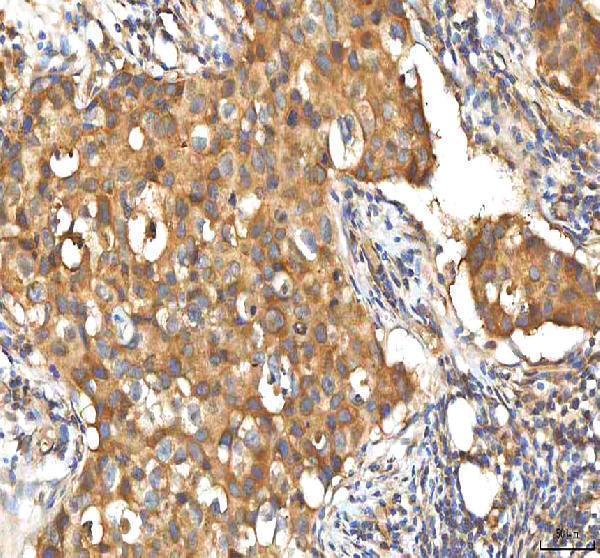
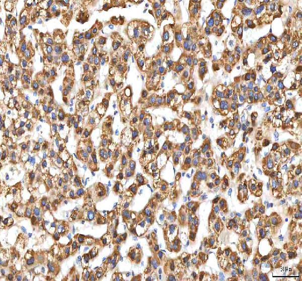
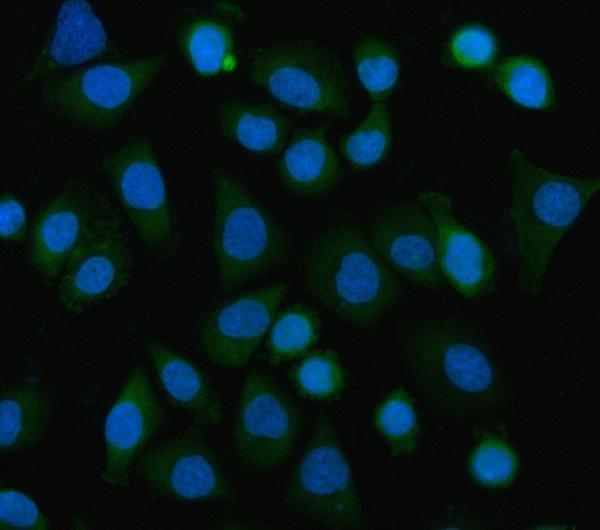


Comprehensive Human Protein Atlas IHC scoring per tissue (reliability: Approved — Medium consistency between antibody staining and RNA expression data.). Rows are taken directly from the HPA tissue chart — click any row's HPA link to view the source.
| Tissue | Cell type | Level | Evidence | Source |
|---|---|---|---|---|
| Adrenal gland | Glandular cells | High | Protein (IHC) | HPA → |
| Appendix | Glandular cells | High | Protein (IHC) | HPA → |
| Bronchus | Respiratory epithelial cells | High | Protein (IHC) | HPA → |
| Cerebellum | Purkinje cells | High | Protein (IHC) | HPA → |
| Cervix | Squamous epithelial cells | High | Protein (IHC) | HPA → |
| Tissue | Cell type | Level | Evidence | Source |
|---|---|---|---|---|
| None in HPA: EIF3B is detected in all 45 scored tissues. Use a no-primary (secondary-only) and an isotype control instead. | ||||
Troubleshoot EIF3B staining in paraffin sections by checking retrieval, cytoplasmic localisation, controls and scoring before interpreting changes in signal.
A04318-1 has real IHC data from human paraffin sections and IF data from SiHa cells (catalog image captions); its listed reactivity covers human, mouse and rat (catalog).
A04318-1 will render with an IHC figure from a human breast cancer paraffin section; its separate IF caption documents SiHa cells (catalog image captions). M04318 will render without an IHC or IF figure; ICC/IF is listed among its applications, with human, mouse and rat reactivity (catalog).
Which to pick: Choose A04318-1 for tissue IHC: its own caption documents staining in a human breast cancer paraffin section after EDTA pH 8.0 retrieval at 2 μg/ml; the fixative is unreported (A04318-1 IHC caption). For IF/ICC, A04318-1 has a SiHa cell IF image at 5 μg/ml, while M04318 is a rabbit monoclonal with ICC/IF listed but no supplied IF image (catalog). Both list human, mouse and rat reactivity, but the supplied tissue IHC images document human samples only (catalog; A04318-1 IHC captions).