This website uses cookies to ensure you get the best experience on our website.


- Table of Contents
Plan EIF3C chromogenic IHC in paraffin sections with a catalog antibody range of 2–5 μg/ml (datasheet A06470-1). Assess cytoplasmic staining against the tissue pattern and consider HPA’s multi-gene targeting caution when scoring (HPA tissue IHC).

Expected localisation, validated protocols, controls and antibodies — the at-a-glance facts below, then the full design guide.
| Expected localisation | General cytoplasmic staining (HPA tissue IHC) | |
| Staining pattern | High cytoplasmic signal in glandular cells (HPA tissue IHC) | |
| Antigen retrieval | EDTA pH 8.0 HIER, heat-mediated (datasheet A06470-1) | |
| Positive control | Appendix+4 more · see all | |
| Negative control | None in HPA (detected in all 45 tissues); use no-primary + isotype controls |
| Fixation | Keep fixation consistent across sections (standard IHC practice; not target-specific) | |
| Caveat | Multi-gene protein targeting may confound scoring (HPA tissue IHC) | |
| Regulation | Low tissue specificity (HPA tissue RNA) | |
| Isoform / epitope | 2 isoforms; epitope coverage is unreported (UniProt) |
The catalog antibody’s IHC-P protocol (datasheet A06470-1) is accompanied by four published EIF3C tissue IHC protocols (PMC6331022; PMC7144925; PMC6389005; PMC9492341).
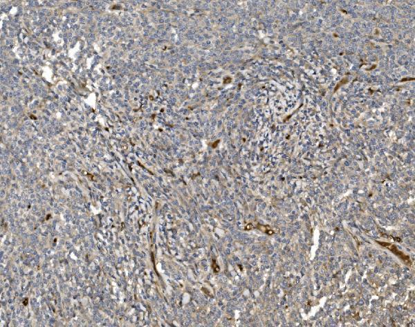
| Sample | Paraffin-embedded human lymphoma tissue; fixative not specified (datasheet A06470-1) |
| Fixation | Image fixative and duration unreported (datasheet A06470-1); verify before use. |
| Sectioning | 4–5 µm sections on charged slides (standard) |
| Deparaffinisation | Xylene, graded ethanol series to water (standard) |
| Antigen retrieval | Heat retrieval: EDTA pH 8.0 (datasheet A06470-1); 20 min, 95–100 °C (standard) |
| Peroxidase block | 3% H2O2, 10 min, room temperature (standard) |
| Blocking | 10% goat serum (datasheet A06470-1) |
| Primary antibody | Rabbit anti-EIF3C, 2-5 μg/ml (datasheet A06470-1) |
| Primary incubation | Overnight at 4 °C (datasheet A06470-1) |
| Detection | Streptavidin-biotin complex (SABC), DAB chromogen (datasheet A06470-1) |
| Counterstain | Hematoxylin, blue, dehydrate and mount (standard) |
| Expected result | EIF3C-positive staining in glandular cells of appendix (HPA tissue IHC: High). HPA tissue profile: General cytoplasmic expression. No signal in the no-primary control. |
EIF3C should show predominantly cytoplasmic staining in paraffin sections (UniProt Q99613: cytoplasm; HPA tissue IHC: general cytoplasmic expression). Expect strong signal in appendix, breast, colon, and duodenal glandular cells, among other HPA high-staining cells (HPA tissue IHC). HPA rates its tissue pattern Supported, with medium agreement between staining and RNA data, and cautions that the antibodies target protein from more than one gene (HPA tissue IHC). EIF3C has no transmembrane segment (UniProt Q99613: topology).
| Clear cytoplasmic chromogen in appendix or colon glandular cells, with nuclei mainly defined by counterstain. | This matches a documented high-staining cell type and the expected compartment (HPA tissue IHC: High in glandular cells; UniProt Q99613: cytoplasm). Assess the pattern in identifiable cells rather than treating overall section darkness as a positive result. |
| Predominantly nuclear, membrane-rim, or extracellular staining with little cytoplasmic signal. | That distribution conflicts with the reported cytoplasmic location (UniProt Q99613; HPA subcellular: cytosol). Review morphology, counterstain, and detection controls before interpreting it as EIF3C; unusual compartment staining alone does not establish a new location. |
| Strong staining confined to cells expected to stain low, while adjacent expected-positive cells show little signal. | Cholangiocytes, cardiomyocytes, myocytes, fibroblasts, and adipocytes are listed as Low, not absent (HPA tissue IHC). Consider off-target binding or endogenous detection activity, but first confirm cell identity and compare staining within the same run. |
| Diffuse chromogen across cells and empty tissue spaces, without a defined cytoplasmic pattern. | This is less persuasive than cell-associated cytoplasmic staining (UniProt Q99613; HPA tissue IHC). As a general IHC assessment, compare a reagent control and inspect washing, blocking, and detection background before assigning cell-level positivity. |
| No detectable cytoplasmic signal in a well-preserved appendix or colon glandular-cell section. | Those cells are reported High (HPA tissue IHC), so an all-negative run warrants a technical check. Absence on one section does not establish biological loss; inspect the antibody, retrieval and detection steps, and an independently stained positive section. |
| Cell and tissue choice | Appendix, breast, colon, and duodenal glandular cells offer documented High staining; liver cholangiocytes and muscle cells are Low comparators, not true negatives (HPA tissue IHC). |
| Compartment and protein topology | EIF3C is cytoplasmic, with no transmembrane segment or signal peptide (UniProt Q99613); HPA ICC-IF supports cytosol. Read cytoplasmic signal in the cell context, without expecting a membrane outline. |
| Antibody evidence and specificity | The listed antibodies have Supported IHC status (HPA antibodies), while tissue reliability is Supported with medium staining–RNA agreement and a multi-gene-target caution (HPA tissue IHC). Validation supports pattern comparison, not attribution of every stained cell to EIF3C alone. |
| Isoforms and epitope | Two isoforms are listed (UniProt Q99613), but the payload gives no antibody epitope or isoform-specific staining evidence. Do not infer which isoform produced a positive IHC signal. |
| IF/ICC Q&A: what pattern should appear? | Expect cytosolic fluorescence (HPA subcellular: Cytosol, supported; UniProt Q99613: cytoplasm). HPA lists ICC-IF images in A-431, U-251MG, U2OS, Hep-G2, and MCF-7; this is a localisation reference, not an IF protocol. |
| Situation | Likely cause | Next action |
|---|---|---|
| Expected-positive glandular cells are blank. | The run may have weak antibody or detection performance; appendix and colon glandular cells are reported High (HPA tissue IHC). | Check section integrity, antibody preparation, retrieval execution, detection reagents, and a known-positive section. Adjust conditions only after locating the failed step; no EIF3C-specific retrieval setting is supplied. |
| Color is strongest in nuclei or along cell borders. | The compartment conflicts with cytoplasmic EIF3C (UniProt Q99613; HPA subcellular: cytosol); counterstain or nonspecific signal may confuse localisation. | Inspect higher-magnification cell boundaries and a detection control. Score only convincing cell-associated cytoplasmic signal; document persistent unexpected staining separately. |
| Low-staining cell types look as dark as expected-positive cells. | HPA lists cholangiocytes, cardiomyocytes, myocytes, fibroblasts, and adipocytes as Low rather than negative (HPA tissue IHC); unexpected intensity also raises a specificity question. | Verify cell identity and compare the positive and low-staining areas on the same slide. Check reagent controls and, if available, an independently validated antibody before assigning the discrepancy to biology. |
| Background obscures cytoplasmic boundaries. | Diffuse color can arise from general chromogenic IHC background; a defined cytoplasmic pattern is expected (HPA tissue IHC; UniProt Q99613). | Compare a reagent control, then review blocking, washing, and detection conditions using standard IHC practice. Reassess signal in intact, identifiable cells after background is controlled. |
| Two IHC antibodies give different distributions. | HPA lists Supported IHC antibodies, but also reports only medium staining–RNA agreement and cautions that antibodies target protein from more than one gene (HPA antibodies; HPA tissue IHC). | Compare each antibody against the same HPA high-staining cells and expected cytoplasmic location (HPA tissue IHC; UniProt Q99613). Report the disagreement rather than combining the signals into one score. |
| A negative section is being treated as proof that EIF3C is absent. | HPA reports low tissue RNA specificity and general cytoplasmic protein expression; its Low cell categories are not negative controls (HPA tissue IHC). | Confirm that expected-positive cells stained in the same run, review morphology and controls, and describe the result as undetected under the tested conditions unless independent evidence supports absence. |


Comprehensive Human Protein Atlas IHC scoring per tissue (reliability: Supported — Medium consistency between antibody staining and RNA expression data. Caution, targets protein from more than one gene.). Rows are taken directly from the HPA tissue chart — click any row's HPA link to view the source.
| Tissue | Cell type | Level | Evidence | Source |
|---|---|---|---|---|
| Appendix | Glandular cells | High | Protein (IHC) | HPA → |
| Breast | Glandular cells | High | Protein (IHC) | HPA → |
| Bronchus | Respiratory epithelial cells | High | Protein (IHC) | HPA → |
| Cerebellum | Purkinje cells | High | Protein (IHC) | HPA → |
| Cerebral cortex | Glial cells | High | Protein (IHC) | HPA → |
| Tissue | Cell type | Level | Evidence | Source |
|---|---|---|---|---|
| None in HPA: EIF3C is detected in all 45 scored tissues. Use a no-primary (secondary-only) and an isotype control instead. | ||||
Troubleshoot EIF3C staining in paraffin sections by checking cytoplasmic localisation, retrieval conditions and antibody specificity (UniProt Q99613; HPA tissue IHC).
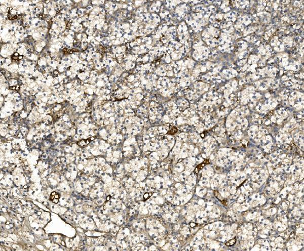
The IHC-validated antibody has images from human lymphoma and renal carcinoma paraffin sections (catalog image captions). Listed reactivity covers human, mouse and rat (datasheet: reactivity).
A06470-1 is listed for IHC, with images of human lymphoma and renal carcinoma paraffin sections (datasheet: applications; catalog image captions). Its listed reactivity is human, mouse and rat, while the supplied IHC images show human tissue (datasheet: reactivity; catalog image captions).
Which to pick: Choose A06470-1 for paraffin-section IHC: its images document EDTA retrieval at pH 8.0 and 2 μg/ml primary antibody in human tissue (catalog image captions). No IF/ICC application or IF image is supplied for A06470-1, so an IF/ICC choice is unsupported (datasheet: applications; catalog: IF image alts). For cross-species work, A06470-1 lists human, mouse and rat reactivity, but its IHC images show human tissue only; the fixative is unreported (datasheet: reactivity; catalog image captions).