EIF3F / Eukaryotic translation initiation factor 3 subunit F · IHC design guide

Design Immunohistochemistry for EIF3F

Plan EIF3F staining in paraffin sections with the IHC-validated antibody at 1:100–1:300 (datasheet: IHC). Compare the cytoplasmic pattern with HPA tissue IHC, using kidney tubules as a high-staining reference and adipocytes as a non-detected reference (HPA tissue IHC).

Evidence assembled Oct 2026 · For research use; verify linked source records and product datasheet before use
Immunohistochemistry protocol sheet for EIF3F (IHC for EIF3F): expected localisation Cytoplasm (HPA tissue IHC), antibody A30682, validated IHC image, and IHC protocol steps
Printable EIF3F IHC protocol sheet — expected localisation Cytoplasm (HPA tissue IHC), antibody A30682, controls and protocol steps. Open the full EIF3F IHC guide →

EIF3F Immunohistochemistry Experimental Design Guide

Expected localisation, validated protocols, controls and antibodies — the at-a-glance facts below, then the full design guide.

Must know before staining
Expected localisation Cytoplasm (HPA tissue IHC)
Staining pattern Cytoplasmic staining in most tissues (HPA tissue IHC)
Antigen retrieval Citrate pH 6.0 HIER, 95–98 °C, 20 min (rule: cytoplasmic / membrane antigen)
Positive control ⓘ Adrenal gland+4 more · see all
Negative control ⓘ Adipose tissue+1 more · see all
Important caveats
Reasons your staining may differ from the expected pattern.
Fixation Keep fixation consistent across sections (standard IHC practice; not target-specific)
Caveat Medium staining–RNA consistency; verification pending (HPA tissue IHC)
Regulation Low tissue specificity (HPA tissue RNA)
Isoform / epitope No annotated isoforms; chain spans residues 2–357 (UniProt)
Section 1

Recommended EIF3F IHC & IF Protocols

The catalog antibody protocol is accompanied by four published EIF3F IHC methods for gastric, skin, prostate, and mouse xenograft tissue (PMC4046624; PMC2635928; PMC7210466; PMC13459262).

Recommended immunohistochemistry (IHC-P) protocol parameters
SampleParaffin-embedded human brain tissue; fixative not specified (datasheet A30682)
FixationImage fixative and duration unreported (datasheet A30682); verify before use.
Sectioning4–5 µm sections on charged slides (standard)
DeparaffinisationXylene, graded ethanol series to water (standard)
Antigen retrievalHeat-induced epitope retrieval in citrate buffer, pH 6.0, 20 min at 95–98 °C (standard rule: cytoplasmic / membrane antigen)
Peroxidase block3% H2O2, 10 min, room temperature (standard)
Blocking10% normal serum of the secondary host, 30 min, room temperature (standard)
Primary antibodyRabbit anti-EIF3F, 1:100 - 1:300 (datasheet A30682)
Primary incubationOvernight at 4 °C (standard)
DetectionHRP-polymer secondary, DAB chromogen 5–10 min (standard)
CounterstainHematoxylin, blue, dehydrate and mount (standard)
Expected resultEIF3F-positive staining in glandular cells of adrenal gland (HPA tissue IHC: High). HPA tissue profile: Cytoplasmic expression in most tissues. No signal in the no-primary control.
💡Decision noteStart with citrate pH 6.0 retrieval at 95–98 °C for 20 min (page antigen retrieval); the prostate method used 25 min (PMC7210466).
Section 2

What Is the Expected EIF3F Staining Pattern?

EIF3F is a cytoplasmic protein without a transmembrane segment (UniProt O00303: location and topology). Expect cytoplasmic staining across many tissues, including strong staining in hepatocytes, kidney tubule cells and several glandular or squamous epithelial populations (HPA tissue IHC: profile and cell levels). Treat this as a reference pattern with limits: HPA rates the tissue staining Approved, with medium consistency against RNA data and external verification pending (HPA tissue IHC: reliability).

What am I looking at on my slide?
Cytoplasmic chromogen is strong in hepatocytes or kidney tubule cells, with cell boundaries and nuclei still interpretable.This matches the reported compartment and two High cell populations (UniProt O00303: cytoplasm; HPA tissue IHC: liver and kidney). Compare like cell types within the section; a whole-tissue average can hide which cells carry the signal (general IHC practice).
Staining is predominantly nuclear, confined to cell surfaces, or concentrated in extracellular material.These compartments do not match the reported cytoplasmic location or lack of a transmembrane segment (UniProt O00303: location and topology). Check morphology and controls before interpreting the signal as EIF3F; an unexpected compartment may reflect nonspecific staining or a detection artefact (general IHC practice).
Adipocytes or smooth muscle cells stain strongly while expected positive cells on the same run show little signal.HPA reports EIF3F as Not detected in those cell populations (HPA tissue IHC: adipose tissue and smooth muscle). Consider antibody cross-reactivity or endogenous detection activity; verify the cell identity and compare an appropriate reagent or detection control (general IHC practice).
Color spreads across tissue, stroma and empty areas without resolving cytoplasm in individual cells.That distribution cannot be scored as the reported cytoplasmic pattern (HPA tissue IHC: profile). Diffuse deposition can arise from nonspecific reagent binding, inadequate washing or chromogen background; assess a no-primary control and the location of deposit before assigning positivity (general IHC practice).
A liver or kidney section lacks interpretable cytoplasmic staining in hepatocytes or tubule cells.Both are High reference populations (HPA tissue IHC: liver and kidney). A blank result raises a run or reagent question, but one negative section cannot by itself establish biological absence; review tissue preservation, retrieval, primary-antibody conditions and detection controls (general IHC practice).
💡Expected EIF3F appearanceA convincing positive result shows cell-resolved cytoplasmic chromogen, potentially High in hepatocytes or kidney tubule cells (UniProt O00303: cytoplasm; HPA tissue IHC: liver and kidney); isolated nuclear, surface or diffuse extracellular color is suspect (general IHC practice).
How each factor affects the staining
Tissue and cell choiceHPA reports High staining in adrenal and breast glandular cells, cervix and esophagus squamous cells, hepatocytes and kidney tubules; adipocytes and smooth muscle cells are Not detected (HPA tissue IHC: cell levels). Score the named population, not the section as a whole (general IHC practice).
Strength of reference evidenceThe tissue profile is Approved with medium antibody-to-RNA consistency, pending external verification (HPA tissue IHC: reliability). HPA lists one IHC Approved antibody, HPA049250; that status supports use as a reference but does not make every unexpected stain target-specific (HPA antibodies: IHC status; general IHC practice).
Target location and processingEIF3F is cytoplasmic, has no transmembrane segment or signal peptide, and has a recorded chain spanning residues 2–357 (UniProt O00303: location, topology and processing). These entries support a cytoplasmic expectation; they do not establish an extracellular or membrane pattern.
IF/ICC expectation?Use cytoplasm as the location hypothesis from UniProt, but do not claim an HPA image-confirmed IF pattern: HPA supplies no main subcellular location or ICC-IF image cell line here (UniProt O00303: location; HPA subcellular: unavailable fields). IF/ICC requires its own guide.
Fixation and retrieval evidenceNo target-specific fixation sensitivity or retrieval condition is supplied by UniProt or HPA (supplied UniProt O00303 and HPA records). Any retrieval adjustment is a general IHC workflow check, not evidence that fixation changes EIF3F staining (general IHC practice).
Why is my staining missing, weak or wrong?
SituationLikely causeNext action
No staining in hepatocytes or kidney tubule cells.The result conflicts with HPA High reference populations; the failed step is not identified by that comparison alone (HPA tissue IHC: liver and kidney).Check section quality, run controls, the catalog antibody's IHC-P directions, retrieval conditions and detection reagents before calling the sample negative (general IHC practice).
Only nuclei, cell borders or extracellular spaces are colored.The compartment differs from the reported cytoplasm (UniProt O00303: location and topology). Nonspecific staining or detection artefact is possible (general IHC practice).Recheck cell morphology and a no-primary control; score EIF3F only where staining resolves in cytoplasm (general IHC practice).
Adipocytes or smooth muscle cells appear strongly positive.Those populations are Not detected in the HPA tissue survey (HPA tissue IHC: adipose tissue and smooth muscle); cross-reactivity or endogenous detection activity is possible (general IHC practice).Confirm cell identity, inspect a no-primary or detection-only control, and compare a known High population in the run (HPA tissue IHC: cell levels; general IHC practice).
Uniform brown haze obscures cell boundaries.A diffuse deposit is not the cell-resolved cytoplasmic profile (HPA tissue IHC: profile); nonspecific binding, washing or chromogen background may contribute (general IHC practice).Use the no-primary control to locate background, then review blocking, washes and detection exposure under the established IHC workflow (general IHC practice).
Low signal in skeletal-muscle myocytes or caudate neuronal cells.HPA reports Low staining for these named populations (HPA tissue IHC: skeletal muscle and caudate). Weak signal there alone is a poor measure of assay performance.Judge run performance using a High reference population, such as hepatocytes or kidney tubule cells, before changing staining conditions (HPA tissue IHC: liver and kidney; general IHC practice).
An IF/ICC image is being used to settle an unexpected IHC result.The supplied HPA subcellular record has no main location or ICC-IF image cell line (HPA subcellular: unavailable fields), so it cannot independently validate that image's pattern.Interpret the paraffin-section result against HPA tissue IHC and UniProt cytoplasmic location; address IF/ICC validation in its separate guide (HPA tissue IHC: profile; UniProt O00303: location).

Sample controls for EIF3F IHC & IF

🧪Run breast first: its glandular cells should stain (HPA: High in breast glandular cells). Use adipose tissue as the negative tissue, and expect adipocytes within the breast section, where present, to lack staining (HPA: adipocytes Not detected).
Positive control tissue: Adrenal gland (Glandular cells, HPA High)
Negative control tissue: Adipose tissue (HPA Not detected)
ICC-IF cell lines (HPA subcellular resource): HPA carries no ICC-IF cell line for EIF3F; derive a cell-line control from the positive tissue's cell type (Glandular cells) and confirm it by RNA or western blot first.
Technical controls: Include no-primary (secondary-only) and host-species-matched isotype controls for a monoclonal primary, or matched nonimmune IgG for a polyclonal primary; use EIF3F-knockout material if available or a peptide-blocked primary as a biological specificity control (A30682 tissue-IHC caption: peptide block). Quench endogenous peroxidase before chromogenic detection (standard IHC practice).
⚠️Feasibility: The selected A30682 paraffin-section caption does not report a fixative, and the supplied evidence reports no target-specific fixation window or antigen-retrieval dependency (A30682 tissue-IHC caption). Test retrieval on paired sections; the supplied evidence does not establish that frozen sections or IF are easier than paraffin IHC. Breast adipose may autofluoresce in IF, so assess it with a no-primary control (standard IF practice).

HPA tissue IHC evidence for EIF3F

Comprehensive Human Protein Atlas IHC scoring per tissue (reliability: Approved — Medium consistency between antibody staining and RNA expression data. Pending external verification.). Rows are taken directly from the HPA tissue chart — click any row's HPA link to view the source.

Positive expression · recommended positive controls

TissueCell typeLevelEvidenceSource
Adrenal gland Glandular cells High Protein (IHC) HPA →
Breast Glandular cells High Protein (IHC) HPA →
Cervix Squamous epithelial cells High Protein (IHC) HPA →
Endometrium Glandular cells Medium Protein (IHC) HPA →
Esophagus Squamous epithelial cells High Protein (IHC) HPA →

Undetected expression · recommended negative controls

TissueCell typeLevelEvidenceSource
Adipose tissue Adipocytes Not detected Protein (IHC) HPA →
Smooth muscle Smooth muscle cells Not detected Protein (IHC) HPA →
Section 3

Advanced EIF3F IHC Tips

Troubleshoot EIF3F chromogenic IHC by checking cytoplasmic staining, section processing and controls before comparing staining intensity across samples (UniProt O00303 localisation; standard IHC practice).

What retrieval should I try first when EIF3F staining is weak?
Start paraffin-section retrieval in citrate buffer at pH 6.0, 95–98 °C for 20 min (page retrieval specification). Let slides cool in buffer, then process test and control sections with the same antibody incubation and detection steps (standard IHC practice). Assess signal in intact cell cytoplasm, the annotated compartment for EIF3F (UniProt O00303 localisation). If staining remains weak, try a higher-pH heat-retrieval buffer on adjacent sections as a fallback while keeping the other conditions fixed (standard IHC optimisation). Compare signal against background and an omitted-primary control before adopting the alternative (standard IHC practice).
How can I assess whether fixation is affecting EIF3F IHC?
Target-specific fixation sensitivity is unknown because the supplied EIF3F evidence gives no tested fixation conditions (supplied evidence). The catalog image shows paraffin-embedded human brain tissue, but its caption does not state the fixative (A30682 caption). For a controlled comparison, record delay to fixation, fixative and duration for each specimen; 10% neutral buffered formalin is a conventional IHC processing reference (standard IHC practice). Process comparison sections together and apply the specified citrate retrieval at pH 6.0 for 20 min (page retrieval specification; standard IHC practice). Evaluate morphology alongside cytoplasmic signal, without assigning a target-specific effect to fixation alone (UniProt O00303 localisation; standard IHC practice).
Where should convincing EIF3F staining appear in paraffin sections?
Expect predominantly cytoplasmic staining: EIF3F is annotated in the cytoplasm, and tissue IHC reports cytoplasmic expression in most tissues (UniProt O00303 localisation; HPA tissue IHC profile). EIF3F has no annotated transmembrane segment, so a sharp membrane-only pattern needs independent validation (UniProt O00303 topology; standard IHC interpretation). Inspect cell types separately: HPA reports high staining in kidney tubule cells and liver hepatocytes, but low staining in caudate neuronal cells (HPA tissue IHC). Compare intact cells in equivalent regions with an omitted-primary control, using the same counterstain and imaging settings (standard IHC practice). Treat isolated nuclear staining cautiously until corroborated by an independent method (UniProt O00303 localisation; standard IHC practice).
Could EIF3F epitopes or modifications explain uneven staining?
The supplied UniProt record lists 0 isoforms and an MPN domain at residues 92–222 (UniProt O00303 isoforms/domains). It also lists acetylation and phosphorylation sites, including residues 2, 238 and 258 (UniProt O00303 modified residues). Those annotations do not establish that any modification alters binding by the catalog antibody; check its documented immunogen before drawing an epitope conclusion (UniProt O00303 modified residues; standard antibody-validation practice). The A30682 tissue image includes a synthesized-peptide blocking comparison, which supports investigating peptide-sensitive staining in that image (A30682 caption). Compare adjacent sections with identical retrieval and detection, then seek an independent antibody or orthogonal assay if staining differs unexpectedly (standard IHC validation practice).
How should I adapt EIF3F localisation checks to multiplex IF?
Use IF as a separate validation workflow and pair EIF3F with a marker identifying the cell population under study (standard IF practice). HPA reports high EIF3F tissue staining in kidney tubule cells and hepatocytes, which can guide cell-type comparisons without establishing an IF protocol (HPA tissue IHC). Because EIF3F is cytoplasmic and has no transmembrane segment, permeabilise fixed cells or sections to expose intracellular epitopes (UniProt O00303 localisation/topology; standard IF practice). A brief 0.1% detergent treatment for 5–10 min is a conventional starting trial, with morphology checked afterward (standard IF practice). Choose fluorophores after measuring tissue autofluorescence, and include single-colour and omitted-primary controls for channel separation and background assessment (standard IF practice).
How do I reduce diffuse EIF3F chromogenic background?
First distinguish diffuse cytoplasmic staining from background: EIF3F is cytoplasmic, and HPA reports expression across most tissues (UniProt O00303 localisation; HPA tissue IHC profile). Include omitted-primary and detection-only sections to identify staining from secondary reagents or endogenous enzyme activity (standard IHC practice). For peroxidase detection, apply a peroxidase block before DAB development and limit development to a consistent endpoint across slides (standard chromogenic IHC practice). Optimise blocking, washes and antibody concentration with matched sections, changing 1 variable at a time (standard IHC optimisation). Compare intact cells with adipocytes or smooth muscle cells, which HPA reports as not detected, while allowing for tissue-specific background (HPA tissue IHC; standard IHC practice).
What is a defensible way to quantify EIF3F IHC? ⚠ ANSWER MARKED FOR VERIFICATION
Define tissue regions and cell classes before scoring, because HPA reports different EIF3F staining levels among cell populations (HPA tissue IHC; standard IHC scoring practice). Within intact cells, record the percentage positive and cytoplasmic intensity, then calculate an H-score from 0–300 if intensity categories are reproducible (standard IHC scoring practice; UniProt O00303 localisation). Alternatively, report positive cells per mm² when cell counts and sampled area are available (standard image-analysis practice). Normalise to the number of evaluable cells or viable tissue area, and hold retrieval, detection and image thresholds constant across specimens (standard IHC practice). Review representative fields manually and report interobserver or segmentation consistency before interpreting small differences (standard IHC scoring practice).
How can I distinguish true EIF3F staining from artefact?
Prioritise cytoplasmic staining in intact cells, consistent with EIF3F localisation and the HPA tissue pattern (UniProt O00303 localisation; HPA tissue IHC profile). HPA reports high staining in kidney tubule cells and hepatocytes but no detection in adipocytes or smooth muscle cells, providing useful comparison patterns rather than absolute controls (HPA tissue IHC). Treat membrane-only or nuclear-only signal, tissue-edge intensification and staining restricted to necrotic areas as prompts for further controls (UniProt O00303 topology/localisation; standard IHC interpretation). Check omitted-primary and peroxidase-block controls when DAB signal could reflect endogenous enzyme activity (standard chromogenic IHC practice). Weigh any conclusion against HPA's Approved rating, which carries medium RNA–staining consistency and awaits external verification (HPA tissue IHC reliability).
Boster reagents

Best EIF3F / Eukaryotic translation initiation factor 3 subunit F IHC Antibodies

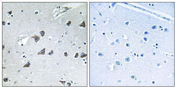
A30682 has an IHC image of paraffin-embedded human brain tissue (catalog IHC image caption). IF/ICC and human, mouse, and rat reactivity are listed, but no IF image is supplied (catalog applications/reactivity/IF image alts).

Real IHC data Immunohistochemistry analysis of paraffin-embedded human brain tissue, using EIF3F Antibody. The picture on the right is blocked with the synthesized peptide.
Anti-EIF3F/Eif3 Epsilon Antibody
Cat # A30682

A30682 will render with an IHC image of paraffin-embedded human brain tissue and a peptide-blocked comparison (catalog IHC image caption). A30682 is also listed for IF/ICC and for human, mouse, and rat, without an IF image in the payload (catalog applications/reactivity/IF image alts).

Which to pick: Choose A30682 for paraffin-section tissue IHC: its own image caption shows human brain tissue, and its listed IHC dilution is 1:100–1:300 (catalog IHC image caption; catalog IHC dilution). For IF/ICC, A30682 lists both applications and an IF dilution of 1:50, although no IF image is supplied (catalog applications/IF dilution/IF image alts). A30682 is the listed cross-species choice for human, mouse, and rat and is rabbit polyclonal; the fixative used for its IHC image is unreported (catalog reactivity/host/dilution_raw/IHC image caption).

Each figure is that product's own IHC / IF validation image from its datasheet.

References

  1. UniProt Consortium. UniProt entry O00303 (EIF3F_HUMAN, Eukaryotic translation initiation factor 3 subunit F).
  2. Human Protein Atlas. EIF3F tissue IHC expression (reliability: Approved).
  3. Human Protein Atlas. EIF3F subcellular location (ICC-IF): Highest expression in HeLa: 52.4 nTPM.
  4. Human Protein Atlas. EIF3F antibody validation summary (1 antibodies).
  5. Decreased expression of eukaryotic initiation factor 3f is an adverse prognostic factor for stage I-III gastric cancer. World journal of surgical oncology 2014 — PMC4046624.
  6. Loss of the eukaryotic initiation factor 3f in melanoma. Molecular carcinogenesis 2008 — PMC2635928.
  7. Targeting eIF3f Suppresses the Growth of Prostate Cancer Cells by Inhibiting Akt Signaling. OncoTargets and therapy 2020 — PMC7210466.
  8. VAV2 drives glycolytic reprogramming in esophageal squamous cell carcinoma via EIF3F-mediated MTA1 deubiquitination. Cancer & metabolism 2026 — PMC13459262.
  9. PubMed PMID:9341143 — UniProt-cited evidence.
  10. PubMed PMID:14702039 — UniProt-cited evidence.
  11. PubMed PMID:15489334 — UniProt-cited evidence.