EIF3H / Eukaryotic translation initiation factor 3 subunit H · IHC design guide

Design Immunohistochemistry for EIF3H

Plan EIF3H chromogenic IHC in paraffin sections around the expected cytoplasmic pattern (HPA tissue IHC). This guide covers fixation, controls and interpretation using the catalog antibody at 2–5 μg/ml (datasheet A05656-1).

Evidence assembled Oct 2026 · For research use; verify linked source records and product datasheet before use
Immunohistochemistry protocol sheet for EIF3H (IHC for EIF3H): expected localisation General cytoplasmic tissue staining (HPA tissue IHC), antibody A05656-1, validated IHC image, and IHC protocol steps
Printable EIF3H IHC protocol sheet — expected localisation General cytoplasmic tissue staining (HPA tissue IHC), antibody A05656-1, controls and protocol steps. Open the full EIF3H IHC guide →

EIF3H Immunohistochemistry Experimental Design Guide

Expected localisation, validated protocols, controls and antibodies — the at-a-glance facts below, then the full design guide.

Must know before staining
Expected localisation General cytoplasmic tissue staining (HPA tissue IHC)
Staining pattern Cytoplasmic signal in glandular and hematopoietic cells (HPA tissue IHC)
Antigen retrieval EDTA pH 8.0 HIER, heat-mediated (datasheet A05656-1)
Positive control ⓘ Appendix+4 more · see all
Negative control ⓘ None in HPA (detected in all 45 tissues); use no-primary + isotype controls
Important caveats
Reasons your staining may differ from the expected pattern.
Fixation Keep paraffin-section fixation consistent (standard IHC practice; not target-specific); Matched tissue-IHC evidence does not establish the fixation claim. Validate the specimen-specific method before use. (selected-SKU IHC image A05656-1)
Caveat Endogenous peroxidase can confound marrow DAB staining (HPA tissue IHC; standard IHC practice)
Regulation No specific expression regulator annotated (UniProt)
Isoform / epitope No alternate isoforms annotated; one 1–352 chain (UniProt)
Section 1

Recommended EIF3H IHC & IF Protocols

Compare the catalog antibody’s IHC-P protocol with published EIF3H staining in ESCC, endometrial tumor TMAs, and gastric xenografts (datasheet A05656-1; PMC7457539; PMC6941158; PMC8606975).

Recommended immunohistochemistry (IHC-P) protocol parameters
SampleParaffin-embedded human colon cancer tissue; fixative not specified (datasheet A05656-1)
FixationImage fixative and duration unreported (datasheet A05656-1); verify before use.
Sectioning4–5 µm sections on charged slides (standard)
DeparaffinisationXylene, graded ethanol series to water (standard)
Antigen retrievalHeat retrieval: EDTA pH 8.0 (datasheet A05656-1); 20 min, 95–100 °C (standard)
Peroxidase block3% H2O2, 10 min, room temperature (standard)
Blocking10% goat serum (datasheet A05656-1)
Primary antibodyRabbit anti-EIF3H, 2-5 μg/ml (datasheet A05656-1)
Primary incubationOvernight at 4 °C (datasheet A05656-1)
DetectionHRP-conjugated secondary, DAB chromogen (datasheet A05656-1)
CounterstainHematoxylin, blue, dehydrate and mount (standard)
Expected resultEIF3H-positive staining in glandular cells of appendix (HPA tissue IHC: High). HPA tissue profile: General cytoplasmic expression. No signal in the no-primary control.
💡Decision noteStart with heat-mediated EDTA pH 8.0 retrieval for the catalog antibody (datasheet A05656-1). The endometrial TMA study reports 30 min HIER without specifying a buffer (PMC6941158).
Section 2

What Is the Expected EIF3H Staining Pattern?

EIF3H should appear predominantly in the cytoplasm of many cell types, consistent with its cytoplasmic assignment and lack of a transmembrane segment (UniProt O15372: subcellular location and topology). In paraffin sections, HPA reports general cytoplasmic staining, including high staining in selected glandular, epithelial, hematopoietic and neuronal cells (HPA: tissue IHC). HPA rates the tissue pattern Approved, pending external verification (HPA: tissue IHC reliability).

What am I looking at on my slide?
Clear cytoplasmic staining in appendix glandular cells, bone marrow hematopoietic cells, or esophageal squamous epithelial cells.This matches reported high staining in those cell populations (HPA: tissue IHC). Judge the signal within the named cells against nearby background; a positive tissue does not imply equally strong staining in every cell (general IHC practice).
Predominantly nuclear, membranous, or extracellular staining, with little cytoplasmic signal.Treat this as discordant with the expected location: UniProt assigns EIF3H to the cytoplasm, HPA describes general cytoplasmic tissue staining, and HPA ICC-IF places it in the cytosol (UniProt O15372: location; HPA: tissue IHC and subcellular). Review controls before interpreting the discordant signal as EIF3H.
Strong staining chiefly in cells outside the HPA-reported positive population within a selected tissue.Possible explanations include antibody cross-reactivity or endogenous detection activity (general IHC practice). Recheck cell identity and controls. HPA identifies high staining in specific populations, such as appendix glandular cells; it does not designate every other cell in that tissue negative (HPA: tissue IHC).
Uniform color across cells and tissue spaces, obscuring cytoplasmic boundaries.This pattern is less interpretable than cell-associated cytoplasmic staining (HPA: tissue IHC profile; general IHC practice). Assess background in a primary-omission control, then review blocking, washes, detection reagent and development time as general chromogenic IHC variables (general IHC practice).
No discernible cytoplasmic signal in a reported high-staining cell population.An absent signal in appendix glandular cells or bone marrow hematopoietic cells conflicts with the reported HPA pattern (HPA: High in those cells). First establish that tissue morphology and the detection control are satisfactory; absence alone does not show that EIF3H is biologically absent (general IHC practice).
💡Expected EIF3H appearanceCall the stain positive when the expected cells show distinct cytoplasmic chromogen above local background, with potentially high staining in HPA-listed populations (HPA: tissue IHC); dominant nuclear or diffuse tissue-wide color is discordant with the reported cytoplasmic pattern (UniProt O15372: location; HPA: tissue IHC).
How each factor affects the staining
Tissue and cell selectionHPA reports low RNA tissue specificity and general cytoplasmic protein expression, while naming high-staining cell populations in eight tissues (HPA: tissue IHC). Ovary stroma, cardiomyocytes and smooth muscle cells are listed as low, not negative; use them only as lower-signal comparators (HPA: tissue IHC).
Antibody evidenceHPA lists HPA023117 and HPA023553 as IHC Approved; its tissue profile is Approved, pending external verification (HPA: antibody validation; tissue IHC reliability). These labels support comparison with the reported pattern but do not independently establish specificity in the current specimen (general IHC practice).
Subcellular interpretationEIF3H has no transmembrane segment or annotated signal peptide, and UniProt assigns it to the cytoplasm (UniProt O15372: topology, processing and location). HPA independently reports cytosol in ICC-IF (HPA: approved subcellular location). These observations support a cytoplasmic call, not a claim about fixation sensitivity.
IF/ICC Q&A: What pattern should IF show?Expect cytosolic fluorescence: HPA reports an approved cytosol location and lists A-431, U-251MG and U2OS ICC-IF images (HPA: subcellular). HPA lists ICC approval for HPA023117; no ICC status is supplied for HPA023553 (HPA: antibodies). This is a location reference, not an IF protocol.
Antigen retrieval and target-specific sensitivityEvaluate retrieval conditions against tissue morphology, controls and signal quality as general paraffin IHC practice (general IHC practice). The supplied UniProt and HPA records give no EIF3H-specific fixation or retrieval response; they cannot predict which condition will improve staining.
Why is my staining missing, weak or wrong?
SituationLikely causeNext action
A reported high-staining population is blank.The cause is unresolved from a blank section alone; detection failure or tissue-processing variation is possible (general IHC practice).Check a detection control and morphology, confirm the expected cell population, then review the antibody dilution and retrieval settings used for this run (general IHC practice; HPA: High in appendix glandular or bone marrow hematopoietic cells).
Nuclei dominate the stain.The distribution disagrees with cytoplasmic tissue staining and cytosolic ICC-IF location (HPA: tissue IHC and subcellular). Its cause cannot be assigned from location alone.Inspect primary-omission and positive-tissue controls; score EIF3H only where a reproducible cell-associated cytoplasmic pattern is distinguishable (general IHC practice; HPA: tissue IHC profile).
Unexpected cells stain strongly.Cross-reactivity, endogenous detection activity, or mistaken cell identification are possibilities (general IHC practice); HPA reports cell-specific high staining rather than a universal cell-level threshold (HPA: tissue IHC).Confirm morphology, compare a reported high-staining population on the same run, and inspect appropriate detection controls before attributing the unexpected signal to EIF3H (general IHC practice; HPA: tissue IHC).
Diffuse chromogen masks cell boundaries.Background from blocking, washing or detection conditions may obscure a localized result (general IHC practice); the expected tissue profile is cytoplasmic (HPA: tissue IHC).Compare the primary-omission control, review blocking and washes, and assess whether development time is excessive for the run (general IHC practice). Interpret only signal separable from background.
A low-staining comparator appears faintly positive.Low is an HPA staining category, not an absence claim: ovarian stroma cells, cardiomyocytes and smooth muscle cells are listed as low (HPA: tissue IHC).Compare its cell-associated cytoplasmic signal with background and a reported high-staining population; do not use faint staining in a low-listed population alone to reject the run (general IHC practice; HPA: tissue IHC).

Sample controls for EIF3H IHC & IF

🧪Run appendix first and expect staining in glandular cells (HPA: High in appendix glandular cells). HPA detects EIF3H in all 45 scored tissues, so there is no supported negative tissue or established negative cell type within the appendix slide; use matched no-primary and isotype sections to assess background (HPA: detected in all 45 scored tissues).
Positive control tissue: Appendix (Glandular cells, HPA High)
Negative control tissue: None in HPA: EIF3H is detected in all 45 scored tissues. Use a no-primary (secondary-only) and an isotype control instead.
ICC-IF cell lines (HPA subcellular resource): HPA ICC-IF images show EIF3H in A-431, U-251MG, U2OS, with annotated localisation: Cytosol (approved) (HPA subcellular).
Technical controls: Include a no-primary, secondary-only section and a rabbit IgG isotype control matched to the primary antibody’s polyclonal or monoclonal format; use EIF3H knockout material or an antigen peptide block, if available, to test target specificity (selected IHC caption: rabbit anti-EIF3H). Quench endogenous peroxidase and check for residual DAB signal in blood-rich or inflammatory areas of appendix; block endogenous biotin if using biotin-based detection (standard IHC practice).
⚠️Feasibility: No target-specific fixation window or fixation effect is reported, and the selected A05656-1 paraffin-section caption does not state a fixative (selected IHC caption: fixative unreported). The caption uses heat-mediated EDTA retrieval at pH 8.0 and 2 μg/ml primary antibody, but it does not establish that retrieval is required (selected IHC caption). Frozen-section or IF ease cannot be ranked from these data; for IF, cytosolic signal is the expected localization, and appendix blood or inflammatory cells may add peroxidase background to chromogenic IHC (HPA: approved cytosol localization; standard IHC practice).

HPA tissue IHC evidence for EIF3H

Comprehensive Human Protein Atlas IHC scoring per tissue (reliability: Approved — Pending external verification.). Rows are taken directly from the HPA tissue chart — click any row's HPA link to view the source.

Positive expression · recommended positive controls

TissueCell typeLevelEvidenceSource
Appendix Glandular cells High Protein (IHC) HPA →
Bone marrow Hematopoietic cells High Protein (IHC) HPA →
Cerebellum Cells in molecular layer High Protein (IHC) HPA →
Cerebral cortex Neuronal cells High Protein (IHC) HPA →
Cervix Squamous epithelial cells High Protein (IHC) HPA →

Undetected expression · recommended negative controls

TissueCell typeLevelEvidenceSource
None in HPA: EIF3H is detected in all 45 scored tissues. Use a no-primary (secondary-only) and an isotype control instead.
Section 3

Advanced EIF3H IHC Tips

Troubleshoot EIF3H staining in paraffin section IHC by checking retrieval, cytoplasmic localisation, controls and cell specific scoring; IF/ICC considerations are summarised separately.

How should I retrieve EIF3H in paraffin sections when staining is weak?
Start with heat mediated retrieval in EDTA at pH 8.0 (datasheet A05656-1). The catalog antibody produced staining in a paraffin embedded human colon cancer section after that retrieval, followed by 2 µg/ml primary antibody overnight at 4°C (datasheet A05656-1). If signal remains weak, compare modest changes in heating time on adjacent sections while keeping the EDTA buffer and detection conditions constant (standard IHC practice). Include a known positive section and a no primary control so increased signal can be judged against tissue damage and background (standard IHC practice).
Could fixation explain weak or patchy EIF3H staining?
The selected colon cancer image identifies a paraffin embedded section but does not report its fixative (datasheet A05656-1). Target specific sensitivity to fixation is therefore unknown; neither the tissue staining profile nor EIF3H topology establishes it (HPA: tissue IHC; UniProt O15372: no transmembrane segment). Record fixative, fixation duration and processing for each specimen, then compare matched sections stained in the same run (standard IHC practice). If staining varies, first check section preservation and retrieval consistency before attributing the difference to EIF3H abundance (standard IHC practice). Use the same reference tissue across runs to reveal processing related drift (standard IHC practice).
Where should convincing EIF3H staining appear in tissue sections?
Expect predominantly cytoplasmic staining: EIF3H is assigned to the cytoplasm, and the tissue IHC profile describes general cytoplasmic expression (UniProt O15372: cytoplasm; HPA: tissue IHC profile). The approved subcellular location is cytosol in ICC/IF images (HPA: subcellular). Inspect cellular boundaries and compare the cytoplasmic signal with a no primary control before scoring densely packed or poorly preserved regions (standard IHC practice). Strong isolated nuclear, membranous or extracellular colour should prompt review of counterstain, pigment and nonspecific detection before being called EIF3H (UniProt O15372: cytoplasm and no transmembrane segment; standard IHC practice).
How do epitope location and modification affect interpretation of EIF3H staining?
The record lists one 352 aa chain and 0 annotated isoforms, so an isoform specific staining claim is unsupported here (UniProt O15372: processing and isoforms). EIF3H has an MPN domain at residues 39–173 and reported phosphorylation at serines 3 and 183 (UniProt O15372: domains and modified residues). The catalog epitope is not specified in the supplied evidence, so these annotations cannot predict whether retrieval exposes or masks antibody binding (datasheet A05656-1: epitope unspecified). Compare retrieval conditions and an independently validated reagent on adjacent sections if epitope accessibility is in doubt (standard IHC practice).
How can I assess EIF3H by multiplex IF alongside this IHC guide?
EIF3H is cytosolic, so an IF/ICC workflow should allow antibody access to the cytosol with a mild permeabilisation step after fixation (HPA: subcellular; UniProt O15372: no transmembrane segment; standard IF practice). Multiplex with a cell identity marker suited to the specimen, such as an epithelial marker when examining glandular cells, and check each channel alone before combining them (HPA: high staining in appendix glandular cells; standard IF practice). Choose a fluorophore in a channel with low tissue autofluorescence, often a far red channel, and include unstained and single label controls (standard IF practice). Fixation performance for this antibody in IF/ICC is not supplied, so optimise it within that assay (supplied evidence: no antibody specific IF/ICC fixation data).
How can I reduce diffuse DAB background without losing EIF3H signal?
The selected IHC image used 10% goat serum blocking, 2 µg/ml rabbit primary overnight at 4°C, and peroxidase based DAB detection (datasheet A05656-1). If background is diffuse, compare a no primary section and titrate primary concentration or incubation time while keeping retrieval fixed (standard IHC practice). Add an endogenous peroxidase block and review washing and DAB development time, especially when colour persists without primary antibody (standard chromogenic IHC practice). Check whether background follows tissue edges, folds or damaged areas rather than the expected cytoplasm before changing the antibody conditions (UniProt O15372: cytoplasm; standard IHC practice).
What is a defensible way to score EIF3H IHC across specimens? ⚠ ANSWER MARKED FOR VERIFICATION
Score cytoplasmic staining within a predefined cell population, because the reported protein location is cytoplasmic and tissue expression varies by cell type (UniProt O15372: cytoplasm; HPA: tissue IHC profile). Record the percentage of positive cells and intensity categories, then calculate an H score from 0–300 if intensity matters (standard IHC scoring practice). Normalise counts to the number of evaluable cells, or positive cell density to analysed tissue area in mm², excluding necrosis and folds (standard image analysis practice). Keep section thickness, illumination, DAB development and scoring thresholds consistent, and compare like cell populations across specimens (standard IHC practice).
How do I distinguish true EIF3H positivity from artefact?
Convincing EIF3H staining should occupy the cytoplasm of preserved cells and be reproducible in matched sections (UniProt O15372: cytoplasm; standard IHC practice). High staining has been reported in appendix glandular cells and bone marrow hematopoietic cells, while ovarian stromal cells are reported low, so assess the cell type before treating a field as positive or negative (HPA: tissue IHC). Colour confined to section edges, folds or necrotic regions suggests a processing artefact; colour in the no primary control suggests detection background, including endogenous peroxidase (standard IHC practice). Treat dominant nuclear or membranous signal cautiously because it disagrees with the recorded cytoplasmic location and lack of a transmembrane segment (UniProt O15372: subcellular location and topology).
Boster reagents

Best EIF3H / Eukaryotic translation initiation factor 3 subunit H IHC Antibodies

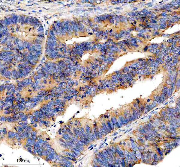
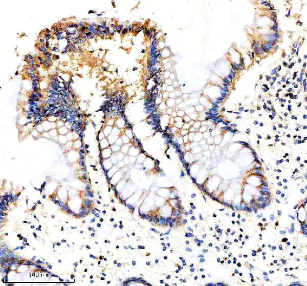
A05656-1 has IHC images from paraffin sections of human colon cancer, human colon, and rat bladder (catalog image captions); no IF/ICC data are supplied (catalog payload).

Real IHC data IHC analysis of EIF3H using anti-EIF3H antibody (A05656-1). EIF3H was detected in a paraffin-embedded section of human colon cancer tissue. Heat mediated antigen retrieval was performed in EDTA buffer (pH 8.0, epitope retrieval solution). The tissue section was blocked with 10% goat serum. The tissue section was then incubated with 2 μg/ml rabbit anti-EIF3H Antibody (A05656-1) overnight at 4°C. Peroxidase Conjugated Goat Anti-rabbit IgG was used as secondary antibody and incubated for 30 minutes at 37°C. The tissue section was developed using HRP Conjugated Rabbit IgG Super Vision Assay Kit (Catalog # SV0002) with DAB as the chromogen.
Anti-EIF3H Antibody ®
Cat # A05656-1

A05656-1 is listed for IHC and reacts with human, mouse, and rat samples (catalog applications and reactivity). Its IHC images show paraffin sections of human colon cancer, human colon, and rat bladder; the fixative is unreported (catalog image captions).

Which to pick: Choose A05656-1 for paraffin-section IHC: its own captions show human colon cancer, human colon, and rat bladder stained with 2 μg/ml primary antibody after EDTA pH 8.0 retrieval (catalog image captions). For cross-species work, the catalog lists human, mouse, and rat reactivity, but its IHC images document human and rat tissues only (catalog reactivity; catalog image captions). No IF/ICC application or image is supplied for A05656-1, so the payload does not support an IF/ICC recommendation (catalog applications; catalog IF images).

Each figure is that product's own IHC / IF validation image from its datasheet.

References

  1. UniProt Consortium. UniProt entry O15372 (EIF3H_HUMAN, Eukaryotic translation initiation factor 3 subunit H).
  2. Human Protein Atlas. EIF3H tissue IHC expression (reliability: Approved).
  3. Human Protein Atlas. EIF3H subcellular location (ICC-IF): Localized to the cytosol..
  4. Human Protein Atlas. EIF3H antibody validation summary (2 antibodies).
  5. EIF3H promotes aggressiveness of esophageal squamous cell carcinoma by modulating Snail stability. Journal of experimental & clinical cancer research : CR 2020 — PMC7457539.
  6. EIF3H Modulates Glycolysis Through LDHA Stabilization in Triple-Negative Breast Cancer. Cancers 2026 — PMC13564525.
  7. The Prognostic Significance of Eukaryotic Translation Initiation Factors (eIFs) in Endometrial Cancer. International journal of molecular sciences 2019 — PMC6941158.
  8. Celastrus orbiculatus Extract Reduces Stemness of Gastric Cancer Stem Cells by Targeting PDCD4 and EIF3H. Integrative cancer therapies 2021 — PMC8606975.
  9. PubMed PMID:9341143 — UniProt-cited evidence.
  10. PubMed PMID:15489334 — UniProt-cited evidence.
  11. PubMed PMID:16286006 — UniProt-cited evidence.