This website uses cookies to ensure you get the best experience on our website.


- Table of Contents
Plan EIF4A1 paraffin IHC around the cytoplasmic staining reported in several tissues (HPA tissue IHC). This guide uses catalog antibody conditions for paraffin sections (datasheet A03922-2) and flags the uncertain reliability of the tissue staining profile (HPA tissue IHC).

Expected localisation, validated protocols, controls and antibodies — the at-a-glance facts below, then the full design guide.
| Expected localisation | Cytoplasmic staining in several tissues (HPA tissue IHC) | |
| Staining pattern | Cytoplasmic staining across several tissue cell types (HPA tissue IHC) | |
| Antigen retrieval | Citrate pH 6 HIER, heat-mediated (datasheet A03922-2) | |
| Positive control | Bone marrow+4 more · see all | |
| Negative control | Adipose tissue+4 more · see all |
| Fixation | Matched tissue-IHC evidence does not establish the fixation claim. Validate the specimen-specific method before use. (selected-SKU IHC image A03922-2) | |
| Caveat | Staining–RNA concordance is low; reliability is uncertain (HPA tissue IHC) | |
| Regulation | Low tissue specificity (HPA tissue RNA) | |
| Isoform / epitope | Three isoforms; epitope effects are undetermined (UniProt) |
The catalog antibody protocol (datasheet: A03922-2) is followed by three published EIF4A1 IHC protocols (PMC9441068; PMC6857661; PMC10195147).
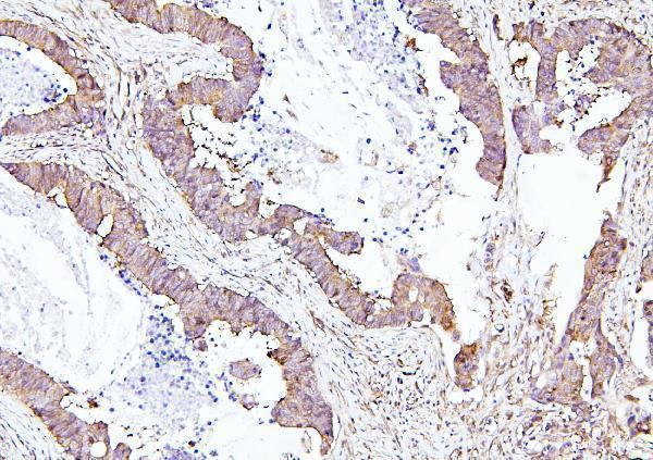
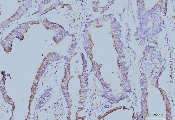
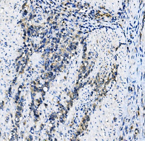
| Sample | Paraffin-embedded human colon cancer tissues; fixative not specified (datasheet A03922-2) |
| Fixation | Image fixative and duration unreported (datasheet A03922-2); verify before use. |
| Sectioning | 4–5 µm sections on charged slides (standard) |
| Deparaffinisation | Xylene, graded ethanol series to water (standard) |
| Antigen retrieval | Heat retrieval: Citrate pH 6, 20 min (datasheet A03922-2) |
| Peroxidase block | 3% H2O2, 10 min, room temperature (standard) |
| Blocking | 10% goat serum (datasheet A03922-2) |
| Primary antibody | Rabbit anti-EIF4A1, 0.5-1μg/ml (datasheet A03922-2) |
| Primary incubation | Overnight at 4 °C (datasheet A03922-2) |
| Detection | Streptavidin-biotin complex (SABC), DAB chromogen (datasheet A03922-2) |
| Counterstain | Hematoxylin, blue, dehydrate and mount (standard) |
| Expected result | EIF4A1-positive staining in hematopoietic cells of bone marrow (HPA tissue IHC: High). HPA tissue profile: Cytoplasmic expression in several tissues. No signal in the no-primary control. |
EIF4A1 is primarily cytoplasmic, with perinuclear, cell membrane and stress granule localisation also annotated (UniProt P60842). In paraffin sections, expect cytoplasmic staining in the cell populations reported by HPA, including colon glandular cells and bone marrow hematopoietic cells (HPA tissue IHC). HPA rates its tissue IHC pattern Uncertain because antibody staining and RNA expression have low consistency; interpret each result with controls (HPA tissue IHC).
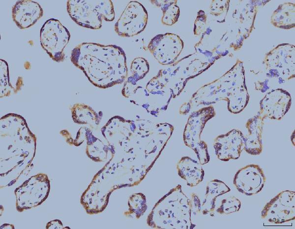
| Cytoplasmic signal in colon glandular cells, placental trophoblastic cells or bone marrow hematopoietic cells, with limited background (HPA tissue IHC). | This matches cell populations scored High by HPA and the expected compartment (HPA tissue IHC; UniProt P60842). Record the fraction and intensity of stained cells separately: a positive population need not make every cell equally dark. HPA's Uncertain reliability means agreement is supportive, not confirmation of antibody specificity (HPA tissue IHC). |
| Predominantly nuclear staining, with little cytoplasmic signal, in an otherwise well-preserved section. | Nuclear enrichment does not match the reported cytoplasmic tissue pattern or approved cytosolic ICC-IF localisation (HPA tissue IHC; HPA subcellular ICC-IF). Consider nonspecific staining, detection background or an interpretation error; compare the positive control and the no-primary control before assigning a nuclear EIF4A1 pattern (general IHC practice). |
| Strong signal in adipocytes or adrenal gland glandular cells while expected positive cells stain weakly (HPA tissue IHC). | HPA reports those specific cell populations as Not detected, so this reversal warrants checking cell identification, cross-reactivity and endogenous detection activity (HPA tissue IHC; general IHC practice). It does not establish that the whole tissue is EIF4A1-negative, and the HPA tissue pattern remains Uncertain (HPA tissue IHC). |
| Diffuse color across nuclei, stroma and extracellular spaces, obscuring cell boundaries. | A widespread haze cannot be scored as the cell-associated cytoplasmic pattern reported for EIF4A1 (HPA tissue IHC). Compare a no-primary control, review blocking and washing, and check whether the detection system produces signal without primary antibody (general IHC practice). Score only interpretable cells after background is controlled. |
| No stain in colon glandular cells or bone marrow hematopoietic cells, despite preserved morphology (HPA tissue IHC). | These are HPA High populations, so a blank result calls for a control-based check of antibody performance, detection and the general IHC workflow (HPA tissue IHC; general IHC practice). A single negative section does not prove EIF4A1 absence; HPA's tissue IHC assessment is itself Uncertain (HPA tissue IHC). |
| Compartment and topology | UniProt places EIF4A1 in cytoplasm and the perinuclear region, and also annotates cell membrane and stress granules; it lists no transmembrane segment (UniProt P60842). Expect the main routine tissue readout to be cytoplasmic (HPA tissue IHC). Membrane-associated signal should be judged with the whole cellular pattern, not treated as proof of an integral membrane protein (UniProt P60842 topology). |
| Choice of tissue and cell population | HPA scores colon, rectum and stomach glandular cells, placental trophoblastic cells, bone marrow hematopoietic cells and tonsillar squamous epithelial cells High; bronchial respiratory epithelial cells and appendix lymphoid tissue are Medium (HPA tissue IHC). Select a documented positive population for a control and score that population rather than averaging the entire section (general IHC practice). |
| Strength of the tissue evidence | The HPA tissue profile says cytoplasmic expression in several tissues, but assigns Uncertain reliability because staining and RNA expression show low consistency and external verification is pending (HPA tissue IHC). Its antibody listing gives CAB011689 an Uncertain IHC status; HPA068286 has ICC approval and no listed IHC status (HPA antibodies). Treat intensity rankings as comparison points, not validated cutoffs. |
| Isoforms and antibody epitope | UniProt lists three isoforms and helicase regions at residues 63–234 and 245–406 (UniProt P60842). The supplied evidence gives no epitope for the antibody being scored, so it cannot establish which isoforms that antibody detects or whether an unusual tissue result reflects isoform selection. Check the antibody's documented target region if interpreting discordant results (general IHC practice). |
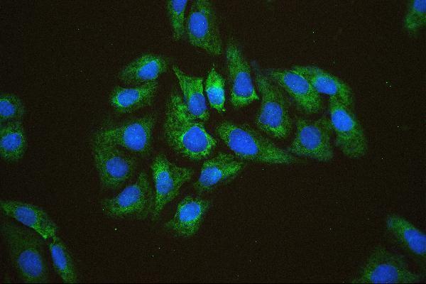
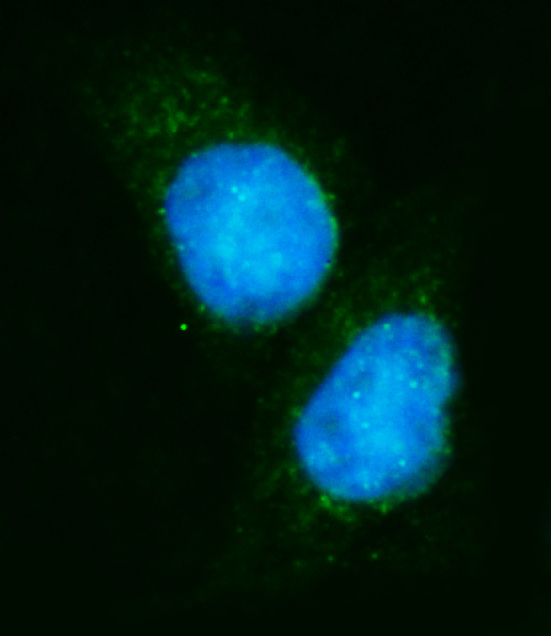
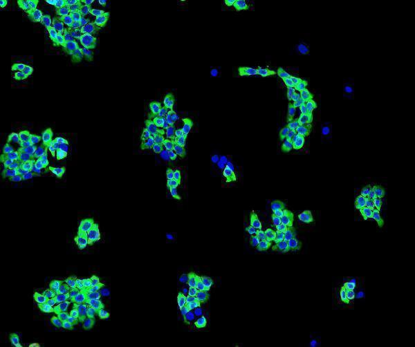
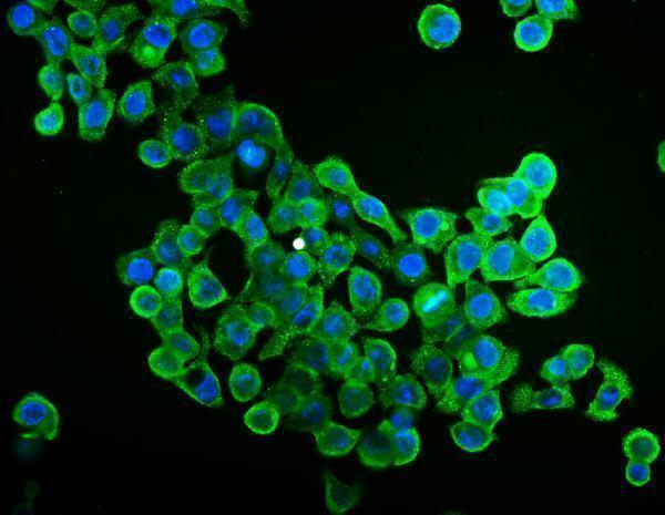
| IF/ICC Q&A: What pattern should fluorescence show? | Predominantly cytosolic signal: HPA approves the main cytosol localisation in ICC-IF images from CACO-2, U-251MG and U2OS (HPA subcellular ICC-IF). HPA cautions that this assignment is based on antibodies targeting proteins from multiple genes, so it is supporting localisation evidence rather than independent validation of this IHC result (HPA subcellular ICC-IF). |
| Antigen retrieval and detection conditions | Retrieval, blocking and detection controls are general paraffin IHC workflow considerations (general IHC practice). The supplied UniProt and HPA records give no EIF4A1-specific retrieval setting or fixation sensitivity; neither tissue intensity nor protein topology establishes one (UniProt P60842; HPA tissue IHC). Evaluate any workflow change against a documented positive population and a no-primary control (general IHC practice). |
| Situation | Likely cause | Next action |
|---|---|---|
| Expected positive cells are blank. | A workflow or detection failure is possible; HPA labels those cells High but the tissue IHC evidence is Uncertain (HPA tissue IHC). | Confirm the intended cell population, then review the IHC-validated antibody's documented conditions, retrieval, detection reagents and positive-control performance (general IHC practice). Avoid calling the specimen negative until the control stains. |
| Only nuclei appear strongly positive. | This conflicts with the cytoplasmic tissue profile and approved cytosol ICC-IF localisation (HPA tissue IHC; HPA subcellular ICC-IF). | Compare with the no-primary control, inspect cytoplasmic staining in a documented positive population, and review counterstain and detection background (general IHC practice). Report unresolved nuclear signal as atypical. |
| Adipocytes stain strongly. | HPA reports adipocytes as Not detected; misidentified cells, cross-reactivity or endogenous detection activity may explain the signal (HPA tissue IHC; general IHC practice). | Verify the stained cell type and compare a no-primary control and a documented HPA High population in the same staining run (HPA tissue IHC; general IHC practice). |
| Brown precipitate or haze covers tissue regardless of cell type. | Diffuse staining is inconsistent with HPA's cell-associated cytoplasmic profile and can arise from detection background (HPA tissue IHC; general IHC practice). | Check the no-primary control; review blocking, washes and chromogen development using the detection system's instructions (general IHC practice). Reassess localisation only where cell boundaries remain clear. |
| A medium or low HPA population looks weaker than a high population. | HPA reports different levels across cell populations: bronchial respiratory epithelial cells are Medium, while colon glandular cells are High (HPA tissue IHC). | Score each named cell population separately and compare intensity within the same staining run (general IHC practice). Keep HPA's Uncertain tissue reliability in the interpretation (HPA tissue IHC). |
| IHC and ICC-IF localisation appear discordant. | HPA ICC-IF supports cytosol localisation but cautions that its assignment draws on antibodies targeting proteins from multiple genes; tissue IHC reliability is Uncertain (HPA subcellular ICC-IF; HPA tissue IHC). | Document the antibody used in each application, compare the compartment seen in each image, and assess each application's controls before inferring a biological difference (general IHC/IF practice). |


Comprehensive Human Protein Atlas IHC scoring per tissue (reliability: Uncertain — Low consistency between antibody staining and RNA expression data. Pending external verification.). Rows are taken directly from the HPA tissue chart — click any row's HPA link to view the source.
| Tissue | Cell type | Level | Evidence | Source |
|---|---|---|---|---|
| Bone marrow | Hematopoietic cells | High | Protein (IHC) | HPA → |
| Colon | Glandular cells | High | Protein (IHC) | HPA → |
| Placenta | Trophoblastic cells | High | Protein (IHC) | HPA → |
| Rectum | Glandular cells | High | Protein (IHC) | HPA → |
| Stomach | Glandular cells | High | Protein (IHC) | HPA → |
| Tissue | Cell type | Level | Evidence | Source |
|---|---|---|---|---|
| Adipose tissue | Adipocytes | Not detected | Protein (IHC) | HPA → |
| Adrenal gland | Glandular cells | Not detected | Protein (IHC) | HPA → |
| Caudate | Glial cells | Not detected | Protein (IHC) | HPA → |
| Cerebellum | Cells in granular layer | Not detected | Protein (IHC) | HPA → |
| Cerebral cortex | Endothelial cells | Not detected | Protein (IHC) | HPA → |
Troubleshoot EIF4A1 staining in paraffin sections by checking retrieval, cytoplasmic localisation, controls and cell-specific scoring before interpreting DAB intensity.
The catalog shows EIF4A1 IHC in human and mouse paraffin sections and IF/ICC in human cells (catalog image captions); all four pictured antibodies list human, mouse and rat reactivity (catalog).
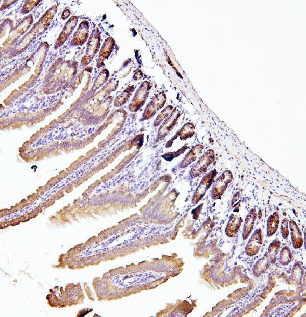
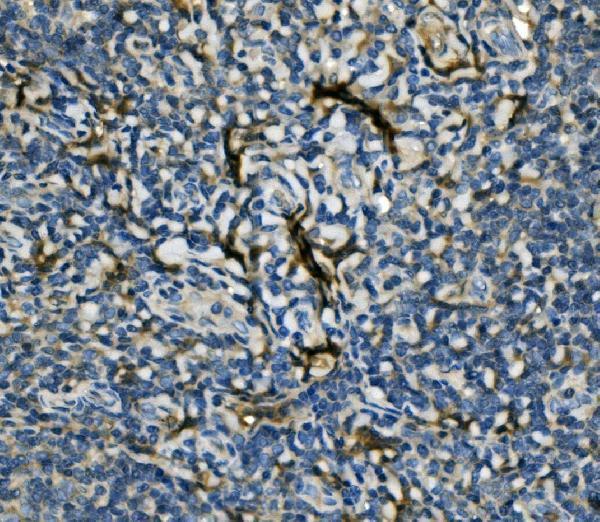
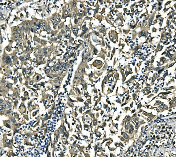
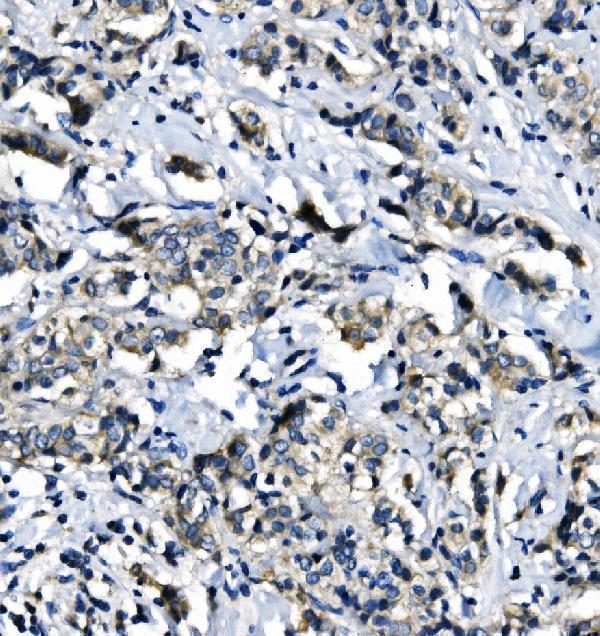
A03922-2 has IHC images from human colon cancer and mouse intestine, while A03922-3 has IHC images from human tonsil and mouse spleen (catalog image captions). M03922-1 has IHC images from human gastric and breast cancers; M03922-2 has images from human placenta, colonic adenocarcinoma and tonsil, plus mouse colon (catalog image captions).
Which to pick: For EIF4A1 tissue IHC, start with A03922-3 for human tonsil or mouse spleen paraffin sections (A03922-3 IHC captions); A03922-2 also detects EIF4A2/3 (catalog title). For IF/ICC, A03922-3 has an A431 cell image and a 2 μg/mL catalog dilution (A03922-3 IF caption; catalog). For human and mouse tissue work, M03922-2 has images in both species and lists rat reactivity, although its pictured sections are paraffin embedded and their fixative is unreported (M03922-2 IHC captions; catalog reactivity).