ELAVL3 / ELAV-like protein 3 · IHC design guide

Design Immunohistochemistry for ELAVL3

Plan chromogenic ELAVL3 IHC in paraffin sections using brain neurons as a positive reference (HPA tissue IHC). This guide covers consistent fixation, the catalog antibody’s 1:50–1:200 IHC dilution (datasheet), and scoring of cytoplasmic and nuclear neuronal staining (HPA tissue IHC).

Evidence assembled Oct 2026 · For research use; verify linked source records and product datasheet before use
Immunohistochemistry protocol sheet for ELAVL3 (IHC for ELAVL3): expected localisation Neuronal cytoplasm and nuclei (HPA tissue IHC), antibody A10061, validated IHC image, and IHC protocol steps
Printable ELAVL3 IHC protocol sheet — expected localisation Neuronal cytoplasm and nuclei (HPA tissue IHC), antibody A10061, controls and protocol steps. Open the full ELAVL3 IHC guide →

ELAVL3 Immunohistochemistry Experimental Design Guide

Expected localisation, validated protocols, controls and antibodies — the at-a-glance facts below, then the full design guide.

Must know before staining
Expected localisation Neuronal cytoplasm and nuclei (HPA tissue IHC)
Staining pattern CNS neurons show cytoplasmic and nuclear staining (HPA tissue IHC)
Antigen retrieval Citrate pH 6.0 HIER, 95–98 °C, 20 min (rule: cytoplasmic / membrane antigen)
Positive control ⓘ Cerebellum+3 more · see all
Negative control ⓘ Adipose tissue+4 more · see all
Important caveats
Reasons your staining may differ from the expected pattern.
Fixation Keep fixation consistent across sections (standard IHC practice; not target-specific); Matched tissue-IHC evidence does not establish the fixation claim. Validate the specimen-specific method before use. (selected-SKU IHC image A10061)
Caveat Signal outside brain may be low due to brain-specific expression (UniProt)
Regulation Brain-specific expression (UniProt)
Isoform / epitope 2 isoforms; epitope coverage is unknown (UniProt)
Section 1

Recommended ELAVL3 IHC & IF Protocols

Compare the catalog antibody’s IHC-P protocol with published ELAVL3 IHC methods for prostate tumor xenografts, human spinal cord, and transgenic embryos or brain sections (PMC10684895; PMC7897311; PMC4690400).

Recommended immunohistochemistry (IHC-P) protocol parameters
SampleParaffin-embedded rat brain tissue; fixative not specified (datasheet A10061)
FixationImage fixative and duration unreported (datasheet A10061); verify before use.
Sectioning4–5 µm sections on charged slides (standard)
DeparaffinisationXylene, graded ethanol series to water (standard)
Antigen retrievalHeat-induced epitope retrieval in citrate buffer, pH 6.0, 20 min at 95–98 °C (standard rule: cytoplasmic / membrane antigen)
Peroxidase block3% H2O2, 10 min, room temperature (standard)
Blocking10% normal serum of the secondary host, 30 min, room temperature (standard)
Primary antibodyRabbit anti-ELAVL3, 1:50-1:200 (datasheet A10061)
Primary incubationOvernight at 4 °C (standard)
DetectionHRP-polymer secondary, DAB chromogen 5–10 min (standard)
CounterstainHematoxylin, blue, dehydrate and mount (standard)
Expected resultELAVL3-positive staining in gLUC cells - cytoplasm/membrane of cerebellum (HPA tissue IHC: High). HPA tissue profile: Cytoplasmic and nuclear expression in neurons in CNS. No signal in the no-primary control.
💡Decision noteStart with the catalog protocol’s citrate pH 6.0 retrieval at 95–98 °C for 20 min (page antigen-retrieval setting); adjust for the sample and antibody used.
Section 2

What Is the Expected ELAVL3 Staining Pattern?

ELAVL3 staining is expected in CNS neurons, with nuclear and cytoplasmic signal in the HPA tissue IHC profile (HPA: Enhanced; cytoplasmic and nuclear expression in CNS neurons). Brain specificity supports this tissue choice (UniProt Q14576: brain specific). ELAVL3 has no transmembrane segment, so a membrane-only pattern would need scrutiny (UniProt Q14576: topology).

What am I looking at on my slide?
Neuronal nuclei and cytoplasm stain in cerebral cortex or hippocampus.This fits the reported compartment and cell pattern; both tissues have high neuronal staining (HPA: tissue IHC, Enhanced). Judge the result by identifiable neurons and their intracellular signal, rather than by the overall darkness of the section (general IHC practice).
Cerebellar GLUC cells show cytoplasmic staining with a membrane-associated appearance.HPA records high staining in these cells as cytoplasm/membrane (HPA: cerebellum tissue IHC). That observation does not establish ELAVL3 as a transmembrane protein (UniProt Q14576: no transmembrane segment). Interpret it alongside the broader nuclear and cytoplasmic CNS neuronal profile (HPA: tissue IHC).
Signal is confined to extracellular spaces or appears as a uniform surface outline without convincing neuronal cytoplasm or nuclei.This departs from the reported CNS neuronal profile (HPA: tissue IHC). Check morphology and background before calling it positive; a misplaced chromogenic deposit or nonspecific staining can mimic a compartment pattern (general IHC practice).
Many non-neuronal cells stain, including cell types HPA reports as not detected.For example, adipocytes in adipose tissue and hematopoietic cells in bone marrow are reported as not detected (HPA: tissue IHC). Review antibody specificity and endogenous detection activity; staining in these cells alone does not establish ELAVL3 expression (general IHC practice).
A known-positive CNS section has no convincing neuronal signal, or the whole slide has diffuse colour.High neuronal staining is reported in cerebral cortex and hippocampus (HPA: tissue IHC). An absent signal therefore calls for a control and detection check; diffuse colour calls for a background check. Neither observation alone proves a biological change in ELAVL3 (general IHC practice).
💡Expected ELAVL3 appearanceCall a section positive when identifiable CNS neurons show clear nuclear and cytoplasmic staining, with strong examples in cortex or hippocampus (HPA: tissue IHC, High); diffuse colour or extracellular-only signal is a suspect positive (general IHC practice).
How each factor affects the staining
Tissue and cell selectionCortex and hippocampus provide high neuronal examples; caudate is reported at medium intensity (HPA: tissue IHC). Adipose adipocytes and bone-marrow hematopoietic cells are reported as not detected (HPA: tissue IHC). Compare the specified cells, since a tissue-level label does not score every cell present.
Antibody evidenceThe listed mouse monoclonal CAB013066 has Enhanced IHC status (HPA: antibodies). HPA describes the tissue profile as highly consistent with RNA expression (HPA: tissue IHC reliability). These support a reference pattern, but do not identify the epitope or establish that every protocol condition works.
Isoforms and epitope coverageUniProt lists two ELAVL3 isoforms and three RNA-recognition motifs (UniProt Q14576: isoforms; domains). No epitope location or isoform coverage is supplied for CAB013066 (HPA: antibody record supplied). Interpret an unexpected absence with that limitation in mind; do not assign it to a particular isoform.
IF/ICC Q: Is a cell-line localisation pattern established?A: No main subcellular location or ICC-IF image-bearing cell line is supplied (HPA: subcellular). SH-SY5Y has the highest listed RNA expression, 43.7 nTPM, but this is not an IF protein-staining validation (HPA: subcellular). Use the separate IF/ICC guide for that application.
Why is my staining missing, weak or wrong?
SituationLikely causeNext action
No staining in cerebral cortex or hippocampal neurons.These neurons are reported as High (HPA: tissue IHC); the slide may reflect an IHC workflow or reagent failure (general IHC practice).Check the positive control, primary-antibody step and chromogenic detection; review the validated IHC-P instructions before changing conditions (general IHC practice).
Weak staining in caudate compared with cortex.Caudate neurons are reported as Medium, whereas cortex neurons are High (HPA: tissue IHC).Compare neuronal compartments and matched controls before treating the difference as assay failure; score each region against its reported level (HPA: tissue IHC; general IHC practice).
Colour covers neurons and surrounding tissue evenly.Diffuse background can arise from nonspecific binding or incomplete washing (general IHC practice).Inspect the no-primary control, blocking and wash steps; retain a positive CNS control while adjusting the workflow (general IHC practice).
Brown signal appears broadly in cells outside the expected neuronal pattern.Endogenous peroxidase activity or nonspecific antibody binding can contribute to chromogenic signal (general IHC practice).Compare a no-primary control and check the endogenous-peroxidase blocking step; assess cell identity against the HPA neuronal profile (HPA: tissue IHC; general IHC practice).
Only extracellular or sharply surface-confined staining is visible.That pattern does not match the reported nuclear and cytoplasmic CNS neuronal expression (HPA: tissue IHC).Recheck focus, morphology and background controls before scoring; do not infer transmembrane localisation from colour at a cell edge (UniProt Q14576: topology; general IHC practice).
Different sections seem to disagree on which cells are positive.The reported levels refer to specified cell types and regions, and can differ by region (HPA: tissue IHC).Record the region, identified cell type and compartment for each section; compare matched positive controls and document any remaining disagreement (general IHC practice).

Sample controls for ELAVL3 IHC & IF

🧪Run cerebellum first: GLUC cells should stain in the cytoplasm/membrane compartment (HPA: High in cerebellar GLUC cells). Use adipose tissue as a negative tissue; adipocytes should lack specific staining (HPA: Not detected in adipocytes). On the cerebellar slide, cells outside the annotated GLUC population should show little or no specific signal; verify their identity before treating them as an internal negative (HPA: GLUC cells are the annotated positive population).
Positive control tissue: Cerebellum (GLUC cells - cytoplasm/membrane, HPA High)
Negative control tissue: Adipose tissue (HPA Not detected)
ICC-IF cell lines (HPA subcellular resource): HPA carries no ICC-IF cell line for ELAVL3; derive a cell-line control from the positive tissue's cell type (GLUC cells - cytoplasm/membrane) and confirm it by RNA or western blot first.
Technical controls: Include a no-primary, secondary-only control and an isotype control matched to the primary antibody’s host species, immunoglobulin class, and monoclonal or polyclonal format; use ELAVL3 knockout tissue as a biological specificity control if available (standard IHC control practice). Block endogenous peroxidase for chromogenic detection and check brain pigment for signal that could be mistaken for staining (standard brain IHC practice).
⚠️Feasibility: A target-specific fixation window or fixation effect is unreported, and the selected rat brain paraffin-section caption does not state its fixative (selected A10061 tissue-IHC caption: fixative not stated). The caption uses microwave retrieval in 10 mM PBS, pH 7.2, with antibody at 1:200; it does not establish whether retrieval is required under other conditions (selected A10061 tissue-IHC caption). Relative ease of frozen-section staining or IF is unreported; brain autofluorescent pigment can complicate IF interpretation (HPA subcellular: no ICC-IF images listed; standard brain IF practice).

HPA tissue IHC evidence for ELAVL3

Comprehensive Human Protein Atlas IHC scoring per tissue (reliability: Enhanced — High consistency between antibody staining and RNA expression data.). Rows are taken directly from the HPA tissue chart — click any row's HPA link to view the source.

Positive expression · recommended positive controls

TissueCell typeLevelEvidenceSource
Cerebellum GLUC cells - cytoplasm/membrane High Protein (IHC) HPA →
Cerebral cortex Neuronal cells High Protein (IHC) HPA →
Hippocampus Neuronal cells High Protein (IHC) HPA →
Caudate Neuronal cells Medium Protein (IHC) HPA →

Undetected expression · recommended negative controls

TissueCell typeLevelEvidenceSource
Adipose tissue Adipocytes Not detected Protein (IHC) HPA →
Adrenal gland Glandular cells Not detected Protein (IHC) HPA →
Appendix Endocrine cells Not detected Protein (IHC) HPA →
Bone marrow Hematopoietic cells Not detected Protein (IHC) HPA →
Breast Adipocytes Not detected Protein (IHC) HPA →
Section 3

Advanced ELAVL3 IHC Tips

Use neuronal staining patterns and matched controls to troubleshoot ELAVL3 chromogenic IHC in paraffin sections (HPA tissue IHC; catalog antibody A10061 caption).

How should I adjust retrieval when ELAVL3 staining is weak?
Start with citrate pH 6.0 heat-induced retrieval at 95–98 °C for 20 min (page retrieval setting). If staining remains weak, compare it with microwave retrieval in 10 mM PBS, pH 7.2, which was used for the selected antibody image in paraffin-embedded rat brain (catalog antibody A10061 caption). Keep antibody dilution and detection conditions matched between sections so retrieval is the variable being assessed (standard IHC practice). Judge improvement by neuronal signal and preserved morphology, including the expected nuclear and cytoplasmic pattern, rather than by overall brown intensity (HPA tissue IHC; standard IHC practice).
Could fixation explain weak or uneven ELAVL3 staining?
The selected image documents paraffin-embedded rat brain but does not state its fixative, so ELAVL3-specific fixation sensitivity is unknown (catalog antibody A10061 caption). Record the fixative, fixation interval, section thickness, and processing history for each specimen before comparing staining intensity (standard IHC practice). When troubleshooting, stain similarly processed sections together and compare neuronal signal in the same anatomical region, since processing differences can complicate interpretation (HPA tissue IHC; standard IHC practice). If morphology or staining varies within a section, inspect folds, damaged areas, and section edges before changing antibody concentration or retrieval conditions (standard IHC practice).
Which cellular pattern should count as ELAVL3 staining?
Expect neuronal nuclear and cytoplasmic staining in CNS tissue, rather than requiring one compartment exclusively (HPA tissue IHC). HPA reports high staining in cerebral cortical and hippocampal neuronal cells and high cerebellar GLUC-cell cytoplasm or membrane staining (HPA tissue IHC). UniProt supplies no subcellular localisation annotation and reports no transmembrane segment, so a membrane-like outline alone should not establish ELAVL3 identity (UniProt Q14576 record). Compare the stained cells with their morphology and location on the counterstained section, and review nuclear and cytoplasmic signals separately when a treatment changes the apparent pattern (HPA tissue IHC; standard IHC practice).
Could isoforms or epitope accessibility change my IHC result?
ELAVL3 has 2 listed isoforms and 3 RNA recognition motifs at residues 39–117, 125–205, and 284–362 (UniProt Q14576 record). The supplied caption does not identify the catalog antibody's epitope, so these annotations cannot establish which isoforms it detects in sections (catalog antibody A10061 caption; UniProt Q14576 record). If staining differs between specimens, check the antibody's documented immunogen or epitope before attributing the difference to isoform expression (standard IHC practice). Keep retrieval and dilution constant during that comparison, and treat isoform-specific interpretation as unconfirmed without direct epitope and isoform evidence (standard IHC practice).
How would I assess ELAVL3 alongside another marker by IF?
Use a neuronal marker in a separate channel to assess whether ELAVL3 signal occurs in the expected cells; the expected CNS pattern includes neuronal nuclei and cytoplasm (HPA tissue IHC). Choose fluorophores after checking background in unstained tissue, placing the weaker signal in a channel with lower tissue autofluorescence (standard IF practice). ELAVL3 has no transmembrane segment, so permeabilisation should permit antibody access to intracellular epitopes; optimise it for the antibody and specimen rather than assuming a membrane-exposed epitope (UniProt Q14576 topology; standard IF practice). Include single-label controls when interpreting overlap, because the supplied catalog image documents chromogenic paraffin IHC rather than IF performance (catalog antibody A10061 caption; standard IF practice).
What should I check when brown signal obscures neurons?
Compare a section without primary antibody with the ELAVL3-stained section to identify detection-related background (standard IHC practice). For peroxidase and DAB detection, check the peroxidase block and inspect whether pigment or endogenous enzyme activity resembles the brown reaction product (standard IHC practice). Reduce nonspecific staining by checking blocking, washes, and the antibody titration while retaining a neuronal positive control in the same staining run (standard IHC practice; HPA tissue IHC). The selected rat-brain image used 1:200, which is a documented starting dilution for that image rather than a guaranteed optimum for every specimen (catalog antibody A10061 caption).
How should I quantify ELAVL3 across paraffin sections? ⚠ ANSWER MARKED FOR VERIFICATION
Score neuronal cells within predefined anatomical regions, using the same inclusion rules and staining run across comparison groups (HPA tissue IHC; standard IHC practice). Report the percentage of positive neurons and, if intensity matters, an H-score on the conventional 0–300 scale (standard IHC practice). Normalise positive-cell counts to the total neurons evaluated, or report positive-cell density per mm² of analysed tissue when cell counts are impractical (standard IHC practice). Record nuclear and cytoplasmic scores separately, since both compartments appear in the reported CNS pattern, and exclude folds or damaged tissue using predefined rules (HPA tissue IHC; standard IHC practice).
How can I distinguish true ELAVL3 signal from artefact?
A credible result follows neuronal morphology and the reported nuclear or cytoplasmic CNS pattern, with strong reference staining in cerebral cortex or hippocampus (HPA tissue IHC). Diffuse staining of unrelated cells, an isolated membrane-like outline, or signal confined to section edges warrants review before calling it ELAVL3 (HPA tissue IHC; UniProt Q14576 topology; standard IHC practice). Compare suspicious areas with the primary-antibody omission control and inspect necrotic or damaged tissue for trapped chromogen (standard IHC practice). With peroxidase and DAB detection, persistent brown signal in the control suggests an endogenous enzyme or detection artefact rather than target-specific staining (standard IHC practice).
Boster reagents

Best ELAVL3 / ELAV-like protein 3 IHC Antibodies

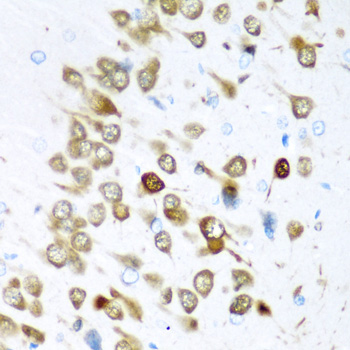
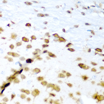
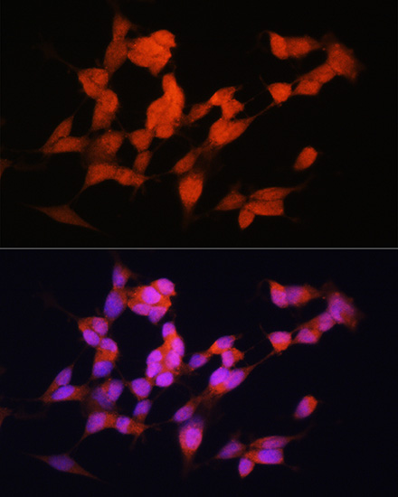
A10061 has IHC images from paraffin-embedded rat and mouse brain (catalog IHC captions) and an IF image from SH-SY5Y cells (catalog IF caption); listed reactivity is Human, Mouse, and Rat (catalog).

Real IHC data Immunohistochemistry of paraffin-embedded rat brain using ELAVL3 antibody at dilution of 1:200 .Perform microwave antigen retrieval with 10 mM PBS buffer pH 7.2 before commencing with IHC staining protocol.
Anti-ELAV-like protein 3 ELAVL3 Antibody
Cat # A10061

A10061 will render with its rat-brain paraffin-section IHC figure at 1:200 (catalog IHC caption); a mouse-brain paraffin-section IHC image is also reported at 1:200 (catalog IHC caption). Its IF image uses SH-SY5Y cells at 1:100 with DAPI nuclear staining (catalog IF caption), and its listed applications include IHC, IF, and ICC (catalog).

Which to pick: Choose A10061 for paraffin-section IHC based on its rat- and mouse-brain IHC captions; the fixative is unreported in those captions (catalog IHC captions). For IF/ICC, A10061 lists both applications and provides an IF image from SH-SY5Y cells (catalog applications; catalog IF caption). A10061 is a rabbit antibody listed as reactive with Human, Mouse, and Rat, making it the catalog choice for work across those species; the supplied IHC images show rat and mouse tissue (catalog host and reactivity; catalog IHC captions).

Each figure is that product's own IHC / IF validation image from its datasheet.

References

  1. UniProt Consortium. UniProt entry Q14576 (ELAV3_HUMAN, ELAV-like protein 3).
  2. Human Protein Atlas. ELAVL3 tissue IHC expression (reliability: Enhanced).
  3. Human Protein Atlas. ELAVL3 subcellular location (ICC-IF): Highest expression in SH-SY5Y: 43.7 nTPM.
  4. Human Protein Atlas. ELAVL3 antibody validation summary (1 antibodies).
  5. The ELAVL3/MYCN positive feedback loop provides a therapeutic target for neuroendocrine prostate cancer. Nature communications 2023 — PMC10684895.
  6. Cross-Comparison of Human iPSC Motor Neuron Models of Familial and Sporadic ALS Reveals Early and Convergent Transcriptomic Disease Signatures. Cell systems 2021 — PMC7897311.
  7. Transcriptomes of post-mitotic neurons identify the usage of alternative pathways during adult and embryonic neuronal differentiation. BMC genomics 2015 — PMC4690400.
  8. Physical activity stimulates neurogenesis via sensory neuron activity in postembryonic zebrafish. iScience 2026 — PMC13503103.
  9. PubMed PMID:15057824 — UniProt-cited evidence.
  10. PubMed PMID:15489334 — UniProt-cited evidence.
  11. PubMed PMID:7511893 — UniProt-cited evidence.