ELK1 / ETS domain-containing protein Elk-1 · IHC design guide

Design Immunohistochemistry for ELK1

Plan ELK1 staining in paraffin sections around its predominantly nuclear tissue pattern (HPA tissue IHC). Use the catalog antibody at 1:50 with chromogenic detection (datasheet M01426) and compare staining with an appropriate control section (standard IHC practice).

Evidence assembled Oct 2026 · For research use; verify linked source records and product datasheet before use
Immunohistochemistry protocol sheet for ELK1 (IHC for ELK1): expected localisation Predominantly nuclear in tissue (HPA tissue IHC), antibody M01426, validated IHC image, and IHC protocol steps
Printable ELK1 IHC protocol sheet — expected localisation Predominantly nuclear in tissue (HPA tissue IHC), antibody M01426, controls and protocol steps. Open the full ELK1 IHC guide →

ELK1 Immunohistochemistry Experimental Design Guide

Expected localisation, validated protocols, controls and antibodies — the at-a-glance facts below, then the full design guide.

Must know before staining
Expected localisation Predominantly nuclear in tissue (HPA tissue IHC)
Staining pattern Glandular cells show abundant nuclear staining (HPA tissue IHC)
Antigen retrieval EDTA pH 8.0 HIER, heat-mediated (datasheet M01426)
Positive control ⓘ Cerebellum+4 more · see all
Negative control ⓘ Adipose tissue+4 more · see all
Important caveats
Reasons your staining may differ from the expected pattern.
Fixation Keep fixation consistent across sections. (standard IHC practice; not target-specific)
Caveat Staining has medium RNA concordance; verify independently (HPA tissue IHC)
Regulation MAPK/JNK stimulation; abundance effect unknown (UniProt)
Isoform / epitope Two isoforms; epitope coverage is unspecified (UniProt)
Section 1

Recommended ELK1 IHC & IF Protocols

The catalog antibody’s IHC-P protocol uses heat retrieval (datasheet: M01426). The published protocols below cover urothelial, bladder, bone, and cervical cancer tissue sections (PMC5877638; PMC4745768; PMC7028725; PMC5463062).

Recommended immunohistochemistry (IHC-P) protocol parameters
SampleParaffin-embedded mouse brain tissue; fixative not specified (datasheet M01426)
FixationImage fixative and duration unreported (datasheet M01426); verify before use.
Sectioning4–5 µm sections on charged slides (standard)
DeparaffinisationXylene, graded ethanol series to water (standard)
Antigen retrievalHeat retrieval: EDTA pH 8.0 (datasheet M01426); 20 min, 95–100 °C (standard)
Peroxidase block3% H2O2, 10 min, room temperature (standard)
Blocking10% goat serum (datasheet M01426)
Primary antibodyRabbit monoclonal (clone CHG-5) anti-ELK1, 1:50 (datasheet M01426)
Primary incubationOvernight at 4 °C (datasheet M01426)
DetectionHRP-conjugated secondary, DAB chromogen (datasheet M01426)
CounterstainHematoxylin, blue, dehydrate and mount (standard)
Expected resultELK1-positive staining in granular cells - nucleus of cerebellum (HPA tissue IHC: High). HPA tissue profile: Nuclear expression, most abundant in glandular cells. No signal in the no-primary control.
💡Decision noteStart with EDTA pH 8.0 heat retrieval (datasheet: M01426). The bladder TMA protocol reports retrieval but gives no buffer or heating conditions (PMC4745768).
Section 2

What Is the Expected ELK1 Staining Pattern?

ELK1 is a nuclear transcription factor with no transmembrane segment (UniProt P19419: function, subcellular location, topology). In paraffin-section IHC, expect staining chiefly in nuclei, with glandular cells prominent overall (HPA: nuclear expression, most abundant in glandular cells). The HPA tissue profile has Enhanced reliability, but antibody staining and RNA show medium consistency and external verification is pending (HPA: tissue reliability).

What am I looking at on my slide?
Distinct nuclear staining in cerebellar granular cells, with little surrounding stain.This fits a strong positive: HPA reports High staining in granular-cell nuclei (HPA: cerebellum). Compare nuclear signal with the counterstain and nearby background when judging whether the chromogen occupies the expected compartment (standard IHC practice).
Predominantly cytoplasmic or membrane-like staining, with weak or absent nuclear staining.Treat this as a localisation mismatch that warrants checking specificity and detection background; ELK1 is nuclear and has no transmembrane segment (UniProt P19419: subcellular location, topology). Do not assign a cytoplasmic pattern to ELK1 solely from strong chromogen (standard IHC interpretation).
Strong staining in adipocytes or skeletal-muscle myocytes while the expected nuclear pattern is absent.HPA reports ELK1 as Not detected in those cells (HPA: adipose tissue, skeletal muscle). Consider antibody cross-reactivity or endogenous detection activity, then compare a no-primary control and a known-positive section processed alongside it (standard IHC practice).
Diffuse chromogen across tissue, stroma, and empty areas, obscuring cell boundaries.A broad haze cannot establish ELK1 localisation; the expected pattern is nuclear (UniProt P19419: subcellular location; HPA: tissue profile). Check whether background persists without primary antibody, then review blocking, washes, and detection conditions (standard IHC practice).
No nuclear signal in cerebellar granular cells on a test section.This conflicts with HPA's High nuclear staining in that cell population (HPA: cerebellum). First confirm that tissue morphology and the detection run are interpretable; then review antibody concentration and retrieval conditions without assuming a documented ELK1-specific fixation effect (standard IHC practice).
💡Expected ELK1 appearanceCall the result positive when nuclei stain clearly in an expected population, especially cerebellar granular cells with High nuclear staining (HPA: cerebellum); predominant membrane-like stain or diffuse haze without nuclear definition is suspect (UniProt P19419: topology, subcellular location; standard IHC interpretation).
How each factor affects the staining
Tissue and cell contextHPA reports High staining in cerebellar granular-cell nuclei and ovarian stromal cells, Medium staining in several glandular populations, and no detected staining in listed adipocytes and myocytes (HPA: tissue IHC). Judge the named cell population, since a tissue-wide score can conceal mixed patterns (standard IHC interpretation).
Validation strengthThe tissue profile is Enhanced, with medium staining–RNA consistency and external verification pending (HPA: tissue reliability). Individual antibodies differ: HPA036084 is IHC Enhanced and CAB003808 is IHC Supported; HPA064381 has no IHC status listed (HPA: antibodies). Match conclusions to the antibody actually used.
Isoforms and modified residuesUniProt lists two isoforms and several MAPK1-associated phosphorylation sites (UniProt P19419: isoforms, modified residues). Neither record identifies the antibody epitope or demonstrates isoform-specific IHC staining, so do not use staining intensity to assign an isoform or phosphorylation state.
Retrieval and detectionRetrieval, blocking, and chromogenic detection are adjustable parts of a paraffin-section IHC workflow (standard IHC practice). The supplied HPA and UniProt records provide no ELK1-specific retrieval setting or fixation-sensitivity result; establish conditions with a positive section and a no-primary control (standard IHC practice).
IF/ICC Q: Where should ELK1 appear?A: In the nucleoplasm; HPA rates that location Enhanced and lists ICC-IF images from HEK293, MCF-7, U2OS, A-431, U-251MG, PC-3, and SiHa cells (HPA: subcellular). This location supports interpretation of IF images but supplies no IF protocol or expected intensity for a particular experiment.
Why is my staining missing, weak or wrong?
SituationLikely causeNext action
The positive section has no nuclear chromogen.A run-level detection problem or unsuitable staining conditions are possible (standard IHC practice); HPA reports High nuclear staining in cerebellar granular cells (HPA: cerebellum).Verify tissue and counterstain quality, the primary and detection steps, and the positive control; then optimise dilution or retrieval using the same control (standard IHC practice). Do not infer an ELK1-specific fixation effect from this result.
Chromogen fills nuclei and surrounding tissue uniformly.Excess background can hide the nuclear pattern expected for ELK1 (UniProt P19419: subcellular location; standard IHC practice).Compare a no-primary section, then review primary concentration, blocking, washes, and detection development (standard IHC practice). Score ELK1 only where nuclear boundaries remain distinguishable.
Adipocytes or skeletal-muscle myocytes stain strongly.That conflicts with Not detected calls for these populations (HPA: adipose tissue, skeletal muscle); cross-reactivity or endogenous detection activity is possible (standard IHC interpretation).Check a no-primary control and the positive section, review endogenous-activity blocking for the detection system, and seek agreement with a separately validated antibody if available (standard IHC practice; HPA: antibodies).
A glandular region appears negative despite the overall glandular profile.HPA patterns vary by organ: appendix and colon glandular cells are Medium, while duodenal glandular cells and pancreatic exocrine glandular cells are Not detected (HPA: tissue IHC).Identify the organ and cell population before calling assay failure; compare with a documented positive population in the same run (HPA: tissue IHC; standard IHC practice).
Placental syncytiotrophoblast staining appears broader than a nuclear outline.HPA labels this population High at the cell-body level, while its overall ELK1 tissue profile is nuclear (HPA: placenta, tissue profile). The cell-body label alone does not resolve subcellular location.Inspect nuclear counterstain alignment and background controls before assigning compartment or specificity (standard IHC practice). Report the observed distribution separately from the HPA cell-body intensity call.
Two antibodies give different tissue patterns.The supplied IHC validation differs by antibody, and HPA's tissue profile has only medium staining–RNA consistency (HPA: antibodies, tissue reliability).Record which antibody produced each pattern, compare the same positive and negative cell populations under matched conditions, and avoid treating the profile's Enhanced label as equivalent validation for every antibody (HPA: antibodies; standard IHC practice).

Sample controls for ELK1 IHC & IF

🧪Run cerebellum first and score ELK1 staining in granular-cell nuclei (HPA: High in cerebellar granular-cell nuclei). Use lymph node germinal center cells as the negative tissue comparator (HPA: Not detected in lymph node germinal center cells); on the cerebellar slide, cells lacking nuclear staining can serve as background comparators, but no other cerebellar cell type is established as ELK1-negative by the supplied HPA row (HPA: High in granular-cell nuclei).
Positive control tissue: Cerebellum (Granular cells - nucleus, HPA High)
Negative control tissue: Adipose tissue (HPA Not detected)
ICC-IF cell lines (HPA subcellular resource): HPA ICC-IF images show ELK1 in HEK293, MCF-7, U2OS, A-431, U-251MG, PC-3, SiHa, with annotated localisation: Nucleoplasm (enhanced) (HPA subcellular).
Technical controls: Include a no-primary, secondary-only control; a concentration-matched rabbit IgG control matched to the primary antibody’s clonality; and an ELK1 knockout specimen or validated peptide block as a biological specificity control (selected M01426 caption: rabbit primary and peroxidase-conjugated anti-rabbit secondary; standard IHC practice). Quench endogenous peroxidase before HRP/DAB detection and check cerebellar pigment against the no-primary slide (selected M01426 caption: HRP/DAB detection; standard IHC practice).
⚠️Feasibility: A target-specific fixation window or fixation effect is unreported, and the selected M01426 paraffin-section caption does not state the fixative (selected M01426 caption: fixative not stated). The reported IHC conditions use heat retrieval in EDTA at pH 8.0 and primary antibody at 1:50 overnight at 4°C; retrieval dependence and whether frozen sections or IF are easier are unreported (selected M01426 caption). For IF, assess cerebellar autofluorescence with an unstained control and expect nucleoplasmic signal where ELK1 is detected (HPA subcellular: nucleoplasm; standard IF practice).

HPA tissue IHC evidence for ELK1

Comprehensive Human Protein Atlas IHC scoring per tissue (reliability: Enhanced — Medium consistency between antibody staining and RNA expression data. Pending external verification.). Rows are taken directly from the HPA tissue chart — click any row's HPA link to view the source.

Positive expression · recommended positive controls

TissueCell typeLevelEvidenceSource
Cerebellum Granular cells - nucleus High Protein (IHC) HPA →
Ovary Ovarian stroma cells High Protein (IHC) HPA →
Placenta Syncytiotrophoblasts - cell body High Protein (IHC) HPA →
Adrenal gland Glandular cells Medium Protein (IHC) HPA →
Appendix Glandular cells Medium Protein (IHC) HPA →

Undetected expression · recommended negative controls

TissueCell typeLevelEvidenceSource
Adipose tissue Adipocytes Not detected Protein (IHC) HPA →
Bone marrow Hematopoietic cells Not detected Protein (IHC) HPA →
Duodenum Glandular cells Not detected Protein (IHC) HPA →
Liver Cholangiocytes Not detected Protein (IHC) HPA →
Lymph node Germinal center cells Not detected Protein (IHC) HPA →
Section 3

Advanced ELK1 IHC Tips

Troubleshoot ELK1 staining in paraffin sections by assessing nuclear signal, tissue context, retrieval and controls before comparing samples.

Which retrieval condition should I start with for ELK1 in paraffin sections?
Start with heat-mediated antigen retrieval in EDTA at pH 8.0 for this catalog antibody (datasheet M01426). The documented mouse brain section used that condition before 10% goat-serum blocking and a 1:50 primary incubation overnight at 4°C (caption M01426). If nuclear staining is weak, compare a cautious adjustment in heating duration or a different retrieval buffer on matched sections, while keeping detection and exposure conditions fixed (standard IHC practice). Include a known positive section and a primary-omission control so that increased signal can be distinguished from increased background (HPA: high nuclear staining in cerebellar granular cells; standard IHC practice).
How should I troubleshoot weak ELK1 staining when fixation history varies?
Target-specific sensitivity to fixation is unknown from the supplied evidence; the product image identifies a paraffin-embedded section but does not state its fixative (caption M01426). Record the fixative, fixation interval and processing history for every specimen, then compare sections processed alike before changing antibody concentration (standard IHC practice). In routinely fixed paraffin material, excessive crosslinking can reduce accessible epitopes, so assess the documented EDTA pH 8.0 retrieval condition first (standard IHC practice; datasheet M01426). If fixation histories differ, report that limitation with the staining results and use matched control sections to separate specimen processing from assay performance (standard IHC practice).
What staining compartment should count as ELK1 positive?
Prioritize signal within nuclei: ELK1 is annotated as nuclear, and independent subcellular imaging places it in the nucleoplasm (UniProt P19419 subcellular; HPA subcellular: nucleoplasm). Its lack of a transmembrane segment gives no basis for interpreting a membrane rim as expected ELK1 localisation (UniProt P19419 topology). Compare nuclear DAB signal with a nuclear counterstain at the same focal plane, especially where crowded cells make cytoplasm and nuclei difficult to separate (standard IHC practice). HPA reports high nuclear staining in cerebellar granular cells, providing a tissue context for checking the compartment call (HPA tissue IHC: cerebellum). Score diffuse cytoplasmic or membrane staining separately until specificity is established (standard IHC practice).
Could isoforms or modification state explain inconsistent ELK1 staining?
ELK1 has 2 annotated isoforms, while the supplied product caption does not identify the antibody epitope or establish isoform selectivity (UniProt P19419 isoforms; caption M01426). Consequently, do not assign a staining difference to isoform switching without an epitope map and an independent isoform-specific measurement (standard IHC interpretation). The record lists MAPK1-associated phosphorylation at residues 324, 336, 353, 363 and 368, but supplies no evidence that these changes alter this antibody’s staining (UniProt P19419 modified residues). Compare matched retrieval and detection conditions first, and describe the result as ELK1 immunoreactivity rather than phosphorylation or isoform abundance unless those states are independently validated (standard IHC practice).
How should I adapt the localisation check to multiplex IF?
For a separate IF assay, multiplex ELK1 with a marker identifying the expected cell population and a nuclear stain; cerebellar granular cells offer a documented nuclear-positive context (HPA tissue IHC: cerebellum). Choose a fluorophore channel after inspecting unstained tissue for autofluorescence, and use single-stain controls to assess bleed-through before judging colocalisation (standard IF practice). Because ELK1 is nuclear and has no transmembrane segment, permeabilisation must permit antibody access to the nucleus rather than target a membrane-facing epitope (UniProt P19419 subcellular and topology; standard IF practice). Optimize fixation and permeabilisation for the IF specimen itself; the catalog tissue caption reports paraffin embedding and does not specify a fixative (caption M01426).
What should I change when DAB background obscures ELK1 nuclei?
First check primary-omission and detection-only controls to locate nonspecific reagent staining, then inspect whether background follows tissue edges, folds or damaged areas (standard IHC practice). For this catalog antibody, the documented section used 10% goat serum before 1:50 primary antibody overnight at 4°C (caption M01426). Recheck blocking, washes and antibody concentration on matched sections, changing one condition at a time while preserving the documented retrieval baseline (standard IHC practice; datasheet M01426: EDTA pH 8.0). Block endogenous peroxidase before HRP/DAB detection as a general chromogenic IHC step; the caption documents an HRP-linked detection workflow with DAB (standard IHC practice; caption M01426).
How should I score ELK1 across paraffin sections? ⚠ ANSWER MARKED FOR VERIFICATION
Define the cell population and nuclear-positive threshold before reviewing outcome groups, then score only interpretable nuclei in comparable tissue regions (standard IHC practice; UniProt P19419 subcellular: nucleus). Report the percentage of positive cells and, when intensity is reproducible, an H-score from 0–300 using intensity categories 0–3 (standard IHC practice). Normalize counts to the number of eligible nuclei in each region; if reporting positive-cell density, use area in mm² and state the region boundaries (standard IHC practice). Keep retrieval, DAB development and counterstaining consistent, and exclude folds, necrosis and section edges from the prespecified analysis (standard IHC practice).
How can I distinguish genuine ELK1 staining from artefact?
A credible ELK1 result has nuclear staining in intact cells, consistent with its annotated nuclear location and the HPA nucleoplasmic signal (UniProt P19419 subcellular; HPA subcellular: nucleoplasm). Check the cell type as well as the compartment: HPA reports high staining in cerebellar granular-cell nuclei but no detection in adipocytes, providing contextual comparators rather than universal controls (HPA tissue IHC: cerebellum and adipose tissue). Treat staining confined to section edges, necrotic areas or the primary-omission control as suspicious, and investigate endogenous peroxidase when DAB appears without primary antibody (standard IHC practice). Interpret intensity cautiously because HPA rates its tissue staining evidence as enhanced with medium RNA–protein consistency and pending external verification (HPA tissue IHC: reliability).
Boster reagents

Best ELK1 / ETS domain-containing protein Elk-1 IHC Antibodies

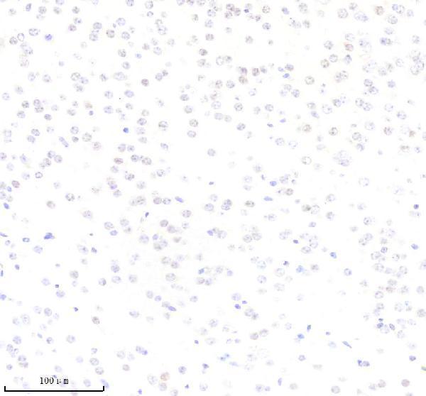
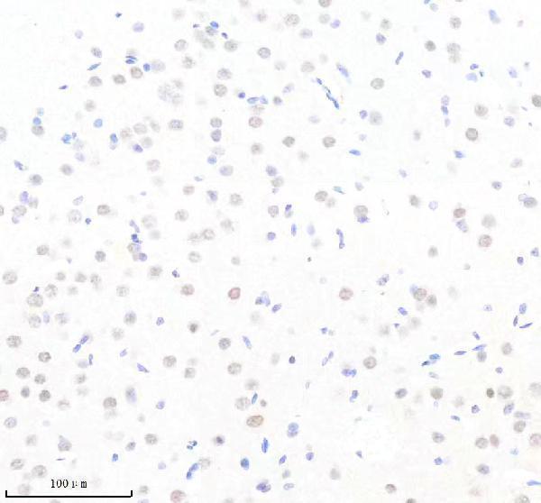
M01426 has real ELK1 IHC images from mouse and rat brain paraffin sections (catalog IHC captions); IF/ICC is listed for human, mouse and rat samples, without an IF image (catalog applications; reactivity; image list).

Real IHC data IHC analysis of ELK1 using anti-ELK1 antibody (M01426). ELK1 was detected in a paraffin-embedded section of mouse brain tissue. Heat mediated antigen retrieval was performed in EDTA buffer (pH 8.0, epitope retrieval solution). The tissue section was blocked with 10% goat serum. The tissue section was then incubated with 1:50 rabbit anti-ELK1 Antibody (M01426) overnight at 4°C. Peroxidase Conjugated Goat Anti-rabbit IgG was used as secondary antibody and incubated for 30 minutes at 37°C. The tissue section was developed using HRP Conjugated Rabbit IgG Super Vision Assay Kit (Catalog # SV0002) with DAB as the chromogen.
Anti-ELK1 Rabbit Monoclonal Antibody
Cat # M01426

M01426 will render with its mouse brain paraffin-section IHC figure (card image caption). Its additional IHC caption shows rat brain paraffin sections, while IF/ICC is listed without an IF image and human reactivity is listed without a human image (catalog IHC captions; applications; reactivity; image list).

Which to pick: For tissue IHC, choose M01426: its rabbit monoclonal antibody has a mouse brain paraffin-section IHC figure using EDTA pH 8.0 retrieval and 1:50 primary dilution (catalog host and clone; card image caption). For IF/ICC, M01426 is the listed option, although no IF image is supplied (catalog applications; image list). For cross-species work, M01426 lists human, mouse and rat reactivity, with IHC captions for mouse and rat brain; the paraffin-section captions do not report the fixative (catalog reactivity; IHC captions).

Each figure is that product's own IHC / IF validation image from its datasheet.

References

  1. UniProt Consortium. UniProt entry P19419 (ELK1_HUMAN, ETS domain-containing protein Elk-1).
  2. Human Protein Atlas. ELK1 tissue IHC expression (reliability: Enhanced).
  3. Human Protein Atlas. ELK1 subcellular location (ICC-IF): Localized to the nucleoplasm..
  4. Human Protein Atlas. ELK1 antibody validation summary (3 antibodies).
  5. Expression of Phospho-ELK1 and Its Prognostic Significance in Urothelial Carcinoma of the Upper Urinary Tract. International journal of molecular sciences 2018 — PMC5877638.
  6. ELK1 is up-regulated by androgen in bladder cancer cells and promotes tumor progression. Oncotarget 2015 — PMC4745768.
  7. Targeted overexpression of the long noncoding RNA ODSM can regulate osteoblast function in vitro and in vivo. Cell death & disease 2020 — PMC7028725.
  8. LncRNA-TCONS_00026907 is involved in the progression and prognosis of cervical cancer through inhibiting miR-143-5p. Cancer medicine 2017 — PMC5463062.
  9. PubMed PMID:2539641 — UniProt-cited evidence.
  10. PubMed PMID:9795224 — UniProt-cited evidence.
  11. PubMed PMID:10231026 — UniProt-cited evidence.