ELOVL5 / Very long chain fatty acid elongase 5 · IHC design guide

Design Immunohistochemistry for ELOVL5

Use adrenal zona fasciculata and sebaceous cells as positive tissue controls for ELOVL5 paraffin IHC (HPA tissue IHC). Assess cytoplasmic staining (HPA tissue IHC) with the IHC-validated antibody at 1:100–1:300 (datasheet: A30674).

Evidence assembled Oct 2026 · For research use; verify linked source records and product datasheet before use
Immunohistochemistry protocol sheet for ELOVL5 (IHC for ELOVL5): expected localisation Cytoplasmic tissue staining (HPA tissue IHC); ER membrane expected (UniProt), antibody A30674, validated IHC image, and IHC protocol steps
Printable ELOVL5 IHC protocol sheet — expected localisation Cytoplasmic tissue staining (HPA tissue IHC); ER membrane expected (UniProt), antibody A30674, controls and protocol steps. Open the full ELOVL5 IHC guide →

ELOVL5 Immunohistochemistry Experimental Design Guide

Expected localisation, validated protocols, controls and antibodies — the at-a-glance facts below, then the full design guide.

Must know before staining
Expected localisation Cytoplasmic tissue staining (HPA tissue IHC); ER membrane expected (UniProt)
Staining pattern Cytoplasmic staining in glandular and sebaceous cells (HPA tissue IHC)
Antigen retrieval Citrate pH 6.0 HIER, 95–98 °C, 20 min (rule: cytoplasmic / membrane antigen)
Positive control ⓘ Adrenal gland+4 more · see all
Negative control ⓘ Adipose tissue+4 more · see all
Important caveats
Reasons your staining may differ from the expected pattern.
Fixation Matched tissue-IHC evidence does not establish the fixation claim. Validate the specimen-specific method before use. (selected-SKU IHC image A30674)
Caveat RNA and staining show medium consistency (HPA tissue IHC)
Regulation Adrenal and testis expression is enriched (UniProt)
Isoform / epitope 3 isoforms; epitope map and sidedness are unreported (UniProt)
Section 1

Recommended ELOVL5 IHC & IF Protocols

The catalog antibody’s IHC-P protocol is accompanied by published ELOVL5 chromogenic IHC methods for colorectal and renal specimens (PMC5607170; PMC13457864).

Recommended immunohistochemistry (IHC-P) protocol parameters
SampleParaffin-embedded human brain tissue; fixative not specified (datasheet A30674)
FixationImage fixative and duration unreported (datasheet A30674); verify before use.
Sectioning4–5 µm sections on charged slides (standard)
DeparaffinisationXylene, graded ethanol series to water (standard)
Antigen retrievalHeat-induced epitope retrieval in citrate buffer, pH 6.0, 20 min at 95–98 °C (standard rule: cytoplasmic / membrane antigen)
Peroxidase block3% H2O2, 10 min, room temperature (standard)
Blocking10% normal serum of the secondary host, 30 min, room temperature (standard)
Primary antibodyRabbit anti-ELOVL5, 1:100 - 1:300 (datasheet A30674)
Primary incubationOvernight at 4 °C (standard)
DetectionHRP-polymer secondary, DAB chromogen 5–10 min (standard)
CounterstainHematoxylin, blue, dehydrate and mount (standard)
Expected resultELOVL5-positive staining in cells in zona fasciculata of adrenal gland (HPA tissue IHC: High). HPA tissue profile: Cytoplasmic expression in several tissues. Highly expressed in mammary, adrenal and sebaceous glands, and testis. No signal in the no-primary control.
💡Decision noteStart with citrate pH 6.0 retrieval at 95–98 °C for 20 min (page antigen-retrieval guidance); adjust against the published methods.
Section 2

What Is the Expected ELOVL5 Staining Pattern?

ELOVL5 is an endoplasmic reticulum membrane protein with 7 transmembrane segments (UniProt Q9NYP7 topology). In paraffin section IHC, expect cytoplasmic staining in selected glandular and epithelial cells, particularly adrenal zona fasciculata cells, breast glandular cells, and skin sebaceous cells (HPA tissue IHC). HPA rates its tissue IHC profile Enhanced, while reporting only medium consistency between antibody staining and RNA expression (HPA tissue IHC reliability).

What am I looking at on my slide?
Distinct cytoplasmic staining in adrenal zona fasciculata cells or breast glandular cells.This fits the reported High staining in those cell populations (HPA tissue IHC). Interpret the stained cells and compartment together; nearby unstained cells do not invalidate a cell-specific pattern.
Predominantly nuclear staining, with little cytoplasmic signal.A predominantly nuclear result conflicts with the endoplasmic reticulum membrane location (UniProt Q9NYP7 subcellular location; HPA subcellular ICC-IF). Recheck the staining pattern against a known-positive tissue before calling it ELOVL5.
Strong staining mainly in cardiomyocytes or adipocytes.HPA reports ELOVL5 as Not detected in these cells (HPA tissue IHC). Such staining raises concern for nonspecific binding or chromogenic detection activity; compare no-primary and known-positive controls (general IHC practice).
Broad chromogen deposition obscures the boundaries of stained cells.Diffuse background prevents a reliable cell-specific call (general IHC practice). Check no-primary staining and review blocking, antibody concentration, washing, and detection steps (general IHC practice).
No signal in adrenal zona fasciculata cells despite an otherwise readable section.These cells are reported High by HPA (HPA tissue IHC). First investigate assay performance with controls; an absent signal alone does not establish that the specimen lacks ELOVL5 (general IHC practice).
💡Expected ELOVL5 appearanceCall a convincing positive when High cytoplasmic staining marks adrenal zona fasciculata cells, breast glandular cells, or sebaceous cells (HPA tissue IHC); predominantly nuclear staining conflicts with the reported endoplasmic reticulum location (UniProt Q9NYP7 subcellular location).
How each factor affects the staining
Membrane location and image resolutionELOVL5 has 7 transmembrane segments in the endoplasmic reticulum membrane (UniProt Q9NYP7 topology). Chromogenic section IHC may show a broader cytoplasmic pattern because fine endoplasmic reticulum structures are not always resolved (general IHC practice).
Cell choice for interpretationHPA reports High staining in adrenal zona fasciculata, breast glandular, and skin sebaceous cells, but Low staining in cerebellar Purkinje cells (HPA tissue IHC). Score the specified cells rather than assigning one intensity to an entire tissue.
Antibody validationHPA lists IHC as Enhanced for HPA047752 and CAB017042; HPA054197 has no listed IHC status (HPA antibodies). Those ratings describe the listed antibodies and do not establish performance for a different catalog antibody.
Isoforms and epitopeUniProt lists 3 ELOVL5 isoforms (UniProt Q9NYP7 isoforms). The supplied evidence does not locate the catalog antibody's epitope, so isoform coverage and epitope-dependent staining differences cannot be predicted from this record.
IF/ICC Q&A: where should signal appear?Expect mainly endoplasmic reticulum signal in ICC-IF (HPA subcellular ICC-IF). UniProt also describes soma and proximal dendritic localization in Purkinje cells (UniProt Q9NYP7 subcellular location). HPA's ICC-IF finding does not define a protocol for paraffin section IHC.
Why is my staining missing, weak or wrong?
SituationLikely causeNext action
Adrenal zona fasciculata cells are blank.A reported High population is missing (HPA tissue IHC); assay failure is one possibility (general IHC practice).Check a known-positive section and controls, then review retrieval, primary-antibody exposure, and detection steps (general IHC practice). No target-specific retrieval condition is supplied.
Only a weak signal appears in cerebellar Purkinje cells.HPA reports Low staining in Purkinje cells (HPA tissue IHC).Compare with a reported high-staining cell population before judging sensitivity (HPA tissue IHC). Avoid using Purkinje cells alone as the positive control.
Cardiomyocytes or adipocytes stain strongly.Both cell populations are reported Not detected (HPA tissue IHC); nonspecific binding or detection background is possible (general IHC practice).Inspect a no-primary control, review blocking and washing, and compare the same run with a known-positive cell population (general IHC practice).
Brown signal covers nuclei more strongly than cytoplasm.The distribution conflicts with the reported endoplasmic reticulum location (UniProt Q9NYP7 subcellular location; HPA subcellular ICC-IF).Check nuclear counterstain versus chromogen and inspect control sections before scoring nuclear signal as ELOVL5 (general IHC practice).
The entire section has diffuse brown background.Excess detection activity, insufficient washing, or nonspecific antibody binding can obscure cell boundaries (general IHC practice).Review no-primary staining, endogenous peroxidase blocking, antibody concentration, and wash steps; score only cells with interpretable signal (general IHC practice).
Prostate glandular cells stain strongly despite the UniProt tissue summary.HPA reports High prostate glandular-cell staining, while UniProt describes prostate expression as weak (HPA tissue IHC; UniProt Q9NYP7 tissue specificity).Record the cell-specific IHC result and this source difference. Use controls to assess staining quality; do not infer an assay fault from the summaries alone.

Sample controls for ELOVL5 IHC & IF

🧪Run adrenal gland first: cells in the zona fasciculata should stain (HPA: High in cells in zona fasciculata). Use adipose tissue adipocytes as the negative comparator (HPA: Not detected in adipocytes); on the adrenal slide, adjacent cells outside the zona fasciculata should show only background staining, but their negative status is not established by the supplied HPA rows.
Positive control tissue: Adrenal gland (Cells in zona fasciculata, HPA High)
Negative control tissue: Adipose tissue (HPA Not detected)
ICC-IF cell lines (HPA subcellular resource): HPA ICC-IF images show ELOVL5 in A-431, U-251MG, U2OS, ASC52telo, NIH 3T3, with annotated localisation: Endoplasmic reticulum (enhanced) (HPA subcellular).
Technical controls: Include a no-primary, secondary-only control and a host-species-matched isotype control for a monoclonal primary, or matched nonimmune IgG for a polyclonal primary (standard IHC practice). Use a synthesized-peptide competition control as the biological specificity check (selected SKU A30674 caption: peptide block), and block endogenous peroxidase for chromogenic detection on the adrenal section (standard IHC practice).
⚠️Feasibility: No target-specific fixation window or fixation effect is reported, and the fixative in the selected SKU A30674 paraffin-section caption is unreported (selected SKU A30674 caption). Retrieval dependency is unreported; compare retrieval conditions empirically while checking for the expected intracellular ER pattern (UniProt Q9NYP7: ER membrane; HPA subcellular: mainly ER). The supplied evidence does not establish frozen sections or IF as easier than paraffin IHC; assess adrenal background with the no-primary control before scoring (standard IHC practice).

HPA tissue IHC evidence for ELOVL5

Comprehensive Human Protein Atlas IHC scoring per tissue (reliability: Enhanced — Medium consistency between antibody staining and RNA expression data.). Rows are taken directly from the HPA tissue chart — click any row's HPA link to view the source.

Positive expression · recommended positive controls

TissueCell typeLevelEvidenceSource
Adrenal gland Cells in zona fasciculata High Protein (IHC) HPA →
Appendix Glandular cells High Protein (IHC) HPA →
Breast Glandular cells High Protein (IHC) HPA →
Prostate Glandular cells High Protein (IHC) HPA →
Seminal vesicle Glandular cells High Protein (IHC) HPA →

Undetected expression · recommended negative controls

TissueCell typeLevelEvidenceSource
Adipose tissue Adipocytes Not detected Protein (IHC) HPA →
Bone marrow Hematopoietic cells Not detected Protein (IHC) HPA →
Heart muscle Cardiomyocytes Not detected Protein (IHC) HPA →
Lymph node Germinal center cells Not detected Protein (IHC) HPA →
Ovary Follicle cells Not detected Protein (IHC) HPA →
Section 3

Advanced ELOVL5 IHC Tips

Troubleshoot ELOVL5 chromogenic IHC in paraffin sections using retrieval, compartment and cell-type controls; IF/ICC considerations appear in one dedicated entry.

How should I retrieve ELOVL5 when paraffin sections stain weakly?
Start with citrate buffer at pH 6.0, heated to 95–98 °C for 20 min, for chromogenic IHC on paraffin sections (page retrieval setting). Allow sections to cool in buffer before washing, and run a known positive tissue alongside the test section, such as adrenal gland with zona fasciculata staining (HPA: High in zona fasciculata cells). If staining remains weak, vary retrieval time on adjacent sections before trying a different buffer or pH, keeping antibody incubation and detection conditions constant. Judge improvement by cell-associated cytoplasmic signal with limited background, since ELOVL5 is mainly associated with the endoplasmic reticulum (HPA: enhanced ER localisation).
Could fixation explain weak or patchy ELOVL5 staining?
ELOVL5-specific sensitivity to fixation is unknown from the supplied evidence; the catalog image shows paraffin-embedded human brain, but its caption does not state a fixative (A30674 tissue-IHC caption). Record the fixative and fixation duration for each specimen, then compare matched sections processed with the same retrieval, antibody incubation and chromogenic detection conditions. If staining varies across a block, check morphology and compare well-preserved central tissue with edges or damaged areas before changing the antibody concentration. Use a processed positive control, such as adrenal zona fasciculata, to distinguish a run-level failure from a specimen-level issue (HPA: High in zona fasciculata cells).
What staining pattern should count as ELOVL5 localisation?
Expect predominantly cytoplasmic staining compatible with an endoplasmic reticulum membrane protein, rather than scoring diffuse staining in every compartment as equivalent (UniProt Q9NYP7: ER membrane; HPA: cytoplasmic tissue expression). In cerebellar Purkinje cells, evaluate the soma and proximal dendritic tree specifically, although tissue IHC expression there is reported as Low (UniProt Q9NYP7: Purkinje localisation; HPA: Low in Purkinje cells). Compare the pattern with a positive tissue compartment on the same run, such as adrenal zona fasciculata (HPA: High). Treat strong isolated nuclear or extracellular chromogen as a reason to inspect section quality and controls before assigning an ELOVL5 score.
How can epitope choice affect ELOVL5 IHC interpretation?
ELOVL5 has 3 annotated isoforms and 7 transmembrane segments, so the antibody epitope and its accessibility matter when interpreting a negative section (UniProt Q9NYP7: isoforms and topology). Check the catalog antibody's stated immunogen or epitope against the isoforms of interest; an unspecified epitope cannot establish which isoforms contribute to staining. The record lists N-terminal acetylation and phosphoserine at residue 285, but it supplies no evidence that either modification changes this antibody's IHC signal (UniProt Q9NYP7: modified residues). Compare adjacent positive and test sections under the same retrieval and detection conditions before attributing a staining difference to isoform expression.
How should I adapt ELOVL5 interpretation to multiplex IF?
On the separate IF/ICC workflow, pair ELOVL5 with a marker that identifies the expected cell type, such as a Purkinje-cell marker when examining cerebellum (UniProt Q9NYP7: Purkinje soma and proximal dendrites). Choose a fluorophore channel with low tissue autofluorescence, and inspect single-stain and unstained controls before accepting overlap as colocalisation. Because ELOVL5 has 7 transmembrane segments, select and verify permeabilisation with respect to the antibody epitope's membrane-facing side; the supplied evidence does not identify that side (UniProt Q9NYP7: topology). Keep compartment interpretation anchored to the ER-enriched pattern and evaluate each channel separately before merging images (HPA: enhanced ER localisation).
How can I reduce nonspecific chromogenic staining?
First compare a no-primary control with the stained section, then check whether background tracks tissue edges, damaged areas or endogenous pigment rather than cells. For a peroxidase and DAB workflow, use a peroxidase block and appropriate protein blocking as general IHC steps, then adjust washing and antibody concentration one variable at a time. Score only cell-associated cytoplasmic signal consistent with ELOVL5's ER localisation, using a known positive compartment to ensure that reduced background has not removed the target signal (UniProt Q9NYP7: ER membrane; HPA: High in adrenal zona fasciculata). Keep counterstaining consistent across comparisons so faint chromogen remains interpretable.
How should I quantify ELOVL5 across paraffin sections? ⚠ ANSWER MARKED FOR VERIFICATION
Define cell types and regions before scoring, then record either the percentage of positive cells with intensity for an H-score or positive-cell density per mm². Normalise to the number of eligible cells or viable tissue area, respectively, and keep section thickness, retrieval, detection and image thresholds consistent across groups. Score cytoplasmic staining within the selected cells, since HPA reports cytoplasmic expression and enhanced ER localisation (HPA: tissue IHC and subcellular profile). Report the chosen tissue compartment alongside the score: adrenal zona fasciculata is High, whereas cerebellar Purkinje cells are Low in the supplied tissue profile (HPA: tissue IHC).
When is an ELOVL5-positive IHC result convincing?
A convincing result is cell-associated cytoplasmic staining in an expected compartment, supported by a positive control such as adrenal zona fasciculata (HPA: cytoplasmic expression; HPA: High in zona fasciculata cells). The selected brain image includes peptide-blocked staining as a comparison, but its caption does not report a fixative or establish how every tissue should stain (A30674 tissue-IHC caption). Question isolated nuclear deposits, section-edge enhancement, necrotic areas and signal present in a no-primary control before calling a sample positive. Interpret weak brain staining cautiously because neuronal cells and cerebellar Purkinje cells are listed as Low, while ELOVL5 is an ER membrane protein (HPA: tissue IHC; UniProt Q9NYP7: localisation).
Boster reagents

Best ELOVL5 / Very long chain fatty acid elongase 5 IHC Antibodies

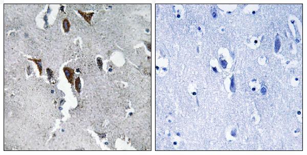
A30674 has a human paraffin-section IHC image with peptide blocking (A30674 image caption). Both antibodies list IF/ICC applications and Human, Mouse and Rat reactivity (catalog applications/reactivity).

Real IHC data Immunohistochemistry analysis of paraffin-embedded human brain tissue, using ELOVL5 Antibody. The picture on the right is blocked with the synthesized peptide.
Anti-ELOVL5 Antibody
Cat # A30674

A30674 is the sole card with its own figure: IHC on paraffin-embedded human brain tissue, with peptide blocking shown (A30674 image caption). M03615 lists IF/ICC and Human, Mouse and Rat reactivity, but has no IHC or IF figure and therefore no card (M03615 catalog applications/reactivity/image alts).

Which to pick: For tissue IHC, choose A30674: its listed IHC dilution is 1:100–1:300, and its image shows paraffin-embedded human brain tissue; the fixative is unreported (A30674 catalog dilution/image caption). For IF/ICC, M03615 is a rabbit monoclonal option with those applications listed, although no IF figure is supplied (M03615 catalog title/applications/image alts). For work across Human, Mouse and Rat, both list reactivity, but A30674 is the IHC option and its tissue image documents human tissue only (catalog applications/reactivity; A30674 image caption).

Each figure is that product's own IHC / IF validation image from its datasheet.

References

  1. UniProt Consortium. UniProt entry Q9NYP7 (ELOV5_HUMAN, Very long chain fatty acid elongase 5).
  2. Human Protein Atlas. ELOVL5 tissue IHC expression (reliability: Enhanced).
  3. Human Protein Atlas. ELOVL5 subcellular location (ICC-IF): Mainly localized to the endoplasmic reticulum..
  4. Human Protein Atlas. ELOVL5 antibody validation summary (3 antibodies).
  5. Methylation associated transcriptional repression of ELOVL5 in novel colorectal cancer cell lines. PloS one 2017 — PMC5607170.
  6. Elovl5 Expression in the Central Nervous System of the Adult Mouse. Frontiers in neuroanatomy 2021 — PMC8116736.
  7. Elovl5 is required for proper action potential conduction along peripheral myelinated fibers. Glia 2021 — PMC8453547.
  8. Co-overexpression of ELOVL2 and ELOVL5 promotes clear cell renal cell carcinoma progression through LIMK1-mediated cytoskeletal reorganization. Scientific reports 2026 — PMC13457864.
  9. PubMed PMID:10970790 — UniProt-cited evidence.
  10. PubMed PMID:11230166 — UniProt-cited evidence.
  11. PubMed PMID:14702039 — UniProt-cited evidence.