This website uses cookies to ensure you get the best experience on our website.


- Table of Contents
Plan chromogenic IHC-P for EMX2 using its mainly nuclear tissue staining pattern (HPA tissue IHC). This guide covers tissue controls, compartment-aware scoring and an IHC-validated antibody with a starting concentration of 2.5 μg/mL (datasheet: A04322-1).

Expected localisation, validated protocols, controls and antibodies — the at-a-glance facts below, then the full design guide.
| Expected localisation | Mainly nuclear in tissues (HPA tissue IHC); also axonal (UniProt) | |
| Staining pattern | Mainly nuclear staining in several tissues (HPA tissue IHC) | |
| Antigen retrieval | Tris-EDTA pH 9.0 HIER, 95–98 °C, 20 min (rule: nuclear antigen) | |
| Positive control | Cerebellum+4 more · see all | |
| Negative control | Adipose tissue+4 more · see all |
| Fixation | Keep fixation consistent across paraffin sections. (standard IHC practice; not target-specific) | |
| Caveat | Cerebellar GLUC cells show cytoplasm/membrane staining (HPA tissue IHC) | |
| Regulation | Expression varies by tissue (HPA tissue IHC) | |
| Isoform / epitope | 2 isoforms; shared epitope status is unknown (UniProt) |
Compare the catalog antibody’s IHC-P protocol with published EMX2 staining protocols for colorectal tissue, lung tissue, and cryosections (PMC5568393; PMC3285542; PMC11062917).
| Sample | Tissue sections; selected-image fixative not specified (standard IHC workflow) |
| Fixation | Image fixative and duration unreported (datasheet A04322); verify before use. |
| Sectioning | 4–5 µm sections on charged slides (standard) |
| Deparaffinisation | Xylene, graded ethanol series to water (standard) |
| Antigen retrieval | Heat-induced epitope retrieval in Tris-EDTA buffer, pH 9.0, 20 min at 95–98 °C (standard rule: nuclear antigen) |
| Peroxidase block | 3% H2O2, 10 min, room temperature (standard) |
| Blocking | 10% normal serum of the secondary host, 30 min, room temperature (standard) |
| Primary antibody | Rabbit anti-EMX2, 5 μg/mL (datasheet A04322) |
| Primary incubation | Overnight at 4 °C (standard) |
| Detection | HRP-polymer secondary, DAB chromogen 5–10 min (standard) |
| Counterstain | Hematoxylin, blue, dehydrate and mount (standard) |
| Expected result | EMX2-positive staining in gLUC cells - cytoplasm/membrane of cerebellum (HPA tissue IHC: Medium). HPA tissue profile: Mainly nuclear expression in several tissues. No signal in the no-primary control. |
EMX2 is primarily a nuclear protein (UniProt Q04743; HPA tissue IHC). In paraffin sections, look for staining in the specific cells HPA reports, including epididymal glandular cells, kidney collecting ducts and ovarian follicle cells (HPA tissue IHC). HPA rates tissue staining reliability Enhanced, with medium consistency between staining and RNA data (HPA tissue IHC). EMX2 has no transmembrane segment (UniProt Q04743 topology).
| Nuclear staining in epididymal glandular cells, kidney collecting ducts or ovarian follicle cells. | This fits the predominant EMX2 pattern; HPA reports Medium staining in each of these cell populations (HPA tissue IHC). Compare positive cells with adjacent structures rather than scoring a whole tissue as uniformly positive (general IHC practice). |
| Broad cytoplasmic or membrane staining replaces nuclear staining across a section. | Treat this as a possible artefact because the overall tissue pattern is mainly nuclear (HPA tissue IHC). Check context before rejecting it: HPA reports cytoplasm/membrane staining in cerebellar GLUC cells, while UniProt reports EMX2 in olfactory axons (HPA tissue IHC; UniProt Q04743). |
| Strong staining appears in an HPA-listed negative cell population, such as adipocytes in adipose tissue. | HPA reports EMX2 as Not detected in those adipocytes (HPA tissue IHC). Consider antibody cross-reactivity or endogenous detection activity; inspect the no-primary control and whether signal follows the expected cell boundaries (general IHC practice). |
| Diffuse colour covers both cells and surrounding tissue, obscuring nuclei. | This is background rather than an interpretable compartment pattern (general IHC practice). Review the no-primary control, blocking, wash steps and chromogen development before assigning EMX2 positivity; HPA's mainly nuclear pattern cannot validate diffuse staining (HPA tissue IHC). |
| No signal appears in a section containing HPA-listed Medium-positive cells. | A negative run is inconclusive until the detection system and tissue quality are checked (general IHC practice). Confirm that the relevant cell population is present, then review the IHC antibody dilution and antigen-retrieval conditions; HPA's reported Medium level is a reference pattern, not a guarantee for every preparation (HPA tissue IHC). |
| Compartment and anatomical context | HPA describes mainly nuclear tissue expression and approved nucleoplasmic ICC-IF localisation; its cerebellar GLUC-cell observation and UniProt's olfactory-axon report are context-specific exceptions (HPA tissue IHC; HPA subcellular; UniProt Q04743). |
| Choice of reference cells | HPA reports Medium staining in epididymal glandular cells, kidney collecting ducts and ovarian follicle cells, but Low staining in cerebral-cortex neurons; use the stated cell population and level when judging a slide (HPA tissue IHC). |
| Antibody evidence | HPA lists HPA065294 as IHC Enhanced and ICC Approved, and HPA003497 as IHC Supported; these are antibody-specific validation statuses, so assess the antibody actually used (HPA antibodies). |
| RNA and protein agreement | HPA calls tissue IHC reliability Enhanced yet describes only medium consistency with RNA expression. Cervix has tissue-enhanced RNA specificity while its glandular cells are Not detected by IHC; do not substitute RNA for a slide-level positive control (HPA tissue IHC). |
| Protein form | UniProt records two EMX2 isoforms, no signal peptide or propeptide, and one chain spanning residues 1–252. Without antibody epitope data, these facts cannot establish isoform coverage or a required antigen-retrieval condition (UniProt Q04743). |
| Situation | Likely cause | Next action |
|---|---|---|
| Nuclei are clear but most EMX2 signal is cytoplasmic. | The compartment disagrees with the mainly nuclear tissue pattern and approved nucleoplasmic ICC-IF localisation (HPA tissue IHC; HPA subcellular). | Check a no-primary control and review detection background; identify the stained cells before judging an exception, since cerebellar GLUC cells show cytoplasm/membrane staining (general IHC practice; HPA tissue IHC). |
| Unexpected cells stain more strongly than the intended positive population. | Cross-reactivity or endogenous detection activity is possible (general IHC practice); HPA reports some cell populations as Not detected (HPA tissue IHC). | Compare cell identity with HPA's cell-level reference, then use a no-primary control and appropriate endogenous-activity control for the detection chemistry (HPA tissue IHC; general IHC practice). |
| The section has diffuse brown background. | Nonspecific binding, incomplete washing or detection activity may obscure localisation (general IHC practice). | Review blocking and washes, inspect the no-primary control, and limit chromogen development as appropriate; score EMX2 only where cell and compartment boundaries remain readable (general IHC practice). |
| A section expected to contain Medium-positive cells has no signal. | The expected cells may be absent, or the staining workflow may have failed; HPA documents Medium staining only in specified cell populations (HPA tissue IHC). | Confirm the target cells on the counterstain, verify detection controls, and review the antibody's IHC dilution and antigen-retrieval instructions (general IHC practice). |
| Cerebral-cortex neurons look weak or negative beside a stronger control. | HPA reports Low staining in cerebral-cortex neurons, despite listing cerebral cortex as a tissue specificity in UniProt (HPA tissue IHC; UniProt Q04743). | Avoid using cortical neurons alone to declare a failed run; compare with an HPA-listed Medium-positive cell population under the same scoring criteria (HPA tissue IHC; general IHC practice). |
| Q: Should IF/ICC show the same compartment pattern? | HPA approves nucleoplasmic ICC-IF localisation for EMX2, while tissue IHC is mainly nuclear (HPA subcellular; HPA tissue IHC). | A: Expect a nucleoplasmic IF/ICC pattern where EMX2 is detected; interpret IF/ICC using its separate guide and assay controls (HPA subcellular; general IF practice). |


Comprehensive Human Protein Atlas IHC scoring per tissue (reliability: Enhanced — Medium consistency between antibody staining and RNA expression data.). Rows are taken directly from the HPA tissue chart — click any row's HPA link to view the source.
| Tissue | Cell type | Level | Evidence | Source |
|---|---|---|---|---|
| Cerebellum | GLUC cells - cytoplasm/membrane | Medium | Protein (IHC) | HPA → |
| Endometrium | Cells in endometrial stroma | Low | Protein (IHC) | HPA → |
| Epididymis | Glandular cells | Medium | Protein (IHC) | HPA → |
| Fallopian tube | Non-ciliated cells | Medium | Protein (IHC) | HPA → |
| Kidney | Collecting ducts | Medium | Protein (IHC) | HPA → |
| Tissue | Cell type | Level | Evidence | Source |
|---|---|---|---|---|
| Adipose tissue | Adipocytes | Not detected | Protein (IHC) | HPA → |
| Adrenal gland | Glandular cells | Not detected | Protein (IHC) | HPA → |
| Appendix | Glandular cells | Not detected | Protein (IHC) | HPA → |
| Bone marrow | Hematopoietic cells | Not detected | Protein (IHC) | HPA → |
| Breast | Adipocytes | Not detected | Protein (IHC) | HPA → |
Troubleshoot EMX2 staining in paraffin sections by checking retrieval, nuclear localisation, cell identity and controls before comparing staining across samples.
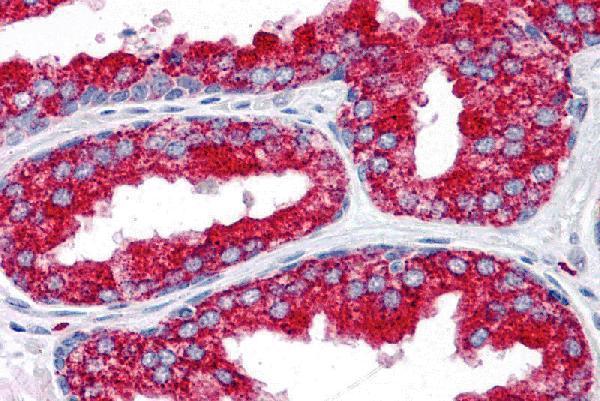
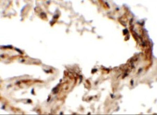
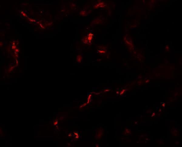
Three anti-EMX2 antibodies have human tissue IHC images; one also has a human tissue IF image (catalog image captions). Human and mouse reactivity is listed for A04322-2 (catalog reactivity).
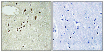
A04322 has a human prostate IHC image, and A04322-1 has human lung IHC and IF images (catalog image captions). A04322-2 has a paraffin-embedded human brain IHC image and lists Human and Mouse reactivity; its image does not establish mouse IHC performance (catalog image caption; catalog reactivity).
Which to pick: For paraffin-section IHC, choose A04322-2: its human brain caption explicitly specifies paraffin embedding and 1:100 dilution; the fixative is unreported (A04322-2 IHC image caption). For human tissue IF, choose A04322-1 at a starting concentration of 20 μg/mL; its IF image shows human lung tissue, while ICC validation is unreported (A04322-1 catalog applications; IF image caption; catalog dilution). For a mouse sample, A04322-2 is the only listed mouse-reactive option, although its IHC image is human (catalog reactivity; A04322-2 IHC image caption).