This website uses cookies to ensure you get the best experience on our website.


- Table of Contents
Plan EN1 IHC-P around the nuclear signal reported in eccrine sweat glands (HPA tissue IHC). The catalog antibody has a listed IHC-P dilution of 1:25 (datasheet A05395).

Expected localisation, validated protocols, controls and antibodies — the at-a-glance facts below, then the full design guide.
| Expected localisation | Nuclear staining in eccrine sweat glands (HPA tissue IHC) | |
| Staining pattern | Distinct nuclear signal in eccrine sweat glands (HPA tissue IHC) | |
| Antigen retrieval | Citrate pH 6 HIER, heat-mediated (datasheet A05395) | |
| Positive control | Skin | |
| Negative control | Adipose tissue+4 more · see all |
| Fixation | Keep fixation consistent across paraffin sections (standard IHC practice; not target-specific) | |
| Caveat | Sections lacking eccrine glands may miss the signal (HPA tissue IHC) | |
| Regulation | Expression regulation unannotated (UniProt) | |
| Isoform / epitope | No isoforms or processing annotated; nuclear target (UniProt) |
The catalog antibody uses citrate pH 6 retrieval (datasheet A05395). Published chromogenic IHC protocols cover salivary adenoid cystic carcinoma and breast TMA or cell-block sections (PMC3208084; PMC10829235; PMC5902237).
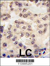
| Sample | FFPE human lung carcinoma tissue (datasheet A05395) |
| Fixation | Image formalin-fixed; duration unreported (datasheet A05395); verify before use. |
| Sectioning | 4–5 µm sections on charged slides (standard) |
| Deparaffinisation | Xylene, graded ethanol series to water (standard) |
| Antigen retrieval | Heat retrieval: Citrate pH 6 (datasheet A05395); 20 min, 95–100 °C (standard) |
| Peroxidase block | 3% H2O2, 10 min, room temperature (standard) |
| Blocking | 10% normal serum of the secondary host, 30 min, room temperature (standard) |
| Primary antibody | Rabbit anti-EN1, 1:25 (datasheet A05395) |
| Primary incubation | 1 hours at 37°C (datasheet A05395) |
| Detection | HRP-polymer secondary, DAB chromogen 5–10 min (standard) |
| Counterstain | Hematoxylin, blue, dehydrate and mount (standard) |
| Expected result | EN1-positive staining in secretory cells of skin (HPA tissue IHC: Medium). HPA tissue profile: Distinct nuclear expression in eccrine sweat glands. No signal in the no-primary control. |
EN1 is a nuclear protein with no transmembrane segment (UniProt Q05925). In tissue IHC, expect distinct nuclear staining in eccrine sweat gland secretory cells in skin; HPA reports medium staining in those cells (HPA tissue IHC). HPA rates the tissue IHC evidence Enhanced, while noting medium consistency between antibody staining and RNA expression data (HPA tissue IHC).
| Distinct nuclear chromogen in eccrine sweat gland secretory cells, with little staining elsewhere on the section. | This matches the reported skin pattern and medium staining level (HPA tissue IHC). Score the stained cells and their nuclei, since a positive gland does not establish uniform EN1 staining across skin (HPA tissue IHC). |
| Strong cytoplasmic or membrane staining dominates while gland cell nuclei remain unstained. | The compartment conflicts with nuclear EN1 localization (UniProt Q05925) and the nuclear tissue pattern (HPA tissue IHC). Treat it as suspect; inspect controls and detection background before assigning it to EN1 (standard IHC practice). |
| Prominent staining appears in adipocytes or other cells outside the reported positive gland population. | HPA reports EN1 as not detected in adipocytes in adipose tissue and breast, while its stated positive skin population is eccrine secretory cells (HPA tissue IHC). Check for antibody cross-reactivity or endogenous detection activity (standard IHC practice). |
| Diffuse chromogen covers tissue, including areas without a distinct nuclear pattern. | Diffuse background cannot establish the reported cell-specific nuclear pattern (HPA tissue IHC). Compare with a control omitting primary antibody, then review blocking, washing and detection conditions (standard IHC practice). |
| No gland cell nuclear signal appears in a skin section containing identifiable eccrine sweat glands. | This misses the reported positive population, whose staining level is medium (HPA tissue IHC). Check that the glands are present and that staining and detection controls worked before interpreting the result as absent EN1 (standard IHC practice). |
| Tissue and cell selection | Skin eccrine secretory cells provide the reported positive tissue IHC pattern; adipocytes in adipose tissue and breast are reported as not detected (HPA tissue IHC). Select comparison areas by their identified cells, since these observations do not assign one staining status to every cell in an organ (HPA tissue IHC). |
| IHC evidence and RNA agreement | HPA labels tissue IHC reliability Enhanced but describes only medium consistency between antibody staining and RNA expression data (HPA tissue IHC). Its group-enriched RNA tissues include adipose tissue, brain, skeletal muscle and skin; that RNA classification does not predict a positive chromogenic result in each sampled cell type (HPA tissue IHC). |
| Protein location and processing | UniProt places EN1 in the nucleus, records no transmembrane segment, signal peptide or propeptide, and lists one chain spanning residues 1–392 (UniProt Q05925). These annotations support scoring nuclear signal; they do not specify an IHC epitope or an antigen retrieval condition (UniProt Q05925). |
| Antibody validation scope | HPA lists HPA073141 as Enhanced for IHC and CAB014884 as Supported for ICC, with no IHC status supplied for CAB014884 (HPA antibodies). Interpret each validation label within its stated assay; the ICC status alone does not validate chromogenic paraffin-section staining (HPA antibodies). |
| IF/ICC Q&A: should its fine nuclear pattern set the IHC scoring rule? | HPA ICC-IF reports a mainly nucleoli-rim signal (approved) plus nucleoplasm (supported), with images in A-431, U-251MG and U2OS (HPA subcellular). Tissue IHC reports distinct nuclear staining in eccrine secretory cells (HPA tissue IHC); assess the finer ICC-IF pattern within that assay. |
| Situation | Likely cause | Next action |
|---|---|---|
| Eccrine glands are visible, but their secretory cell nuclei show no chromogen. | The expected tissue-positive population has been missed (HPA tissue IHC); the slide alone cannot distinguish absent signal from a failed staining run (standard IHC practice). | Confirm gland morphology and compare a successfully stained positive control processed in the same run; then review primary antibody application, retrieval and detection steps (standard IHC practice). |
| Cytoplasmic staining is stronger than nuclear staining in gland cells. | The dominant compartment disagrees with nuclear EN1 and HPA's nuclear tissue pattern (UniProt Q05925; HPA tissue IHC); nonspecific primary or detection staining is possible (standard IHC practice). | Compare a primary-omission control and assess whether any distinct nuclear signal remains after background is accounted for; interpret the compartment before scoring intensity (standard IHC practice). |
| Adipocytes stain prominently in a comparison section. | Adipocytes are reported as not detected in adipose tissue and breast (HPA tissue IHC). Cross-reactivity or endogenous chromogenic activity could produce an unexpected signal (standard IHC practice). | Verify cell identity, check a primary-omission control and review the detection system's endogenous-activity blocking steps; do not treat the signal as confirmed EN1 from color alone (standard IHC practice). |
| Nearly every structure carries weak, hazy chromogen. | Broad haze obscures the distinct gland cell nuclear pattern reported for EN1 (HPA tissue IHC); excess detection background or inadequate washing are general possibilities (standard IHC practice). | Compare background controls, inspect wash and blocking steps, and use the catalog antibody's IHC-P instructions for antibody working conditions (standard IHC practice). |
| The result varies between sections or batches. | Section content or run conditions may differ (standard IHC practice); HPA's Enhanced rating still comes with only medium staining-to-RNA consistency (HPA tissue IHC). | Compare sections with identifiable eccrine glands and the same control tissue, then document nuclear intensity and the proportion of stained secretory cells separately (standard IHC practice). |
| Fine nucleoli-rim staining is absent from an otherwise nuclear-positive IHC section. | The finer nucleoli-rim assignment comes from ICC-IF, whereas the tissue IHC description is distinct nuclear expression in eccrine glands (HPA subcellular; HPA tissue IHC). | Judge the paraffin-section IHC result against its tissue pattern and controls; use the separate IF/ICC guide when evaluating the ICC-IF subnuclear pattern (HPA tissue IHC; HPA subcellular). |


Comprehensive Human Protein Atlas IHC scoring per tissue (reliability: Enhanced — Medium consistency between antibody staining and RNA expression data.). Rows are taken directly from the HPA tissue chart — click any row's HPA link to view the source.
| Tissue | Cell type | Level | Evidence | Source |
|---|---|---|---|---|
| Skin | Secretory cells | Medium | Protein (IHC) | HPA → |
| Tissue | Cell type | Level | Evidence | Source |
|---|---|---|---|---|
| Adipose tissue | Adipocytes | Not detected | Protein (IHC) | HPA → |
| Adrenal gland | Glandular cells | Not detected | Protein (IHC) | HPA → |
| Appendix | Glandular cells | Not detected | Protein (IHC) | HPA → |
| Bone marrow | Hematopoietic cells | Not detected | Protein (IHC) | HPA → |
| Breast | Adipocytes | Not detected | Protein (IHC) | HPA → |
Troubleshoot EN1 staining by checking retrieval, nuclear localisation, cell identity and controls before interpreting signal intensity.
The IHC-validated anti-EN1 antibody has human paraffin-section images and human cell IF images (catalog images: A05395); its listed reactivity also includes mouse (catalog: A05395).
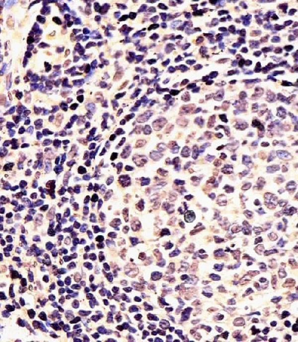
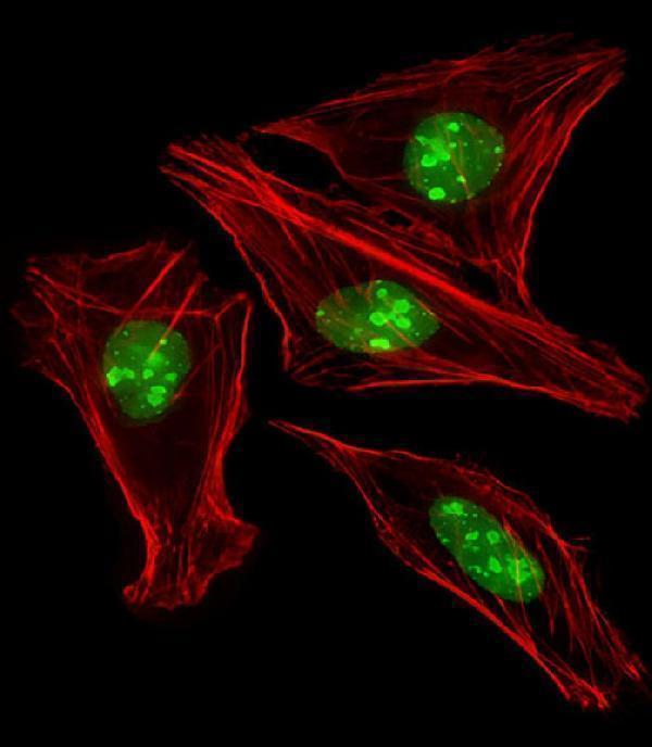
A05395 will render with an IHC figure showing formalin-fixed, paraffin-embedded human lung carcinoma tissue stained by DAB detection (A05395 IHC image caption). Its application list includes IHC-P and IF, and its IF images show human U-2 OS and U251 cells (catalog: A05395 applications; A05395 IF image captions).
Which to pick: Choose A05395 for tissue IHC: its own images show human lung carcinoma after formalin fixation and paraffin embedding, plus human tonsil sections after formaldehyde fixation and paraffin embedding (A05395 IHC image captions). Choose the same rabbit polyclonal antibody for IF/ICC because IF is listed and human U-2 OS and U251 cell images are supplied (catalog: A05395 host, clonality and applications; A05395 IF image captions). For cross-species work, A05395 lists human and mouse reactivity, although the supplied IHC and IF images show human samples (catalog: A05395 reactivity; A05395 IHC and IF image captions).