This website uses cookies to ensure you get the best experience on our website.


- Table of Contents
Plan ENO2 chromogenic IHC in paraffin sections using cytoplasmic CNS staining as a reference (HPA tissue IHC). Compare neuronal and pancreatic endocrine staining with the catalog antibody’s validated IHC conditions (HPA tissue IHC; datasheet A02930).

Expected localisation, validated protocols, controls and antibodies — the at-a-glance facts below, then the full design guide.
| Expected localisation | Cytoplasmic tissue staining (HPA tissue IHC); membrane translocation is possible (UniProt) | |
| Staining pattern | Cytoplasmic staining in CNS, peripheral nerves and islets (HPA tissue IHC) | |
| Antigen retrieval | Citrate pH 6 HIER, heat-mediated (datasheet A02930) | |
| Positive control | Caudate+4 more · see all | |
| Negative control | Adipose tissue+4 more · see all |
| Fixation | Keep fixation consistent across sections. (standard IHC practice; not target-specific) | |
| Caveat | Pancreatic endocrine cells stain less strongly than CNS neurons (HPA tissue IHC) | |
| Regulation | Enolase dimers vary with cell type and development (UniProt) | |
| Isoform / epitope | 2 isoforms; check epitope coverage across variants (UniProt) |
The catalog antibody protocol and four published ENO2 IHC protocols provide starting conditions for paraffin sections (datasheet A02930; PMC8140915; PMC8253290; PMC9367517; PMC3390755).
| Sample | Paraffin-embedded human lung cancer tissue; fixative not specified (datasheet A02930) |
| Fixation | Image fixative and duration unreported (datasheet A02930); verify before use. |
| Sectioning | 4–5 µm sections on charged slides (standard) |
| Deparaffinisation | Xylene, graded ethanol series to water (standard) |
| Antigen retrieval | Heat retrieval: Citrate pH 6, 20 min (datasheet A02930) |
| Peroxidase block | 3% H2O2, 10 min, room temperature (standard) |
| Blocking | 10% goat serum (datasheet A02930) |
| Primary antibody | Rabbit anti-ENO2, 0.5-1μg/ml (datasheet A02930) |
| Primary incubation | Overnight at 4 °C (datasheet A02930) |
| Detection | Streptavidin-biotin complex (SABC), DAB chromogen (datasheet A02930) |
| Counterstain | Hematoxylin, blue, dehydrate and mount (standard) |
| Expected result | ENO2-positive staining in neuronal cells of caudate (HPA tissue IHC: High). HPA tissue profile: Cytoplasmic expression in CNS, peripheral nerves and pancreatic islets. No signal in the no-primary control. |
In paraffin-section IHC, expect ENO2 mainly in the cytoplasm of CNS neurons, cells in the cerebellar granular layer, peripheral nerves, and pancreatic islets (HPA tissue IHC: Enhanced; cytoplasmic profile). Neuronal staining is high in the sampled CNS regions, while pancreatic endocrine-cell staining is medium (HPA tissue IHC). ENO2 can also reach the cell membrane despite having no transmembrane segment (UniProt P09104: location and topology).
| Strong cytoplasmic staining in cortical, hippocampal, or caudate neurons. | This matches the High neuronal staining reported in these regions (HPA tissue IHC). Judge the result by its cellular distribution as well as intensity: the expected signal occupies neuronal cytoplasm (HPA tissue IHC). A membrane edge can be plausible because ENO2 can translocate to the plasma membrane (UniProt P09104: location). |
| Predominantly nuclear staining, with little neuronal cytoplasmic signal. | A nuclear-only pattern does not match the reported cytoplasmic tissue profile (HPA tissue IHC) or the cytoplasm and cell-membrane locations (UniProt P09104). Treat it as a possible artefact and review the controls and staining distribution before scoring it as ENO2. Do not reject a membrane-associated rim solely for being outside the cytoplasm (UniProt P09104: location). |
| Strong staining in adipocytes or respiratory epithelial cells. | Those cell types are reported as Not detected in adipose tissue and bronchus, respectively (HPA tissue IHC). If staining persists in them, consider antibody cross-reactivity or, with an enzyme-based detection system, endogenous detection activity (general IHC practice). Compare the suspect cells with a known-positive neuronal region and the assay controls before assigning target-specific signal. |
| Widespread pale color obscures cell boundaries across the section. | Diffuse, poorly localized color is difficult to reconcile with the reported cytoplasmic pattern in CNS, peripheral nerves, and pancreatic islets (HPA tissue IHC). It can reflect nonspecific binding, residual detection activity, or excessive chromogen development (general IHC practice). Interpretation should wait until the background is low enough to distinguish positive cells from surrounding tissue. |
| No signal in a known-positive neuronal region. | Absent staining conflicts with the High neuronal signal reported in caudate, cerebral cortex, and hippocampus (HPA tissue IHC). It does not establish that the specimen lacks ENO2. Check that tissue morphology is preserved, the antibody and detection reagents worked, and the assay conditions follow an IHC-validated protocol (general IHC practice). |
| Cell compartment | Cytoplasm is the principal tissue-IHC pattern (HPA tissue IHC). Membrane-associated signal is biologically plausible because ENO2 can translocate there despite lacking a transmembrane segment (UniProt P09104: location and topology). |
| Cell and tissue context | CNS neuronal cells and cerebellar granular-layer cells are High; pancreatic endocrine cells are Medium (HPA tissue IHC). Compare like cell types when judging intensity. |
| Antibody evidence | The tissue-IHC profile is rated Enhanced for consistency with RNA data (HPA tissue IHC). Individual IHC antibody statuses differ: CAB000063 and CAB073539 are Enhanced; CAB079023 is Supported (HPA antibodies). |
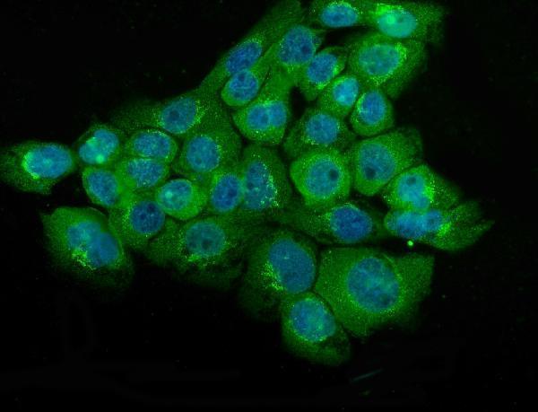
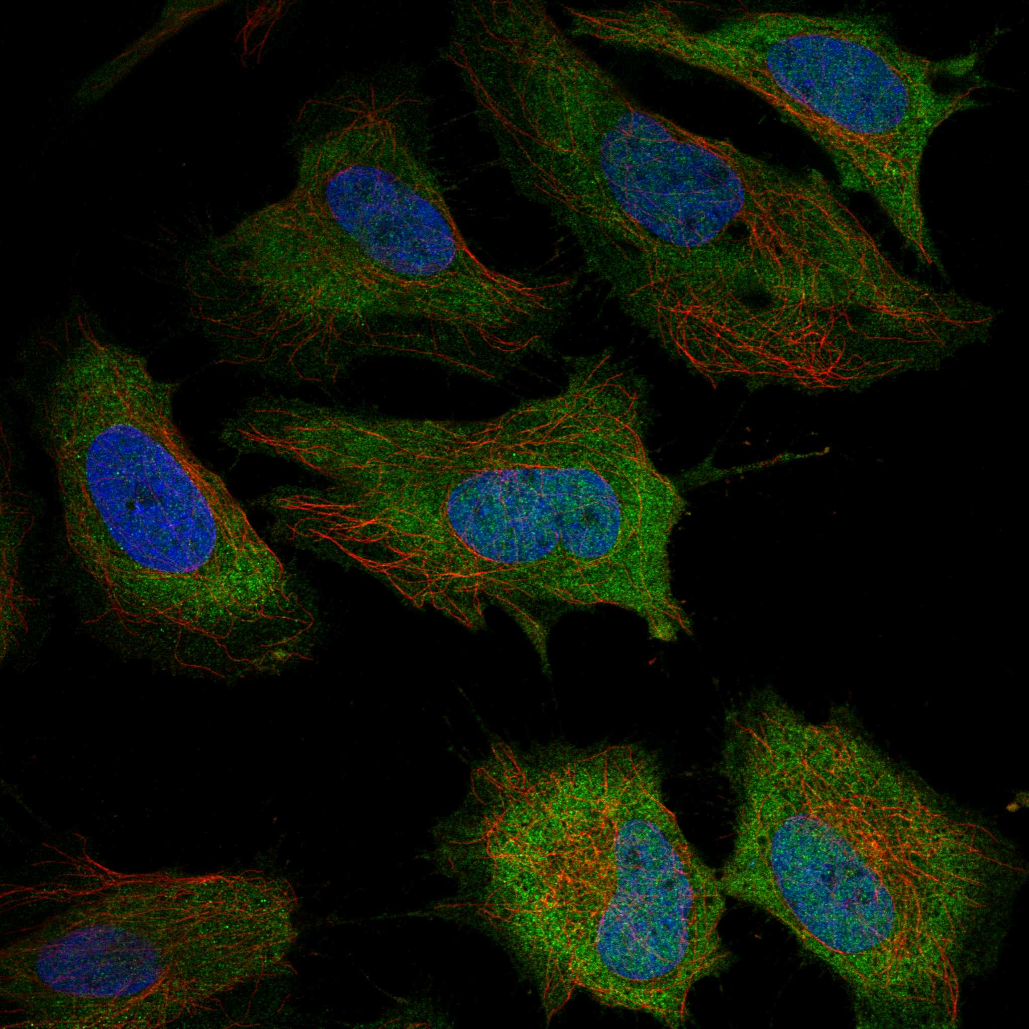
| What should IF/ICC show? | HPA reports cytosol and plasma-membrane localization in ICC-IF, but cautions that its summary uses antibodies targeting proteins from multiple genes (HPA subcellular). Use the separate IF/ICC guide for assay design. |
| Situation | Likely cause | Next action |
|---|---|---|
| Neuronal positive control is blank. | A failed staining or detection step is possible; high neuronal staining is expected in sampled CNS regions (HPA tissue IHC). | Confirm tissue identity and morphology, then check reagent activity, the IHC-validated antibody conditions, and detection controls (general IHC practice). |
| Only nuclei appear positive. | The compartment conflicts with the cytoplasmic tissue profile (HPA tissue IHC) and UniProt locations (UniProt P09104). | Check the counterstain and omission control, then reassess whether any specific neuronal cytoplasmic signal remains (general IHC practice). |
| Adipocytes or bronchial epithelium stain strongly. | These cells are reported as Not detected (HPA tissue IHC); cross-reactivity or endogenous detection activity is possible. | Compare with a neuronal positive control; use primary-antibody omission and detection-system controls to investigate the unwanted color (general IHC practice). |
| Color covers cells and extracellular areas uniformly. | Nonspecific binding or overdevelopment may obscure the expected cell-associated pattern (general IHC practice; HPA tissue IHC: cytoplasmic profile). | Review blocking, washes, antibody concentration, and chromogen development; retain settings that resolve individual positive cells (general IHC practice). |
| Pancreatic endocrine cells stain less strongly than CNS neurons. | That difference agrees with Medium endocrine-cell versus High neuronal staining (HPA tissue IHC). | Assess each compartment against its reported level and cell type; do not require pancreatic cells to match neuronal intensity (HPA tissue IHC). |
| A narrow membrane rim accompanies neuronal cytoplasmic staining. | ENO2 can move to the plasma membrane (UniProt P09104: location); HPA ICC-IF also reports membrane and cytosol, with a multi-gene-antibody caution (HPA subcellular). | Score the tissue-IHC cytoplasmic pattern first (HPA tissue IHC), and use controls before interpreting the rim as target-specific (general IHC practice). |
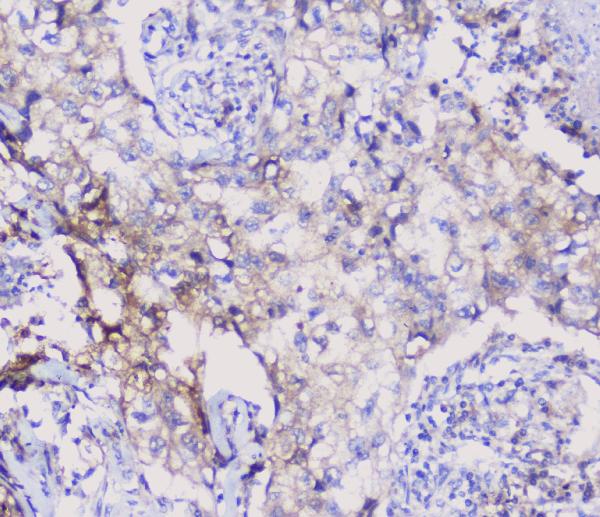

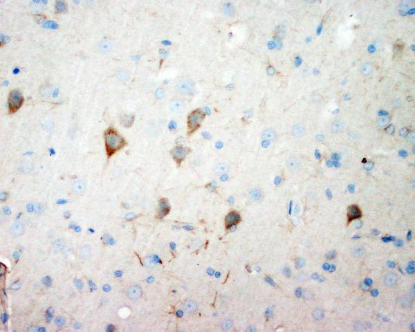
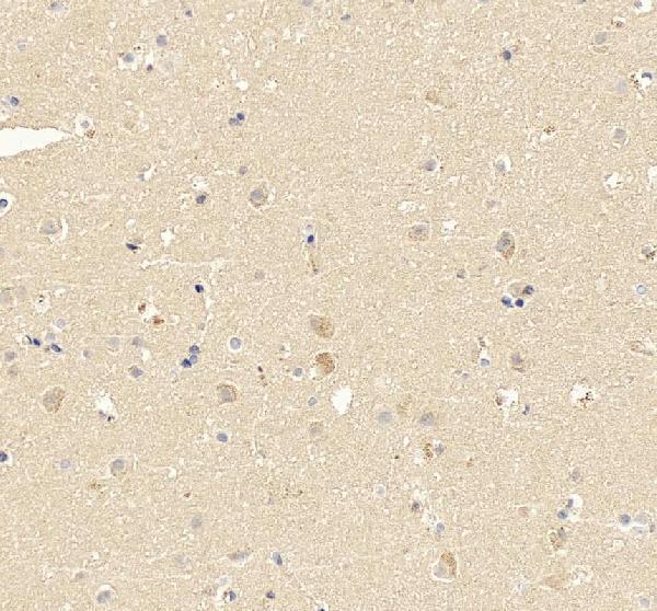


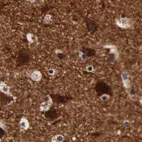
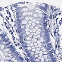
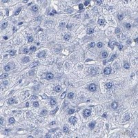
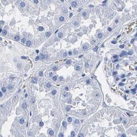

Comprehensive Human Protein Atlas IHC scoring per tissue (reliability: Enhanced — High consistency between antibody staining and RNA expression data.). Rows are taken directly from the HPA tissue chart — click any row's HPA link to view the source.
| Tissue | Cell type | Level | Evidence | Source |
|---|---|---|---|---|
| Caudate | Neuronal cells | High | Protein (IHC) | HPA → |
| Cerebellum | Cells in granular layer | High | Protein (IHC) | HPA → |
| Cerebral cortex | Neuronal cells | High | Protein (IHC) | HPA → |
| Hippocampus | Neuronal cells | High | Protein (IHC) | HPA → |
| Pancreas | Pancreatic endocrine cells | Medium | Protein (IHC) | HPA → |
| Tissue | Cell type | Level | Evidence | Source |
|---|---|---|---|---|
| Adipose tissue | Adipocytes | Not detected | Protein (IHC) | HPA → |
| Adrenal gland | Glandular cells | Not detected | Protein (IHC) | HPA → |
| Appendix | Endocrine cells | Not detected | Protein (IHC) | HPA → |
| Bone marrow | Hematopoietic cells | Not detected | Protein (IHC) | HPA → |
| Breast | Adipocytes | Not detected | Protein (IHC) | HPA → |
Troubleshoot ENO2 staining in paraffin sections by checking retrieval, compartment, cell identity and matched controls (datasheet A02930; HPA tissue IHC; UniProt P09104).
Anti-ENO2 antibodies have IHC images from human and rodent paraffin sections (catalog IHC captions); IF/ICC images show A431 cells (catalog IF captions).
A02930 shows IHC in human lung cancer, human placenta, rat brain and mouse brain, plus IF/ICC in A431 cells (A02930 image captions). PA1061 shows IHC in rat and human brain, while M02930-1 shows IHC in human uterus cancer and IF in A431 cells (PA1061 and M02930-1 image captions).
Which to pick: For paraffin-section IHC, choose A02930 for the broadest pictured tissue and species coverage; PA1061 has pictured rat and human brain, and M02930-1 has pictured human uterus cancer (each SKU’s IHC captions). For IF/ICC, choose A02930 or rabbit monoclonal M02930-1, both listed for IF/ICC with A431 images (catalog applications and IF captions); A02930 has pictured IHC examples in human, mouse and rat (A02930 IHC captions). The paraffin captions do not report a fixative (catalog IHC captions).