This website uses cookies to ensure you get the best experience on our website.


- Table of Contents
Plan ENPP1 staining in paraffin sections using the membranous pattern seen in selected cells (HPA tissue IHC). Catalog antibody A01342-1 is listed for human IHC at 1–2 μg/ml (datasheet A01342-1); consider the secreted form when interpreting staining (UniProt).

Expected localisation, validated protocols, controls and antibodies — the at-a-glance facts below, then the full design guide.
| Expected localisation | Membranous staining in selected cells (HPA tissue IHC) | |
| Staining pattern | Membranous in placental stroma, chondrocytes and glands (HPA tissue IHC) | |
| Antigen retrieval | EDTA pH 8.0 HIER, heat-mediated (datasheet A01342-1) | |
| Positive control | Parathyroid gland+4 more · see all | |
| Negative control | Adipose tissue+4 more · see all |
| Fixation | Keep fixation consistent across sections (standard IHC practice; not target-specific) | |
| Caveat | A secreted form may alter the expected membrane pattern (UniProt) | |
| Regulation | Placenta-enhanced RNA expression (HPA tissue RNA) | |
| Isoform / epitope | No listed isoforms; secreted form lacks the cytoplasmic region (UniProt) |
The catalog antibody protocol is supplemented by three published ENPP1 IHC methods covering rabbit knee cartilage, breast carcinoma tissue microarrays, and histological sections (PMC9276012; PMC3783308; PMC5803363).
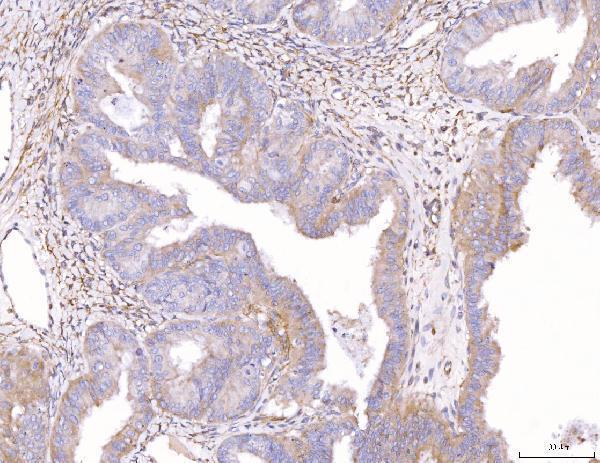
| Sample | Paraffin-embedded human endometrial cancer tissue; fixative not specified (datasheet A01342-1) |
| Fixation | Image fixative and duration unreported (datasheet A01342-1); verify before use. |
| Sectioning | 4–5 µm sections on charged slides (standard) |
| Deparaffinisation | Xylene, graded ethanol series to water (standard) |
| Antigen retrieval | Heat retrieval: EDTA pH 8.0 (datasheet A01342-1); 20 min, 95–100 °C (standard) |
| Peroxidase block | 3% H2O2, 10 min, room temperature (standard) |
| Blocking | 10% goat serum (datasheet A01342-1) |
| Primary antibody | Rabbit anti-ENPP1, 1-2 μg/ml (datasheet A01342-1) |
| Primary incubation | Overnight at 4 °C (datasheet A01342-1) |
| Detection | HRP-conjugated secondary, DAB chromogen (datasheet A01342-1) |
| Counterstain | Hematoxylin, blue, dehydrate and mount (standard) |
| Expected result | ENPP1-positive staining in glandular cells of parathyroid gland (HPA tissue IHC: High). HPA tissue profile: Membranous expression in a subset of cells including placental stroma, chondrocytes, exocrine pancreas, renal tubules, thyroid and parathyroid gland. No signal in the no-primary control. |
ENPP1 is a membrane protein with one transmembrane segment, residues 77–97, and may also be secreted (UniProt P22413 topology; UniProt P22413 processing). In paraffin-section IHC, expect membranous staining in selected cells, including thyroid and parathyroid glandular cells, renal distal tubules and chondrocytes (HPA tissue IHC). HPA rates its tissue pattern Enhanced, while noting medium consistency with RNA expression and a secreted variant that complicates tissue-level comparisons (HPA tissue IHC).
| Distinct membrane staining in thyroid or parathyroid glandular cells, with adjacent cells less stained (HPA tissue IHC). | This fits HPA's High staining in those glandular cells and its description of expression in a subset of cells (HPA tissue IHC). Score the named cell population and its membrane pattern rather than assigning one intensity to the whole section (general IHC practice). |
| Predominantly nuclear staining, without a convincing membrane pattern, in a known-positive cell population. | Nuclear localization does not fit ENPP1's annotated membrane and secreted locations (UniProt P22413 subcellular location; HPA subcellular summary). Treat it as a suspected artefact; check the counterstain and detection controls before scoring it as ENPP1 (general IHC practice). |
| Strong staining throughout HPA-negative cell populations, such as adipocytes or bone-marrow hematopoietic cells (HPA tissue IHC). | HPA reports ENPP1 as Not detected in those named cells (HPA tissue IHC). Widespread staining there raises concern for antibody cross-reactivity or endogenous detection activity; a no-primary control helps distinguish detection background (general IHC practice). |
| Diffuse color across cells and tissue spaces, obscuring individual membrane outlines. | This cannot establish the cell-selective membrane pattern reported by HPA (HPA tissue IHC). Review no-primary staining, blocking, washing and detection conditions; extracellular color alone also needs caution because a secreted ENPP1 form is annotated (general IHC practice; UniProt P22413 processing). |
| No discernible signal in thyroid or parathyroid glandular cells (HPA tissue IHC). | Those cells are useful positive comparators because HPA reports High staining (HPA tissue IHC). A blank comparator makes a negative result elsewhere hard to interpret; review the catalog antibody's IHC-P instructions and the staining controls (general IHC practice). |
| Topology and epithelial polarity (UniProt P22413 topology; UniProt P22413 subcellular location). | ENPP1 has a short cytoplasmic portion, residues 1–76, and an extracellular portion, residues 98–925 (UniProt P22413 topology). UniProt places it on basolateral membranes in polarized epithelia and hepatocytes, so assess membrane position where cell borders are visible (UniProt P22413 subcellular location). |
| Processing and extracellular signal (UniProt P22413 processing). | UniProt annotates a secreted form spanning residues 103–925 and matrix-vesicle targeting in osteoblasts (UniProt P22413 processing; UniProt P22413 subcellular location). Extracellular staining can therefore merit investigation, but it should not replace cell and compartment checks or be assumed specific from position alone (general IHC practice). |
| Cell-specific reference levels (HPA tissue IHC). | HPA reports High glandular-cell staining in thyroid and parathyroid, Medium staining in kidney distal tubules and pancreatic exocrine glandular cells, and Low staining in lung macrophages (HPA tissue IHC). Compare equivalent cells when judging signal, since a faint low-reference population is a weaker positive control (general IHC practice). |
| Interpretive limits of tissue evidence (HPA tissue IHC). | Target-specific fixation effects are not established by the supplied assay evidence. Verify with a matched IHC source before attributing a result to fixation. |
| Antibody-specific validation (HPA antibodies). | HPA lists IHC status as Supported for HPA062066 and Enhanced for CAB032904 (HPA antibodies). These are separate antibody assessments; confirm which reagent generated a reference image before treating its intensity as directly comparable to the catalog antibody (general IHC practice). |
| IF/ICC: should the IHC pattern be expected there? (HPA subcellular summary). | Membrane or secreted localization is a reasonable starting expectation (UniProt P22413 subcellular location; HPA subcellular summary). HPA provides no ICC-IF image cell lines or main-location assignment here, so this IHC tissue record cannot validate an IF/ICC image or establish an IF/ICC staining condition (HPA subcellular summary). |
| Situation | Likely cause | Next action |
|---|---|---|
| Known-positive thyroid or parathyroid cells remain unstained (HPA tissue IHC). | The positive comparator has failed; HPA reports High staining in those cells, but this observation alone does not identify the failed staining step (HPA tissue IHC; general IHC practice). | Check slide integrity, reagent sequence and detection controls, then review the catalog antibody's IHC-P protocol before interpreting other tissue as negative (general IHC practice). |
| Membrane staining is weak in kidney distal tubules (HPA tissue IHC). | HPA reports Medium staining there, so a weaker signal than in its high-staining glandular references may be expected (HPA tissue IHC). | Compare the same cell type across sections and confirm that a high-reference tissue and the detection control worked before adjusting the IHC workflow (HPA tissue IHC; general IHC practice). |
| Broad brown color appears in HPA-negative cells and in the no-primary control (HPA tissue IHC). | Color without primary antibody points to background from the detection workflow; endogenous enzyme activity is one possible cause with enzyme-based chromogenic detection (general IHC practice). | Review the detection system's endogenous-activity blocking step and repeat the no-primary control before calling those cells ENPP1-positive (general IHC practice). |
| Broad staining persists only when primary antibody is added, including adipocytes or bone-marrow hematopoietic cells (HPA tissue IHC). | Those named cell populations are Not detected in HPA; primary-dependent staining raises concern for cross-reactivity or insufficient antibody optimization (HPA tissue IHC; general IHC practice). | Recheck the catalog antibody's IHC-P guidance, assess its staining against a high-reference cell population, and interpret the discordant cells cautiously (HPA tissue IHC; general IHC practice). |
| A strong nuclear pattern dominates while cell membranes are difficult to identify. | The observed compartment conflicts with ENPP1's membrane and secreted annotations; counterstain or nonspecific color may be misleading (UniProt P22413 subcellular location; general IHC practice). | Inspect a no-primary control and the counterstain separately, then score ENPP1 only where the primary-dependent signal has a credible cell and compartment pattern (general IHC practice). |
| Extracellular deposits appear beside weakly stained cells. | A secreted ENPP1 form is annotated, but extracellular color by itself does not identify the deposited material (UniProt P22413 processing; general IHC practice). | Record the deposit separately from membrane staining, compare known-positive cells and controls, and avoid assigning the deposit to ENPP1 from location alone (HPA tissue IHC; general IHC practice). |


Comprehensive Human Protein Atlas IHC scoring per tissue (reliability: Enhanced — Medium consistency between antibody staining and RNA expression data. At least one protein variant secreted, tissue location of RNA and protein might differ and correlation is complex. Antibody staining in cells/structures not annotated, view images.). Rows are taken directly from the HPA tissue chart — click any row's HPA link to view the source.
| Tissue | Cell type | Level | Evidence | Source |
|---|---|---|---|---|
| Parathyroid gland | Glandular cells | High | Protein (IHC) | HPA → |
| Thyroid gland | Glandular cells | High | Protein (IHC) | HPA → |
| Appendix | Germinal center cells | Medium | Protein (IHC) | HPA → |
| Gallbladder | Glandular cells | Medium | Protein (IHC) | HPA → |
| Kidney | Distal tubules | Medium | Protein (IHC) | HPA → |
| Tissue | Cell type | Level | Evidence | Source |
|---|---|---|---|---|
| Adipose tissue | Adipocytes | Not detected | Protein (IHC) | HPA → |
| Adrenal gland | Glandular cells | Not detected | Protein (IHC) | HPA → |
| Bone marrow | Hematopoietic cells | Not detected | Protein (IHC) | HPA → |
| Bronchus | Respiratory epithelial cells | Not detected | Protein (IHC) | HPA → |
| Cerebellum | Cells in granular layer | Not detected | Protein (IHC) | HPA → |
Troubleshoot ENPP1 staining in paraffin sections by checking retrieval, compartment, cell type and controls before interpreting chromogenic signal (UniProt P22413; HPA tissue IHC).
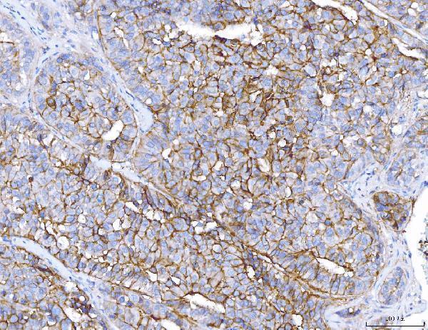
A01342-1 has real IHC images from human paraffin-embedded endometrial cancer, liver cancer, and placenta sections (catalog image captions). No IF/ICC image is supplied (catalog payload).
A01342-1 is listed for human IHC, with staining shown in paraffin-embedded endometrial cancer and liver cancer sections (catalog applications and image captions). Its IHC staining is also shown in human placenta (catalog image caption).
Which to pick: Choose A01342-1 for human paraffin-section IHC; its captions report staining at 2 μg/ml after EDTA retrieval at pH 8.0 (catalog image captions). The captions do not report the fixative (catalog image captions). No listed SKU has IF/ICC validation or nonhuman reactivity, so this payload does not support an IF/ICC or cross-species pick (catalog applications and reactivity).