This website uses cookies to ensure you get the best experience on our website.


- Table of Contents
Plan chromogenic ENPP3 IHC on paraffin sections using the catalog antibody at a 1:100 starting dilution (datasheet: A05615 IHC). This guide highlights strong microvillar staining in enterocytes and proximal tubules (HPA tissue IHC) and the potential for secreted ENPP3 to contribute extracellular signal (UniProt).

Expected localisation, validated protocols, controls and antibodies — the at-a-glance facts below, then the full design guide.
| Expected localisation | Membranous and extracellular tissue staining (HPA tissue IHC) | |
| Staining pattern | Strong microvillar staining in enterocytes and proximal tubules (HPA tissue IHC) | |
| Antigen retrieval | Citrate pH 6.0 HIER, 95–98 °C, 20 min (rule: cytoplasmic / membrane antigen) | |
| Positive control | Duodenum+4 more · see all | |
| Negative control | Adipose tissue+4 more · see all |
| Fixation | Keep fixation consistent across sections (standard IHC practice; not target-specific); assess staining empirically. | |
| Caveat | Secreted ENPP3 may contribute extracellular staining (UniProt) | |
| Regulation | Staining varies by tissue and cell type (HPA tissue IHC) | |
| Isoform / epitope | No isoforms listed; extracellular vs cytoplasmic epitope matters (UniProt) |
The catalog antibody’s IHC-P protocol is followed by published ENPP3 staining methods for heart, renal carcinoma, and ovarian endometriosis sections (PMC12203807; PMC6731023; PMC11293237).
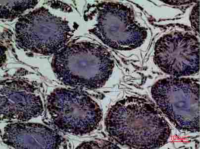
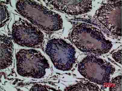
| Sample | Paraffin-embedded Rat Testis tissue; fixative not specified (datasheet A05615) |
| Fixation | Image fixative and duration unreported (datasheet A05615); verify before use. |
| Sectioning | 4–5 µm sections on charged slides (standard) |
| Deparaffinisation | Xylene, graded ethanol series to water (standard) |
| Antigen retrieval | Heat-induced epitope retrieval in citrate buffer, pH 6.0, 20 min at 95–98 °C (standard rule: cytoplasmic / membrane antigen) |
| Peroxidase block | 3% H2O2, 10 min, room temperature (standard) |
| Blocking | 10% normal serum of the secondary host, 30 min, room temperature (standard) |
| Primary antibody | Rabbit anti-ENPP3, 1:100 (datasheet A05615) |
| Primary incubation | Overnight at 4 °C (standard) |
| Detection | HRP-polymer secondary, DAB chromogen 5–10 min (standard) |
| Counterstain | Hematoxylin, blue, dehydrate and mount (standard) |
| Expected result | ENPP3-positive staining in enterocytes - Microvilli of duodenum (HPA tissue IHC: High). HPA tissue profile: Membranous expression and extracellular positivity in several tissues. No signal in the no-primary control. |
ENPP3 is a membrane protein with an extracellular domain spanning residues 31–875; it is also reported as secreted (UniProt O14638 topology; UniProt O14638 subcellular). In paraffin-section IHC, expect apical or surface staining in the specified epithelial cells, including intestinal enterocytes and kidney proximal tubules (HPA: High in enterocyte and proximal-tubule microvilli). HPA rates its tissue IHC reliability Enhanced, while noting that secreted protein can complicate the match between tissue RNA and protein location (HPA: tissue IHC reliability description).
| A crisp luminal rim on duodenal or small-intestinal enterocytes, or on kidney proximal tubules. | This matches the strongest compartment-specific examples: enterocyte and proximal-tubule microvilli are High (HPA: duodenum, small intestine, kidney). Score the relevant epithelial surface, rather than averaging its intensity with the entire section (general IHC scoring practice). |
| Membranous staining with extracellular positivity in other tissue compartments. | Both can be compatible with ENPP3: HPA describes membranous expression and extracellular positivity in several tissues, and UniProt reports membrane-bound and secreted protein (HPA: tissue IHC profile; UniProt O14638 subcellular). Confirm the local cell and compartment before calling a positive result (general IHC interpretation practice). |
| Strong nuclear staining or a predominantly intracellular pattern without the expected surface signal. | This is a compartment mismatch against the reported apical membrane, cell-surface and secreted locations (UniProt O14638 subcellular; HPA: tissue IHC profile). Treat it as questionable staining and compare it with appropriate control sections before assigning ENPP3 expression (general IHC interpretation practice). |
| Signal in cells that HPA lists as not detected, such as adipocytes in adipose tissue. | Investigate cross-reactivity or detection background before interpreting that cell population as ENPP3-positive (HPA: adipose adipocytes Not detected; general IHC troubleshooting practice). HPA's finding applies to the named cells; it does not declare every cell in that tissue negative (HPA: adipose tissue cell-level entry). |
| No visible stain in a section containing intact intestinal enterocytes or kidney proximal tubules. | An absent signal conflicts with HPA's High microvillar staining in those cells (HPA: duodenum, small intestine, kidney). Review section quality and the IHC workflow before interpreting an experimental sample as negative (general IHC troubleshooting practice). |
| Topology and compartment | ENPP3 has one transmembrane segment at residues 12–30 and an extracellular region at 31–875 (UniProt O14638 topology). Use the epithelial surface as the main spatial reference when interpreting the reported apical staining (UniProt O14638 subcellular; HPA: microvillar IHC entries). |
| Secreted protein | UniProt reports ENPP3 in serum and epithelial lumens; HPA reports extracellular positivity (UniProt O14638 subcellular; HPA: tissue IHC profile). Extracellular colour therefore needs anatomical context, and its presence alone cannot identify the producing cell (HPA: reliability description). |
| Tissue and cell selection | Duodenal and small-intestinal enterocyte microvilli and kidney proximal-tubule microvilli are High; endometrial glandular cells are Medium (HPA: tissue IHC entries). Compare the same named cell compartment across samples when judging staining intensity (general IHC scoring practice). |
| Evidence strength | HPA calls the tissue staining Enhanced and lists antibody HPA043772 as IHC Enhanced (HPA: tissue IHC reliability; HPA: antibody validation). Its reliability description also reports medium staining–RNA consistency and warns that secretion complicates tissue-location comparisons (HPA: reliability description). |
| Antigen retrieval and epitope | The supplied UniProt topology defines extracellular orientation, but neither source identifies the catalog antibody's epitope or an ENPP3-specific retrieval response (UniProt O14638 topology; HPA: antibody entry). Record the retrieval conditions used and compare controls when optimizing IHC (general IHC practice). |
| IF/ICC? | HPA's subcellular summary says Secreted, lists no main location and provides no ICC-IF cell-line images (HPA: subcellular record). The IHC microvillar pattern can guide interpretation, but this record does not establish a validated IF/ICC pattern or an IF/ICC protocol (HPA: tissue IHC; HPA: subcellular record). |
| Situation | Likely cause | Next action |
|---|---|---|
| Expected intestinal or kidney microvilli are unstained. | The result disagrees with High staining in those named cells (HPA: duodenum, small intestine, kidney); the failing step cannot be identified from that observation alone (general IHC troubleshooting practice). | Check that the expected cells are present, then inspect the IHC-validated antibody, retrieval, detection reagents and a known-positive control together (general IHC troubleshooting practice). |
| Chromogen coats much of the section, including unrelated structures. | Diffuse colour can arise from nonspecific binding or detection background in chromogenic IHC (general IHC practice); HPA's extracellular positivity does not make every diffuse deposit specific (HPA: tissue IHC profile). | Compare a primary-omission control, review blocking and washes, and assess whether colour tracks recognisable cells or extracellular spaces (general IHC troubleshooting practice). |
| Signal appears mainly nuclear or uniformly cytoplasmic. | That distribution differs from the reported cell-surface, apical and extracellular pattern (UniProt O14638 subcellular; HPA: tissue IHC profile). | Recheck the compartment at higher magnification and compare the same staining run with intestinal or kidney microvilli (HPA: High in those microvilli; general IHC interpretation practice). |
| Adipocytes or adrenal glandular cells show a strong signal. | Those named populations are listed as Not detected (HPA: adipose adipocytes; HPA: adrenal glandular cells); cross-reactivity or detection background is possible (general IHC troubleshooting practice). | Check primary-omission and known-positive controls, then score the stained cell type explicitly rather than assigning positivity to the whole tissue (general IHC scoring practice). |
| A glandular or luminal deposit is difficult to assign to a cell. | ENPP3 can be secreted into epithelial lumens, and HPA reports extracellular positivity (UniProt O14638 subcellular; HPA: tissue IHC profile). Its tissue RNA and protein locations need not coincide (HPA: reliability description). | Record membrane-associated and extracellular staining separately; use cell morphology and a control section when assigning a cellular source (general IHC interpretation practice). |
| An IF/ICC image shows puncta without a clear surface pattern. | HPA supplies no ICC-IF cell-line images or main subcellular location for this record (HPA: subcellular record), so that image cannot be validated against an HPA IF reference. | Treat the puncta as unresolved, assess appropriate IF controls and consult the separate IF/ICC guide for its workflow (general IF interpretation practice). |
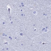


Comprehensive Human Protein Atlas IHC scoring per tissue (reliability: Enhanced — Medium consistency between antibody staining and RNA expression data. At least one protein variant secreted, tissue location of RNA and protein might differ and correlation is complex.). Rows are taken directly from the HPA tissue chart — click any row's HPA link to view the source.
| Tissue | Cell type | Level | Evidence | Source |
|---|---|---|---|---|
| Duodenum | Enterocytes - Microvilli | High | Protein (IHC) | HPA → |
| Kidney | Proximal tubules (microvilli) | High | Protein (IHC) | HPA → |
| Nasopharynx | Ciliated cells (cilia axoneme) | High | Protein (IHC) | HPA → |
| Seminal vesicle | Glandular cells | High | Protein (IHC) | HPA → |
| Small intestine | Enterocytes - Microvilli | High | Protein (IHC) | HPA → |
| Tissue | Cell type | Level | Evidence | Source |
|---|---|---|---|---|
| Adipose tissue | Adipocytes | Not detected | Protein (IHC) | HPA → |
| Adrenal gland | Glandular cells | Not detected | Protein (IHC) | HPA → |
| Appendix | Endocrine cells | Not detected | Protein (IHC) | HPA → |
| Bone marrow | Hematopoietic cells | Not detected | Protein (IHC) | HPA → |
| Breast | Adipocytes | Not detected | Protein (IHC) | HPA → |
Troubleshoot ENPP3 staining by checking apical localisation, extracellular signal, and cell-specific controls before comparing staining intensity across sections.
A05615 has IHC images from paraffin-embedded rat testis and human prostate cancer (catalog image captions); IF is listed as an application, with no IF image supplied (catalog applications and images).
A05615 has IHC images of paraffin-embedded rat testis and human prostate cancer at 1:100 (catalog image captions). It lists IF as an application and Human, Mouse, and Rat reactivity; no IF image is supplied (catalog applications, reactivity, and images).
Which to pick: For tissue IHC, choose A05615 for paraffin sections supported by its rat testis and human prostate cancer captions; the fixative is unreported (catalog image captions). For IF, A05615 lists a 1:50 dilution, but no IF image or ICC validation is supplied (catalog dilution, applications, and images). For work across species, A05615 lists Human, Mouse, and Rat reactivity, while its IHC images show Rat and Human samples only; clone information is unreported (catalog reactivity, image captions, and clone field).