This website uses cookies to ensure you get the best experience on our website.


- Table of Contents
Plan ENTPD5 IHC in paraffin sections using hepatocytes and kidney proximal tubules as positive controls (HPA tissue IHC). This guide covers fixation, antibody dilution and chromogenic scoring with the IHC-validated antibody (datasheet A06908-2).

Expected localisation, validated protocols, controls and antibodies — the at-a-glance facts below, then the full design guide.
| Expected localisation | Cytoplasmic tissue staining (HPA tissue IHC) | |
| Staining pattern | Strong cytoplasmic staining in hepatocytes and proximal tubules (HPA tissue IHC) | |
| Antigen retrieval | EDTA pH 8.0 HIER, heat-mediated (datasheet A06908-2) | |
| Positive control | Appendix+4 more · see all | |
| Negative control | Adipose tissue+4 more · see all |
| Fixation | Keep fixation consistent across sections (standard IHC practice; not target-specific) | |
| Caveat | A secreted variant may shift tissue staining (HPA tissue IHC) | |
| Regulation | No expression regulator annotated (UniProt) | |
| Isoform / epitope | No isoforms annotated; signal peptide 1–24 is cleaved (UniProt) |
The catalog antibody’s IHC-P protocol (datasheet A06908-2) is accompanied by two published ENTPD5 paraffin-section protocols (PMC3743121; PMC4368616).
| Sample | Paraffin-embedded human lung cancer tissue; fixative not specified (datasheet A06908-2) |
| Fixation | Image fixative and duration unreported (datasheet A06908-2); verify before use. |
| Sectioning | 4–5 µm sections on charged slides (standard) |
| Deparaffinisation | Xylene, graded ethanol series to water (standard) |
| Antigen retrieval | Heat retrieval: EDTA pH 8.0 (datasheet A06908-2); 20 min, 95–100 °C (standard) |
| Peroxidase block | 3% H2O2, 10 min, room temperature (standard) |
| Blocking | 10% goat serum (datasheet A06908-2) |
| Primary antibody | Rabbit anti-ENTPD5, 2-5 μg/ml (datasheet A06908-2) |
| Primary incubation | Overnight at 4 °C (datasheet A06908-2) |
| Detection | HRP-conjugated secondary, DAB chromogen (datasheet A06908-2) |
| Counterstain | Hematoxylin, blue, dehydrate and mount (standard) |
| Expected result | ENTPD5-positive staining in endocrine cells of appendix (HPA tissue IHC: High). HPA tissue profile: Cytoplasmic expression in most tissues, highly expressed in liver, kidney, prostate and gastrointestinal mucosa. No signal in the no-primary control. |
Expect mainly cytoplasmic ENTPD5 staining in hepatocytes, kidney proximal tubules, and selected gastrointestinal cells (HPA: tissue IHC, Enhanced reliability). UniProt places ENTPD5 in the endoplasmic reticulum and also annotates it as secreted; it has no transmembrane segment (UniProt O75356: subcellular location and topology). Interpret cell identity and staining distribution together, since a secreted variant can complicate tissue-level agreement between RNA and protein (HPA: reliability description).
| Strong cytoplasmic staining in hepatocytes and kidney proximal tubules, with recognizable tissue structure. | This matches two high-staining cell populations (HPA: High in hepatocytes and proximal tubules). Compare intensity within the intended cell population rather than treating all cells in a positive tissue as equivalent (general IHC practice). |
| Predominantly nuclear staining, or a sharp membrane outline with little cytoplasmic signal. | The distribution conflicts with HPA’s predominantly cytoplasmic tissue profile and UniProt’s ER location and lack of a transmembrane segment (HPA: tissue IHC; UniProt O75356: location and topology). Check the counterstain and antibody specificity before scoring it as ENTPD5 (general IHC practice). |
| Strong reaction product in cells expected to be unstained, while expected positive cells are weak. | For example, strong staining of cardiomyocytes or adipocytes conflicts with their HPA not-detected calls (HPA: heart muscle cardiomyocytes; adipose adipocytes). Cross-reactivity or endogenous detection activity is possible; investigate with appropriate staining controls (general IHC practice). |
| Haze or precipitate extends across cells and surrounding tissue without a coherent cellular pattern. | Diffuse background cannot establish ENTPD5 expression by itself (general IHC practice). Because UniProt also annotates ENTPD5 as secreted, extracellular signal needs careful interpretation rather than automatic dismissal; compare it with cellular staining and controls (UniProt O75356: secreted; general IHC practice). |
| Little or no staining in a section containing hepatocytes or kidney proximal tubules. | That is discordant with the high HPA staining reported for those cells (HPA: liver hepatocytes; kidney proximal tubules). First assess tissue preservation, detection performance, and whether the expected cells are present on the examined section (general IHC practice). |
| Cell population and tissue choice | HPA reports high staining in liver hepatocytes, kidney proximal tubules, and endocrine cells of appendix, colon, and duodenum (HPA: tissue IHC). It reports low staining in lung macrophages and no detection in cardiomyocytes, so these populations have different interpretive value (HPA: tissue IHC). |
| Location and secreted variant | An ER-associated protein can yield cytoplasmic tissue staining, while the secreted annotation makes extracellular signal possible to consider (UniProt O75356: location; HPA: cytoplasmic profile). HPA cautions that RNA and protein locations may differ for a secreted variant (HPA: reliability description). |
| Processing and molecular features | UniProt annotates a signal peptide at residues 1–24, a mature chain at 25–428, and glycosylation sites at 232 and 368 (UniProt O75356: processing and glycosylation). The supplied sources do not locate the antibody epitope, so these features cannot predict its staining sensitivity. |
| Antibody evidence | The HPA tissue profile has Enhanced reliability, and HPA lists antibody HPA002927 as IHC Enhanced (HPA: tissue IHC and antibody validation). That supports using its observed pattern as a comparison, while an individual slide still needs appropriate controls (general IHC practice). |
| IF/ICC Q&A: Is a punctate intracellular IF pattern established? | No defined main ICC-IF location or cell-line images are provided (HPA: subcellular record). UniProt annotates ER and secreted locations, but those annotations alone do not validate a particular IF pattern (UniProt O75356: location; HPA: subcellular record). |
| Situation | Likely cause | Next action |
|---|---|---|
| Known-positive liver or kidney appears negative. | The result conflicts with high staining in hepatocytes and proximal tubules (HPA: tissue IHC); the precise technical cause cannot be identified from this record. | Confirm that those cells are present, inspect section quality, and check antibody, detection, and control performance (general IHC practice). Optimize retrieval only as a general IHC variable; no ENTPD5-specific retrieval or fixation sensitivity is supplied. |
| Signal is mostly nuclear. | A nuclear-dominant result disagrees with the cytoplasmic HPA profile and ER annotation (HPA: tissue IHC; UniProt O75356: location). | Review the unstained or counterstained appearance, compare a no-primary control, and reassess whether the signal follows the expected positive cells (general IHC practice). |
| Unexpected cells stain more strongly than expected positive cells. | This may reflect antibody cross-reactivity or endogenous detection activity (general IHC practice). HPA reports cardiomyocytes, adipocytes, and fibroblasts as not detected (HPA: tissue IHC). | Verify cell identity and include suitable no-primary and detection controls; address endogenous enzyme activity if relevant to the chromogenic system (general IHC practice). |
| Weak staining occurs throughout nearly every compartment. | Widespread low-level reaction product may reflect background rather than a cell-specific pattern (general IHC practice). HPA describes cytoplasmic expression in most tissues, so weak staining alone is not decisive (HPA: tissue IHC). | Compare with no-primary background and a high-staining reference cell population; review blocking, antibody concentration, washing, and detection conditions (general IHC practice). |
| Extracellular material stains, but cellular staining is uncertain. | UniProt annotates ENTPD5 as secreted as well as ER-localized (UniProt O75356: location). HPA warns that a secreted variant can complicate RNA–protein tissue correlation (HPA: reliability description). | Record extracellular and cellular signal separately, compare matched controls, and avoid assigning the extracellular deposit to a producing cell without supporting evidence (general IHC practice). |
| A presumed negative tissue shows scattered positive cells. | A whole-tissue label can obscure cell-specific differences: HPA reports low lung macrophage staining despite low or absent calls in other listed cell populations (HPA: tissue IHC). | Identify the stained cell type before calling the tissue positive or nonspecific; compare that cell type with the corresponding HPA entry and assess controls (HPA: tissue IHC; general IHC practice). |
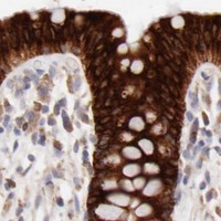

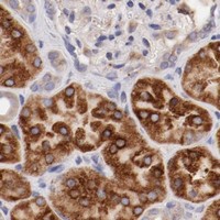
Comprehensive Human Protein Atlas IHC scoring per tissue (reliability: Enhanced — High consistency between antibody staining and RNA expression data. At least one protein variant secreted, tissue location of RNA and protein might differ and correlation is complex.). Rows are taken directly from the HPA tissue chart — click any row's HPA link to view the source.
| Tissue | Cell type | Level | Evidence | Source |
|---|---|---|---|---|
| Appendix | Endocrine cells | High | Protein (IHC) | HPA → |
| Breast | Glandular cells | High | Protein (IHC) | HPA → |
| Bronchus | Ciliated cells (cell body) | High | Protein (IHC) | HPA → |
| Colon | Endocrine cells | High | Protein (IHC) | HPA → |
| Duodenum | Endocrine cells | High | Protein (IHC) | HPA → |
| Tissue | Cell type | Level | Evidence | Source |
|---|---|---|---|---|
| Adipose tissue | Adipocytes | Not detected | Protein (IHC) | HPA → |
| Heart muscle | Cardiomyocytes | Not detected | Protein (IHC) | HPA → |
| Ovary | Ovarian stroma cells | Not detected | Protein (IHC) | HPA → |
| Skeletal muscle | Myocytes | Not detected | Protein (IHC) | HPA → |
| Smooth muscle | Smooth muscle cells | Not detected | Protein (IHC) | HPA → |
Use the catalog antibody’s paraffin-section IHC conditions as the starting point, then check staining against ENTPD5’s reported cell and compartment patterns.
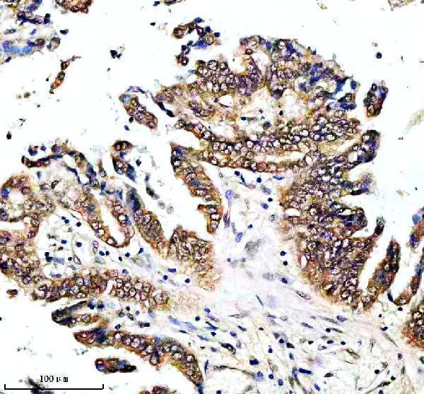
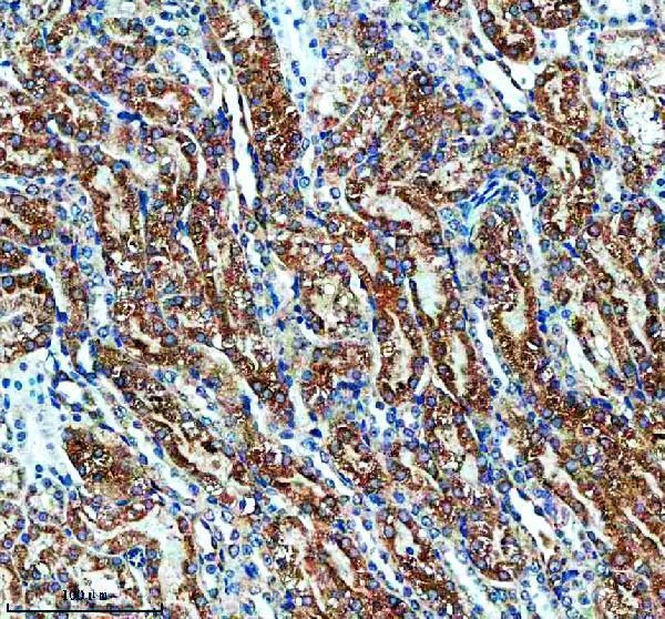
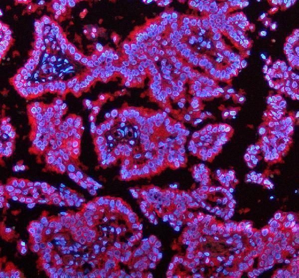
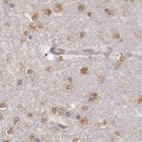
Anti-ENTPD5 IHC images cover paraffin sections of human lung cancer, rat kidney, and mouse adrenal tissue; an IF image covers human lung cancer sections (A06908-2 image captions).
A06908-2 has IHC images from human lung cancer, rat kidney, and mouse adrenal paraffin sections, plus an IF image from human lung cancer paraffin sections (A06908-2 image captions). M06908-2 lists IHC and human, mouse, and rat reactivity, but the payload provides no IHC or IF image for it (M06908-2 catalog applications, reactivity, and image alts).
Which to pick: For tissue IHC, choose A06908-2: its own paraffin-section captions document EDTA retrieval at pH 8.0 and 2 μg/ml primary antibody; the fixative is unreported (A06908-2 IHC image captions). For IF, choose A06908-2 based on its human tissue IF image; ICC validation is unreported (A06908-2 IF image caption and catalog applications). For work across human, mouse, and rat tissues, A06908-2 has IHC images in all three species; M06908-2 is a rabbit monoclonal, clone 21E91, with listed IHC and three-species reactivity but no IHC image in the payload (A06908-2 IHC image captions; M06908-2 catalog clone, applications, reactivity, and image alts).