This website uses cookies to ensure you get the best experience on our website.


- Table of Contents
EPHA4 IHC-P shows high staining in cerebellar molecular-layer cells and cerebral cortical neuropil (HPA tissue IHC). This guide uses those tissue patterns and the catalog antibody's 1:25 IHC-P dilution to plan staining and controls (HPA tissue IHC; datasheet: 1:25).

Expected localisation, validated protocols, controls and antibodies — the at-a-glance facts below, then the full design guide.
| Expected localisation | Cytoplasm and membrane in cerebellum (HPA tissue IHC) | |
| Staining pattern | Cerebellar molecular layer: cytoplasm/membrane (HPA tissue IHC) | |
| Antigen retrieval | Citrate pH 6.0 HIER, 95–98 °C, 20 min (rule: cytoplasmic / membrane antigen) | |
| Positive control | Cerebellum+4 more · see all | |
| Negative control | Adrenal gland+4 more · see all |
| Fixation | Keep fixation consistent across sections (standard IHC practice; not target-specific) | |
| Caveat | Brain signal varies by cell type (HPA tissue IHC) | |
| Regulation | Expression regulation not specified (UniProt) | |
| Isoform / epitope | 2 isoforms; epitope-side coverage is unknown (UniProt) |
The catalog antibody’s IHC-P protocol is followed by published chromogenic IHC protocols for gastric adenocarcinoma, hippocampal tissue, and uveal melanoma (PMC3720259; PMC4149234; PMC9139903).
| Sample | Paraffin-embedded H. testis tissue; fixative not specified (datasheet A01860-1) |
| Fixation | Image fixative and duration unreported (datasheet A01860-1); verify before use. |
| Sectioning | 4–5 µm sections on charged slides (standard) |
| Deparaffinisation | Xylene, graded ethanol series to water (standard) |
| Antigen retrieval | Heat-induced epitope retrieval in citrate buffer, pH 6.0, 20 min at 95–98 °C (standard rule: cytoplasmic / membrane antigen) |
| Peroxidase block | 3% H2O2, 10 min, room temperature (standard) |
| Blocking | 10% normal serum of the secondary host, 30 min, room temperature (standard) |
| Primary antibody | Rabbit anti-EPHA4, 1:25 (datasheet A01860-1) |
| Primary incubation | Overnight at 4 °C (standard) |
| Detection | HRP-polymer secondary, DAB chromogen 5–10 min (standard) |
| Counterstain | Hematoxylin, blue, dehydrate and mount (standard) |
| Expected result | EPHA4-positive staining in molecular layer cells - cytoplasm/membrane of cerebellum (HPA tissue IHC: High). HPA tissue profile: Cytoplasmic expression in several different tissue types, highly abundant in brain. No signal in the no-primary control. |
EPHA4 is a single-pass receptor with an extracellular region and cytoplasmic kinase region; UniProt places it at the cell membrane, neuronal projections, postsynaptic membrane, junctions and early endosomes (UniProt P54764 topology/localisation). In paraffin IHC, expect membrane-associated or cell-body staining in HPA-positive populations, especially brain neuropil, cerebellar molecular layer cells and placental syncytiotrophoblasts (HPA tissue IHC). HPA rates tissue staining reliability Enhanced, with medium consistency against RNA expression (HPA tissue IHC).
| Cerebral cortex neuropil stains strongly; cerebellar molecular layer cells show cytoplasmic/membrane staining. | These are reported High IHC patterns (HPA tissue IHC). Assess the labelled structures and surrounding cells separately; a strong field-wide deposit alone cannot establish the reported cell pattern (general IHC practice). |
| A population shows predominantly nuclear staining, with little convincing membrane or cytoplasmic signal. | Predominantly nuclear staining does not match the listed membrane, projection, junction or endosomal locations (UniProt P54764 localisation). Check morphology, the no-primary control and detection background before interpreting it as EPHA4 (general IHC practice). |
| Strong staining appears in an HPA-undetected cell population, such as adrenal glandular cells or bronchial respiratory epithelial cells. | HPA reports these specific populations as Not detected (HPA tissue IHC). Consider antibody cross-reactivity or endogenous detection activity; compare with an HPA-positive section and appropriate detection controls (general IHC practice). |
| Colour spreads across tissue, edges and spaces without a discernible cell or neuropil pattern. | This is difficult to score as EPHA4 because HPA reports staining in defined cells and neuropil (HPA tissue IHC). Examine no-primary and detection-only controls, washing and chromogen development for background (general IHC practice). |
| No stain is visible in cerebral cortex neuropil or placental syncytiotrophoblasts. | Both are High HPA IHC reference patterns (HPA tissue IHC). First confirm that the expected structures are present and the detection run worked; absence in one section does not establish EPHA4 absence (general IHC practice). |
| Compartment and epitope position | EPHA4 spans the membrane at residues 548–569, with extracellular residues 20–547 and cytoplasmic residues 570–986 (UniProt P54764 topology). The supplied record gives no antibody epitope, so it cannot predict whether retrieval will favour a particular compartment or antibody (catalog antibody record). |
| Activation and intracellular signal | UniProt reports receptor clustering after activation and targeting to early endosomes (UniProt P54764 localisation). Thus a cellular component alongside membrane staining can fit the annotated biology; morphology and the HPA cell pattern remain necessary for interpretation (UniProt P54764 localisation; HPA tissue IHC). |
| Choice of reference tissue | Cortex neuropil, cerebellar molecular layer cells and placental syncytiotrophoblasts are High; colon enterocytes and kidney proximal tubule cell bodies are Medium (HPA tissue IHC). Use the same named population when comparing sections, since tissue-wide positivity would obscure these distinctions (general IHC practice). |
| Antibody evidence and limits | The supplied catalog antibody, CAB028368, is mouse monoclonal and IHC Enhanced (HPA antibodies). HPA describes overall tissue reliability as Enhanced but RNA agreement as medium consistency (HPA tissue IHC); judge a new pattern against controls rather than treating validation status as proof for every cell. |
| IF/ICC pattern? | HPA summarises EPHA4 as membrane-associated in its subcellular record, but supplies no main location or ICC-IF image cell lines (HPA subcellular). The IHC tissue patterns cannot establish an IF-positive cell line or IF protocol; consult the separate IF/ICC guide for that application (HPA tissue IHC; HPA subcellular). |
| Situation | Likely cause | Next action |
|---|---|---|
| Known-positive brain or placenta is blank. | The slide may lack the named structure, or the staining run may have failed; HPA reports High signal only in specified populations (HPA tissue IHC; general IHC practice). | Confirm structure on the counterstain, check a concurrent positive-control section and review the run's retrieval, antibody and detection steps (general IHC practice). |
| Only faint signal appears in a nominally positive section. | The sampled population may differ from the HPA-positive one; staining levels also vary from High to Medium across listed populations (HPA tissue IHC). | Identify the precise cell type before changing conditions; compare it with a concurrent High reference and then review the run's validated settings (HPA tissue IHC; general IHC practice). |
| Nuclei dominate the stain. | A nuclear-dominant pattern is unsupported by the supplied EPHA4 localisation annotations (UniProt P54764 localisation); nonspecific or detection signal is possible (general IHC practice). | Inspect a no-primary control and compare membrane and cytoplasmic detail in an HPA-positive population before scoring the nuclei (general IHC practice; HPA tissue IHC). |
| Adrenal glandular cells or bronchial respiratory epithelium stain strongly. | HPA lists those populations as Not detected (HPA tissue IHC). Cross-reactivity or endogenous detection activity is a possibility, not a diagnosis from one slide (general IHC practice). | Run matched no-primary and detection controls, inspect localisation, and compare a positive reference population on the same staining run (general IHC practice; HPA tissue IHC). |
| Brown colour is diffuse or concentrated at tissue edges. | A deposit without the reported cell or neuropil pattern is hard to attribute to EPHA4 (HPA tissue IHC); excess detection background is possible (general IHC practice). | Review control slides, washing and chromogen development, then score only resolvable cellular or neuropil staining (general IHC practice; HPA tissue IHC). |
| The expected compartment changes between samples. | UniProt lists both membrane sites and early endosomes, with clustering after activation (UniProt P54764 localisation); the record gives no sample-specific mechanism for a shift. | Record membrane and cellular staining separately, verify the named HPA-positive cell population, and avoid assigning activation from localisation alone (UniProt P54764 localisation; HPA tissue IHC). |
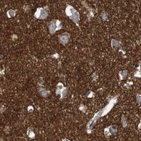


Comprehensive Human Protein Atlas IHC scoring per tissue (reliability: Enhanced — Medium consistency between antibody staining and RNA expression data.). Rows are taken directly from the HPA tissue chart — click any row's HPA link to view the source.
| Tissue | Cell type | Level | Evidence | Source |
|---|---|---|---|---|
| Cerebellum | Molecular layer cells - cytoplasm/membrane | High | Protein (IHC) | HPA → |
| Cerebral cortex | Neuropil | High | Protein (IHC) | HPA → |
| Placenta | Syncytiotrophoblasts - cell body | High | Protein (IHC) | HPA → |
| Bone marrow | Hematopoietic cells | Medium | Protein (IHC) | HPA → |
| Colon | Enterocytes | Medium | Protein (IHC) | HPA → |
| Tissue | Cell type | Level | Evidence | Source |
|---|---|---|---|---|
| Adrenal gland | Glandular cells | Not detected | Protein (IHC) | HPA → |
| Breast | Adipocytes | Not detected | Protein (IHC) | HPA → |
| Bronchus | Respiratory epithelial cells | Not detected | Protein (IHC) | HPA → |
| Duodenum | Endocrine cells | Not detected | Protein (IHC) | HPA → |
| Endometrium | Cells in endometrial stroma | Not detected | Protein (IHC) | HPA → |
Troubleshoot EPHA4 staining in paraffin sections by checking retrieval, antibody controls, expected localisation and cell type before interpreting chromogenic signal.
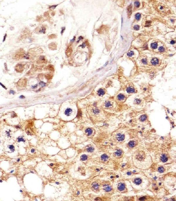
A01860-1 has real IHC data from a human paraffin-embedded testis section (image caption); the catalog lists human and mouse reactivity (catalog: reactivity).
A01860-1 will render with an IHC image of a human paraffin-embedded testis section stained at 1:25 with DAB detection (image caption). Its listed applications are IHC-P and WB, and its listed reactivity is human and mouse (catalog: applications and reactivity).
Which to pick: Choose A01860-1 for paraffin-section IHC: its own image shows staining of a human testis section (image caption). The caption does not report the fixative (image caption). For mouse tissue, A01860-1 has listed mouse reactivity but no mouse IHC image in the payload (catalog: reactivity; image caption); no IF/ICC application or IF image is listed for this SKU (catalog: applications and images).