EPHA7 / Ephrin type-A receptor 7 · IHC design guide

Design Immunohistochemistry for EPHA7

Plan chromogenic EPHA7 IHC in paraffin sections using adipocytes or pancreatic exocrine cells as high-staining controls (HPA tissue IHC). Start within the catalog antibody’s 1:200–1:1000 IHC dilution range (M03943 datasheet) and assess membrane and cytoplasmic staining (HPA tissue IHC).

Evidence assembled Oct 2026 · For research use; verify linked source records and product datasheet before use
Immunohistochemistry protocol sheet for EPHA7 (IHC for EPHA7): expected localisation Membrane and cytoplasm (HPA tissue IHC), antibody M03943, validated IHC image, and IHC protocol steps
Printable EPHA7 IHC protocol sheet — expected localisation Membrane and cytoplasm (HPA tissue IHC), antibody M03943, controls and protocol steps. Open the full EPHA7 IHC guide →

EPHA7 Immunohistochemistry Experimental Design Guide

Expected localisation, validated protocols, controls and antibodies — the at-a-glance facts below, then the full design guide.

Must know before staining
Expected localisation Membrane and cytoplasm (HPA tissue IHC)
Staining pattern Membranous and cytoplasmic staining in most tissues (HPA tissue IHC)
Antigen retrieval Citrate pH 6.0 HIER, 95–98 °C, 20 min (rule: cytoplasmic / membrane antigen)
Positive control ⓘ Adipose tissue+4 more · see all
Negative control ⓘ Caudate+3 more · see all
Important caveats
Reasons your staining may differ from the expected pattern.
Fixation Keep fixation consistent across tissue sections (standard IHC practice; not target-specific)
Caveat Tissue staining awaits external verification (HPA tissue IHC)
Regulation Widely expressed; regulation unspecified (UniProt)
Isoform / epitope 5 isoforms; check extracellular versus cytoplasmic epitope (UniProt)
Section 1

Recommended EPHA7 IHC & IF Protocols

The catalog antibody’s IHC-P protocol (datasheet) is accompanied by four published EPHA7 tissue-staining protocols (PMC4068196; PMC4491497; PMC2292196; PMC11541825).

Recommended immunohistochemistry (IHC-P) protocol parameters
SampleParaffin-embedded human Breast tissues; fixative not specified (datasheet M03943)
FixationImage fixative and duration unreported (datasheet M03943); verify before use.
Sectioning4–5 µm sections on charged slides (standard)
DeparaffinisationXylene, graded ethanol series to water (standard)
Antigen retrievalHeat-induced epitope retrieval in citrate buffer, pH 6.0, 20 min at 95–98 °C (standard rule: cytoplasmic / membrane antigen)
Peroxidase block3% H2O2, 10 min, room temperature (standard)
Blocking10% normal serum of the secondary host, 30 min, room temperature (standard)
Primary antibodyMouse monoclonal (clone 6C8G7) anti-EPHA7, 1:200-1:1000 (datasheet M03943)
Primary incubationOvernight at 4 °C (standard)
DetectionHRP-polymer secondary, DAB chromogen 5–10 min (standard)
CounterstainHematoxylin, blue, dehydrate and mount (standard)
Expected resultEPHA7-positive staining in adipocytes of adipose tissue (HPA tissue IHC: High). HPA tissue profile: Membrane and cytoplasmic expresssion in most tissues, including parathyroid gland. No signal in the no-primary control.
💡Decision noteStart with citrate pH 6.0 HIER at 95–98 °C for 20 min (page retrieval); follow article-specific retrieval when reproducing its specimen workflow (PMC4491497; PMC11541825).
Section 2

What Is the Expected EPHA7 Staining Pattern?

EPHA7 is a cell-membrane receptor with an extracellular region and a cytoplasmic kinase region (UniProt Q15375 topology). In paraffin-section IHC, expect membrane and cytoplasmic staining in many tissues (HPA: tissue IHC profile), including adipocytes, pancreatic exocrine glandular cells, and smooth muscle cells with High staining (HPA: tissue IHC). The tissue profile is Approved but pending external verification (HPA: reliability).

What am I looking at on my slide?
Membrane-associated staining with some cytoplasmic signal in adipocytes, pancreatic exocrine glandular cells, or smooth muscle cells.This fits the reported compartments and High cell-specific staining (HPA: tissue IHC). Compare cells within the section; a High category does not specify an intensity threshold for an individual slide.
Predominantly nuclear staining, without convincing membrane-associated staining.A nuclear-dominant pattern is unexpected for a cell-membrane receptor (UniProt Q15375: subcellular location; HPA: membrane and cytoplasmic tissue IHC profile). Check controls and whether the signal follows cell boundaries before calling it EPHA7.
Strong staining in cardiomyocytes or glial cells of the caudate or hippocampus.Those specified cells are Not detected in the HPA tissue IHC record (HPA: cardiomyocytes; caudate and hippocampal glial cells). Assess cross-reactivity and endogenous detection activity before assigning the signal to EPHA7; the category does not make the entire tissue a negative control.
Diffuse chromogen across tissue and background areas, obscuring cell boundaries.The distribution cannot establish the reported membrane and cytoplasmic pattern (HPA: tissue IHC profile). Inspect a no-primary control and review blocking, washing, and detection conditions (general IHC practice).
No signal in pancreatic exocrine glandular cells or adipocytes.An absent result conflicts with High staining reported for those cells (HPA: pancreas; adipose tissue). Check tissue preservation, antibody performance, retrieval, and detection with appropriate controls (general IHC practice); one failed section cannot establish biological absence.
💡Expected EPHA7 appearanceCall a section positive when identifiable adipocytes, pancreatic exocrine glandular cells, or smooth muscle cells show membrane and cytoplasmic staining consistent with their High HPA categories; nuclear-dominant signal or strong staining in HPA Not detected cells warrants control review (HPA: tissue IHC profile and cell-specific levels; UniProt Q15375: cell membrane).
How each factor affects the staining
Antibody-accessible regionEPHA7 spans the membrane at residues 556–576, with extracellular residues 28–555 and cytoplasmic residues 577–998 (UniProt Q15375 topology). The supplied record gives no antibody epitope, so it cannot predict which region the IHC antibody recognizes.
Isoform coverageFive EPHA7 isoforms are listed (UniProt Q15375: isoforms 1–5). The supplied record does not establish which isoforms the IHC antibody detects; interpret a negative section without assuming that every isoform was tested.
Cell-specific reference patternHPA reports High staining in several cell populations but Not detected staining in selected others (HPA: tissue IHC). Use the named cells when comparing results; a negative category for one cell population does not describe every cell in its tissue.
Strength of reference evidenceThe tissue profile is Approved, pending external verification, and an IHC antibody is listed as Approved (HPA: tissue reliability; CAB010496 IHC status). Neither supplied status is an Enhanced IHC validation claim; resolve unexpected staining with controls.
Why is my staining missing, weak or wrong?
SituationLikely causeNext action
Positive-control cells remain unstained.A failed section or detection step is possible when cells reported High show no signal (HPA: pancreatic exocrine glandular cells; adipocytes).Confirm the expected cells are present, then review antigen retrieval, primary-antibody use, and chromogenic detection alongside a working control (general IHC practice).
Signal appears mainly in nuclei.That compartment conflicts with EPHA7 cell-membrane localization and the reported membrane and cytoplasmic tissue profile (UniProt Q15375; HPA: tissue IHC).Compare a no-primary control, inspect the counterstain separately, and require convincing membrane-associated staining before assigning positivity (general IHC practice).
Cardiomyocytes stain strongly.Cardiomyocytes are reported Not detected (HPA: heart muscle tissue IHC); cross-reactivity or endogenous detection activity is possible.Examine a no-primary control and the detection reagents; score the specific cell population rather than treating all heart-muscle cells as equivalent (general IHC practice).
Weak cellular signal is lost in diffuse brown background.Nonspecific binding, endogenous detection activity, or incomplete washing can obscure localization (general IHC practice).Review no-primary and reagent controls, blocking, washing, and detection development; interpret only signal with identifiable cellular boundaries (general IHC practice).
Two sampled tissues seem to disagree in intensity.HPA categories describe particular cell types, and reported levels vary by cell population (HPA: tissue IHC).Record the scored cell type and compartment in each section. Compare with the corresponding HPA cell-level entry rather than applying one tissue-wide intensity expectation.
Does an ICC/IF principal-piece signal establish the paraffin-section IHC pattern?HPA reports an approved principal-piece localization in sperm by ICC/IF (HPA: subcellular ICC/IF); it is a different preparation and readout.Use the membrane and cytoplasmic tissue profile to assess IHC sections (HPA: tissue IHC). Treat the sperm ICC/IF observation as application-specific evidence, not an IHC control.

Sample controls for EPHA7 IHC & IF

🧪Run pancreas first: exocrine glandular cells should stain (HPA: High in pancreatic exocrine glandular cells). Use heart muscle as the negative tissue; its cardiomyocytes should lack specific staining (HPA: Not detected in cardiomyocytes). On the positive slide, treat cells without convincing membrane-associated staining as candidate internal negatives, without assuming a particular cell type is negative (UniProt Q15375: cell membrane).
Positive control tissue: Adipose tissue (Adipocytes, HPA High)
Negative control tissue: Caudate (HPA Not detected)
ICC-IF cell lines (HPA subcellular resource): HPA ICC-IF images show EPHA7 in Sperm, with annotated localisation: Principal piece (approved) (HPA subcellular).
Technical controls: Include a no-primary, secondary-only control; an immunoglobulin isotype control matched to the primary antibody’s host species and clonality; and an EPHA7 knockout biological negative (standard IHC practice). Quench endogenous peroxidase in pancreatic sections before chromogenic detection, and check for endogenous biotin background if using avidin–biotin detection (standard IHC practice).
⚠️Feasibility: No target-specific fixation window or antigen-retrieval effect is reported in the supplied evidence; optimize retrieval empirically for paraffin sections (catalog caption: paraffin-section IHC). The selected M03943 tissue-IHC caption does not report a fixative (catalog caption: fixative not stated). No matched evidence establishes that frozen sections or IF/ICC are easier; in pancreatic sections, endogenous peroxidase can create misleading chromogenic signal (standard IHC practice).

HPA tissue IHC evidence for EPHA7

Comprehensive Human Protein Atlas IHC scoring per tissue (reliability: Approved — Pending external verification.). Rows are taken directly from the HPA tissue chart — click any row's HPA link to view the source.

Positive expression · recommended positive controls

TissueCell typeLevelEvidenceSource
Adipose tissue Adipocytes High Protein (IHC) HPA →
Duodenum Glandular cells High Protein (IHC) HPA →
Epididymis Glandular cells High Protein (IHC) HPA →
Pancreas Exocrine glandular cells High Protein (IHC) HPA →
Seminal vesicle Glandular cells High Protein (IHC) HPA →

Undetected expression · recommended negative controls

TissueCell typeLevelEvidenceSource
Caudate Glial cells Not detected Protein (IHC) HPA →
Heart muscle Cardiomyocytes Not detected Protein (IHC) HPA →
Hippocampus Glial cells Not detected Protein (IHC) HPA →
Testis Cells in seminiferous ducts Not detected Protein (IHC) HPA →
Section 3

Advanced EPHA7 IHC Tips

Troubleshoot EPHA7 staining in paraffin sections by checking retrieval, cell identity, membrane localization, and controls before assigning biological meaning.

How should I optimize EPHA7 antigen retrieval in paraffin sections?
Use citrate pH 6.0 heat-induced retrieval at 95–98 °C for 20 min before chromogenic IHC (page retrieval setting). Compare retrieved and unretrieved serial sections while keeping antibody concentration, detection, and development time identical, so changes in signal can be attributed to retrieval (standard IHC practice). Evaluate membrane and cytoplasmic staining separately because EPHA7 spans the membrane at residues 556–576 and has extracellular and cytoplasmic regions (UniProt Q15375 topology; HPA tissue IHC). If staining remains weak, test another retrieval buffer as a fallback on matched sections, checking whether signal improves without increased background or tissue damage (standard IHC practice).
Does fixation alter EPHA7 staining in paraffin IHC?
The selected M03943 image shows AEC staining in paraffin-embedded human breast tissue, but its caption does not identify the fixative (catalog caption M03943). EPHA7-specific fixation sensitivity is therefore unknown; document fixative and processing history for each specimen instead of assigning a target-specific fixation effect (catalog caption M03943; standard IHC practice). If specimens differ in processing, compare at least two matched sections with identical retrieval and detection, and assess tissue preservation alongside staining (standard IHC practice). Treat loss of signal as inconclusive until section quality, positive-control staining, and cell composition have been checked under the same run conditions (standard IHC practice).
Where should convincing EPHA7 staining appear within cells?
Prioritize staining at cell boundaries because EPHA7 is a cell-membrane receptor with a single transmembrane segment at residues 556–576 (UniProt Q15375 topology). Cytoplasmic staining can also fit the reported tissue-IHC pattern, which describes membrane and cytoplasmic expression across many tissues (HPA tissue IHC). Compare compartments within a defined cell population, such as pancreatic exocrine glandular cells or smooth muscle cells, rather than pooling unlike cells (HPA tissue IHC; standard IHC practice). Predominantly nuclear staining warrants review against no-primary controls, morphology, and an independently validated reagent before it is called EPHA7 (UniProt Q15375 subcellular location; standard IHC practice).
How could EPHA7 isoforms or epitope position change my IHC readout?
EPHA7 has five listed isoforms, so establish which forms the chosen antibody detects before treating staining as total EPHA7 (UniProt Q15375 isoforms; standard IHC practice). The mature receptor begins at residue 28; its extracellular region spans 28–555, while its cytoplasmic region spans 577–998 (UniProt Q15375 processing and topology). Extracellular glycosylation sites at residues 343 and 410, and cytoplasmic phosphorylation sites including 608 and 614, make epitope mapping relevant to interpretation without proving an effect on this antibody (UniProt Q15375 modifications). If the epitope is undisclosed, avoid claims about isoform coverage or modification-dependent staining and compare a second validated epitope where available (standard IHC practice).
How should I adapt the EPHA7 readout for multiplex IF?
For multiplex IF, pair EPHA7 with an independently validated marker of the expected cell population, such as pancreatic exocrine glandular cells, and include single-stain controls (HPA tissue IHC; standard IF practice). Select spectrally separated fluorophores after inspecting tissue autofluorescence; a far-red channel may improve contrast if the specimen is bright at shorter wavelengths (standard IF practice). If the antibody recognizes the extracellular region at residues 28–555, compare staining without detergent; access to a cytoplasmic epitope at 577–998 generally requires controlled permeabilisation (UniProt Q15375 topology; standard IF practice). Confirm epitope location and IF performance independently because the selected product caption establishes paraffin-section chromogenic staining only (catalog caption M03943).
What controls help separate EPHA7 signal from chromogenic background?
Run a no-primary control through the complete detection sequence and compare it with the antibody-stained section at the same chromogen development time (standard IHC practice). For peroxidase-based detection, include a peroxidase block and inspect pigment, necrosis, and tissue edges before assigning brown DAB deposits to EPHA7 (standard IHC practice). DAB is a general chromogenic option; the selected M03943 paraffin-section caption specifically reports AEC staining (catalog caption M03943). Recheck antibody titration, blocking, and washing if diffuse color obscures the expected membrane or cytoplasmic pattern, especially when the no-primary control is also colored (HPA tissue IHC; standard IHC practice).
How should I quantify EPHA7 staining across heterogeneous sections? ⚠ ANSWER MARKED FOR VERIFICATION
Define the cell population and compartment before scoring, because EPHA7 tissue IHC reports both membrane and cytoplasmic staining (HPA tissue IHC; standard IHC practice). Record the percentage of positive viable cells and staining intensity separately, or calculate an H-score from 0–300 using intensity categories 0–3 (standard IHC practice). For spatial analyses, report positive-cell density per mm² of viable tissue or a prespecified region, with consistent thresholds and chromogen exposure across sections (standard IHC practice). Normalize comparisons to the number or area of the relevant cell type, since differing proportions of glandular, stromal, and damaged tissue can change a whole-section average (standard IHC practice).
When is an EPHA7-positive IHC pattern biologically credible?
Give greatest weight to reproducible membrane or cytoplasmic staining in morphologically intact cells, consistent with EPHA7 topology and the reported tissue-IHC pattern (UniProt Q15375 topology; HPA tissue IHC). Pancreatic exocrine glandular cells and smooth muscle cells are reported as high, whereas cardiomyocytes and caudate glial cells are reported as not detected (HPA tissue IHC). Use those contrasts as context, while recognizing that the HPA tissue-IHC reliability is Approved with external verification pending (HPA tissue IHC reliability). Distrust isolated nuclear staining, edge-only color, necrotic deposits, or signal reproduced in a no-primary control; inspect endogenous enzyme activity when using enzyme-based detection (standard IHC practice).
Boster reagents

Best EPHA7 / Ephrin type-A receptor 7 IHC Antibodies

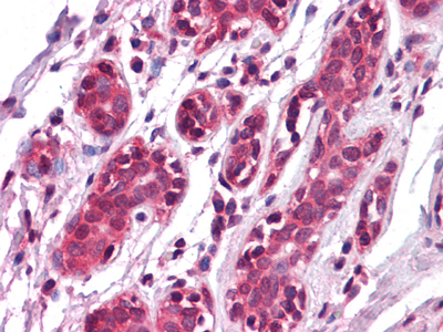
IHC figure evidence covers paraffin-embedded human breast tissue stained with AEC (M03943 IHC image caption). IF is listed for human samples, with no IF figure supplied (M03943 applications/reactivity/IF image alts).

Real IHC data Immunohistochemistry analysis of paraffin-embedded human Breast tissues with AEC staining using EphA7 Monoclonal Antibody.
Anti-Ephrin type-A receptor 7 EphA7 Monoclonal Antibody
Cat # M03943

M03943 is the sole rendered card; its IHC figure shows paraffin-embedded human breast tissue stained with AEC (M03943 IHC image caption). It also lists human reactivity and IF, but provides no IF figure (M03943 applications/reactivity/IF image alts).

Which to pick: For tissue IHC, choose M03943, a mouse monoclonal antibody with a paraffin-section image; the caption does not report the fixative (M03943 host/clone/IHC image caption). For IF, M03943 lists human reactivity and a 1:50 dilution; ICC is not listed, so confirm suitability before using it for ICC (M03943 applications/reactivity/IF dilution). For mouse or rat tissue IHC, consider A03943, a rabbit antibody listing IHC and reactivity in both species, though it has no IHC image (A03943 host/applications/reactivity/IHC image alts).

Each figure is that product's own IHC / IF validation image from its datasheet.

References

  1. UniProt Consortium. UniProt entry Q15375 (EPHA7_HUMAN, Ephrin type-A receptor 7).
  2. Human Protein Atlas. EPHA7 tissue IHC expression (reliability: Approved).
  3. Human Protein Atlas. EPHA7 subcellular location (ICC-IF): Localized to the principal piece..
  4. Human Protein Atlas. EPHA7 antibody validation summary (2 antibodies).
  5. Complementary expression of EphA7 and SCO-spondin during posterior commissure development. Frontiers in neuroanatomy 2014 — PMC4068196.
  6. Low EphA7 Expression Correlated with Lymph Node Metastasis and Poor Prognosis of Patients with Esophageal Squamous Cell Carcinoma. Acta histochemica et cytochemica 2015 — PMC4491497.
  7. Increased expression of EphA7 correlates with adverse outcome in primary and recurrent glioblastoma multiforme patients. BMC cancer 2008 — PMC2292196.
  8. Intra-BLA alteration of interneurons' modulation of activity in rats, reveals a dissociation between effects on anxiety symptoms and extinction learning. Neurobiology of stress 2024 — PMC11541825.
  9. PubMed PMID:7898931 — UniProt-cited evidence.
  10. PubMed PMID:14702039 — UniProt-cited evidence.
  11. PubMed PMID:14574404 — UniProt-cited evidence.