This website uses cookies to ensure you get the best experience on our website.


- Table of Contents
Plan chromogenic ERAP2 IHC in paraffin sections with the human-reactive, IHC-validated antibody (datasheet A04269-1). Use lung macrophages as a high-staining reference and assess cytoplasmic signal by cell type (HPA tissue IHC).

Expected localisation, validated protocols, controls and antibodies — the at-a-glance facts below, then the full design guide.
| Expected localisation | Cytoplasmic tissue staining (HPA tissue IHC) | |
| Staining pattern | Cytoplasmic staining in several tissues, including immune cells (HPA tissue IHC) | |
| Antigen retrieval | EDTA pH 8.0 HIER, heat-mediated (datasheet A04269-1) | |
| Positive control | Lung+4 more · see all | |
| Negative control | Cerebellum+4 more · see all |
| Fixation | Keep fixation consistent across paraffin sections (standard IHC practice; not target-specific) | |
| Caveat | A secreted variant may alter tissue protein distribution (HPA tissue IHC) | |
| Regulation | Higher expression in spleen and leukocytes (UniProt) | |
| Isoform / epitope | 4 isoforms; antibody epitope coverage is unspecified (UniProt; datasheet A04269-1) |
Compare the catalog antibody’s IHC-P protocol (datasheet A04269-1) with four published ERAP2 IHC methods (PMC5617457; PMC8725995; PMC7076169; PMC5939618).
| Sample | Paraffin-embedded human lung cancer tissue; fixative not specified (datasheet A04269-1) |
| Fixation | Image fixative and duration unreported (datasheet A04269-1); verify before use. |
| Sectioning | 4–5 µm sections on charged slides (standard) |
| Deparaffinisation | Xylene, graded ethanol series to water (standard) |
| Antigen retrieval | Heat retrieval: EDTA pH 8.0 (datasheet A04269-1); 20 min, 95–100 °C (standard) |
| Peroxidase block | 3% H2O2, 10 min, room temperature (standard) |
| Blocking | 10% goat serum (datasheet A04269-1) |
| Primary antibody | Rabbit anti-ERAP2, 2-5μg/ml (datasheet A04269-1) |
| Primary incubation | Overnight at 4 °C (datasheet A04269-1) |
| Detection | Streptavidin-biotin complex (SABC), DAB chromogen (datasheet A04269-1) |
| Counterstain | Hematoxylin, blue, dehydrate and mount (standard) |
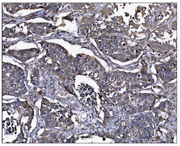
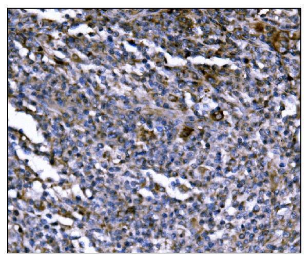
| Expected result | ERAP2-positive staining in macrophages of lung (HPA tissue IHC: High). HPA tissue profile: Cytoplasmic expression in several tissues, including immune cells. No signal in the no-primary control. |
In paraffin-section IHC, expect cytoplasmic ERAP2 staining, especially in lung macrophages and skin Langerhans cells (HPA: High in both; cytoplasmic tissue profile). ERAP2 has an ER membrane anchor with a lumenal region spanning residues 41–960 (UniProt Q6P179 topology). HPA rates tissue IHC reliability Enhanced, while reporting medium consistency with RNA and a secreted protein variant (HPA: reliability description).
| Strong cytoplasmic stain in lung macrophages or skin Langerhans cells (HPA: High in both). | This matches the strongest listed tissue IHC observations (HPA: tissue IHC). Judge the cells and compartment together; HPA describes a cytoplasmic profile across several tissues, including immune cells (HPA: tissue IHC profile). |
| Predominantly nuclear stain, with little cytoplasmic signal in expected cells (HPA: cytoplasmic tissue profile). | Question specificity or slide processing: nuclear dominance does not match the reported tissue pattern (HPA: tissue IHC profile). Inspect the counterstain, detection controls and antibody conditions before assigning ERAP2 positivity (general IHC practice). |
| Strong stain in cells listed as undetected, such as pancreatic exocrine glandular cells (HPA: Not detected). | Treat this as discordant with that HPA observation, not proof of cross-reactivity (HPA: tissue IHC). Compare tissue context and negative controls; endogenous detection activity can mimic chromogenic signal (general IHC practice). |
| Diffuse stain across cells and tissue spaces, obscuring boundaries (general IHC practice). | High background limits cell-level interpretation (general IHC practice). Review no-primary controls and blocking. Interpret extracellular signal cautiously because HPA reports at least one secreted ERAP2 variant (HPA: reliability description). |
| No stain in lung macrophages or skin Langerhans cells (HPA: High in both). | First check assay performance with the expected positive cells on the same run (HPA: tissue IHC; general IHC practice). An absent signal alone cannot establish ERAP2 absence when the run's positive control also fails (general IHC practice). |
| Cell compartment (UniProt Q6P179 topology). | ERAP2 has one membrane segment at 21–40 and a lumenal region at 41–960 (UniProt Q6P179 topology). Tissue IHC is reported as cytoplasmic; it does not resolve the ER membrane directly (HPA: tissue IHC profile; general IHC practice). |
| Secreted variant and RNA agreement (HPA: reliability description). | HPA reports at least one secreted protein variant and medium staining–RNA consistency (HPA: reliability description). Do not require cellular protein staining to mirror local RNA distribution in every tissue (HPA: reliability description). |
| Antibody-specific IHC evidence (HPA: antibody validation). | CAB025618 is IHC Enhanced; HPA034498 is IHC Supported (HPA: antibody validation). These ratings apply to those antibodies, so record the antibody used when comparing staining patterns (HPA: antibody validation; general IHC practice). |
| Isoforms and glycosylation (UniProt Q6P179). | UniProt lists 4 isoforms and 5 glycosylation sites (UniProt Q6P179). The supplied records do not link either feature to a particular IHC pattern or retrieval response; avoid assigning such effects. |
| IF/ICC Q&A: Should Golgi signal match tissue IHC? (HPA: subcellular ICC-IF; tissue IHC). | HPA calls the Golgi the main approved ICC-IF location, with images from RT-4, U-251MG and U2OS (HPA: subcellular ICC-IF). Tissue IHC reports cytoplasmic staining; compare each assay against its own observation (HPA: tissue IHC). |
| Situation | Likely cause | Next action |
|---|---|---|
| The positive tissue control is blank (HPA: High in lung macrophages and skin Langerhans cells). | A failed staining run is possible (general IHC practice). | Check antibody application, retrieval and detection steps against the run record, then repeat with the positive control (general IHC practice). Do not infer target-specific retrieval sensitivity from these records. |
| Nuclei dominate the signal (HPA: cytoplasmic tissue profile). | This compartment disagrees with reported tissue staining (HPA: tissue IHC profile). | Check a no-primary control and the detection setup; interpret only cell-associated cytoplasmic staining that survives those checks (general IHC practice; HPA: tissue IHC profile). |
| Chromogen appears in listed undetected cells (HPA: pancreatic exocrine cells, Not detected). | Cross-reactivity or endogenous detection activity is possible (general IHC practice). | Compare no-primary and detection controls, then reassess cell identity and antibody-specific validation (general IHC practice; HPA: antibody validation). |
| Diffuse chromogen masks cell boundaries (general IHC practice). | Background from blocking or detection conditions is possible (general IHC practice). | Review blocking, washing and detection controls before scoring cells (general IHC practice). Do not automatically discard localized extracellular signal: HPA reports a secreted variant (HPA: reliability description). |
| Stain seems weak compared with local RNA (HPA: medium staining–RNA consistency). | Protein and RNA locations can differ when a variant is secreted (HPA: reliability description). | Score the observed cells against HPA tissue IHC and the run control; avoid treating RNA abundance as an IHC intensity standard (HPA: tissue IHC; reliability description). |
| Two antibodies give different cell patterns (general IHC practice). | Their HPA IHC validation differs: CAB025618 is Enhanced and HPA034498 is Supported (HPA: antibody validation). | Document each antibody and compare its cell pattern with HPA tissue observations and controls before assigning ERAP2 staining (HPA: antibody validation; tissue IHC; general IHC practice). |


Comprehensive Human Protein Atlas IHC scoring per tissue (reliability: Enhanced — Medium consistency between antibody staining and RNA expression data. At least one protein variant secreted, tissue location of RNA and protein might differ and correlation is complex.). Rows are taken directly from the HPA tissue chart — click any row's HPA link to view the source.
| Tissue | Cell type | Level | Evidence | Source |
|---|---|---|---|---|
| Lung | Macrophages | High | Protein (IHC) | HPA → |
| Skin | Langerhans | High | Protein (IHC) | HPA → |
| Adipose tissue | Adipocytes | Medium | Protein (IHC) | HPA → |
| Adrenal gland | Glandular cells | Medium | Protein (IHC) | HPA → |
| Appendix | Glandular cells | Medium | Protein (IHC) | HPA → |
| Tissue | Cell type | Level | Evidence | Source |
|---|---|---|---|---|
| Cerebellum | Cells in granular layer | Not detected | Protein (IHC) | HPA → |
| Pancreas | Exocrine glandular cells | Not detected | Protein (IHC) | HPA → |
| Parathyroid gland | Glandular cells | Not detected | Protein (IHC) | HPA → |
| Salivary gland | Glandular cells | Not detected | Protein (IHC) | HPA → |
| Smooth muscle | Smooth muscle cells | Not detected | Protein (IHC) | HPA → |
Use the catalog antibody’s tissue IHC conditions as a starting point, then assess staining by cell type and compartment (datasheet A04269-1; HPA tissue IHC).
A04269-1 has real IHC data from human paraffin-embedded lung cancer and lymphoma sections (A04269-1 IHC captions). No IF data are provided (catalog: A04269-1 image records).
A04269-1 is listed for human IHC and has images from paraffin-embedded human lung cancer and lymphoma sections (catalog: A04269-1 applications/reactivity; A04269-1 IHC captions). No IF image or IF/ICC application is listed for A04269-1 (catalog: A04269-1 image records/applications).
Which to pick: For tissue IHC, choose A04269-1: its own captions document paraffin sections with EDTA pH 8.0 retrieval and 2 μg/ml primary antibody (A04269-1 IHC captions). No SKU in the payload supports an IF/ICC or cross-species recommendation; A04269-1 has no listed IF/ICC application, is human-reactive, and has no reported clone (catalog: A04269-1 applications/reactivity/clone). The fixative is unreported (A04269-1 IHC captions).