This website uses cookies to ensure you get the best experience on our website.


- Table of Contents
Plan ERBB4 paraffin-section IHC using the nuclear and cytoplasmic tissue pattern as a starting point (HPA tissue IHC). This guide covers fixation consistency, compartment scoring, and how isoforms and cleavage affect epitope interpretation (UniProt).

Expected localisation, validated protocols, controls and antibodies — the at-a-glance facts below, then the full design guide.
| Expected localisation | Nuclear and cytoplasmic tissue staining (HPA tissue IHC) | |
| Staining pattern | Widespread nuclear and cytoplasmic staining (HPA tissue IHC) | |
| Antigen retrieval | EDTA pH 8.0 HIER, heat-mediated (datasheet A00296-1) | |
| Positive control | Adipose tissue+4 more · see all | |
| Negative control | Cerebral cortex+3 more · see all |
| Fixation | Keep fixation consistent across sections. (standard IHC practice; not target-specific) | |
| Caveat | Cleaved 4ICD may change staining by epitope location (UniProt) | |
| Regulation | NRG1 activation drives internalization (UniProt) | |
| Isoform / epitope | 4 isoforms; JM-A cleavage makes epitope location matter (UniProt) |
Compare the catalog antibody’s IHC-P protocol (datasheet A00296-1) with 4 published ERBB4 chromogenic IHC protocols (PMC3382207; PMC4103820; PMC2864910; PMC2811086).
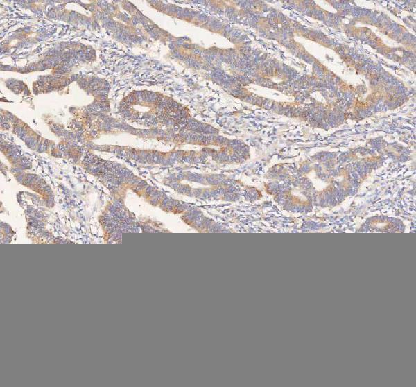
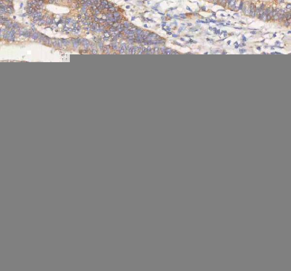
| Sample | Paraffin-embedded human colon adenocarcinoma tissue; fixative not specified (datasheet A00296-1) |
| Fixation | Image fixative and duration unreported (datasheet A00296-1); verify before use. |
| Sectioning | 4–5 µm sections on charged slides (standard) |
| Deparaffinisation | Xylene, graded ethanol series to water (standard) |
| Antigen retrieval | Heat retrieval: EDTA pH 8.0 (datasheet A00296-1); 20 min, 95–100 °C (standard) |
| Peroxidase block | 3% H2O2, 10 min, room temperature (standard) |
| Blocking | 10% goat serum (datasheet A00296-1) |
| Primary antibody | Rabbit anti-ERBB4, 2-5 μg/ml (datasheet A00296-1) |
| Primary incubation | Overnight at 4 °C (datasheet A00296-1) |
| Detection | HRP-conjugated secondary, DAB chromogen (datasheet A00296-1) |
| Counterstain | Hematoxylin, blue, dehydrate and mount (standard) |
| Expected result | ERBB4-positive staining in adipocytes of adipose tissue (HPA tissue IHC: Medium). HPA tissue profile: Ubiquitous nuclear and cytoplasmic expression. No signal in the no-primary control. |
ERBB4 is a cell-surface receptor with one transmembrane segment; its intracellular domain can also localise to the nucleus or mitochondrion (UniProt Q15303 topology; UniProt Q15303 function). In paraffin-section IHC, expect nuclear and cytoplasmic staining in many cell types, including breast glandular cells and lung alveolar cells at medium intensity (HPA: ubiquitous nuclear and cytoplasmic profile; HPA: Medium in these cells). HPA rates its tissue IHC reliability Enhanced, with external verification pending (HPA: reliability).
| Nuclear and cytoplasmic signal in breast glandular cells, with a medium-strength example (HPA: Breast glandular cells Medium). | This agrees with the reported tissue IHC profile (HPA: ubiquitous nuclear and cytoplasmic expression). Nuclear signal is biologically plausible because a processed ERBB4 intracellular domain can enter the nucleus (UniProt Q15303 function). |
| Predominantly membranous signal in an otherwise plausible positive cell (UniProt Q15303 topology). | Membrane localisation fits the full-length receptor, while HPA tissue IHC reports a broader nuclear and cytoplasmic profile (UniProt Q15303 subcellular location; HPA: tissue IHC profile). Judge the distribution alongside a validated positive section; membrane-only staining does not by itself prove an artefact (general IHC practice). |
| Staining appears in a cell population reported as undetected, such as heart cardiomyocytes (HPA: Heart muscle cardiomyocytes Not detected). | Check cell identity and staining controls before calling ERBB4 positive: nonspecific antibody binding or endogenous chromogen activity can mimic signal (general IHC practice). A tissue-level UniProt expression statement does not establish staining in each constituent cell type (UniProt Q15303 tissue specificity; HPA: cardiomyocytes Not detected). |
| Uniform colour coats the section, including tissue spaces, with little cell-specific pattern (general IHC practice). | Treat this as background until controls clarify it; excess primary antibody, incomplete blocking, or insufficient washing are general IHC causes (general IHC practice). It does not resemble the reported nuclear and cytoplasmic cellular profile (HPA: tissue IHC profile). |
| No signal appears in breast glandular cells that serve as the positive comparator (HPA: Breast glandular cells Medium). | First investigate the IHC workflow and antibody performance using an appropriate positive section (general IHC practice). A negative result in one section cannot establish ERBB4 absence, especially given HPA's medium RNA–staining consistency and pending external verification (HPA: reliability description). |
| Epitope location and receptor topology (UniProt Q15303 topology). | The extracellular region spans residues 26–651, the membrane segment 652–675, and the cytoplasmic region 676–1308 (UniProt Q15303 topology). Interpret nuclear signal in light of the antibody's stated epitope, if known (UniProt Q15303 processing; general IHC practice). |
| Proteolytic processing (UniProt Q15303 function). | JM-A isoforms can yield a soluble intracellular domain that enters the nucleus or mitochondrion (UniProt Q15303 function). An antibody to an extracellular epitope may not report that intracellular fragment; verify epitope coverage before attributing nuclear staining to it (UniProt Q15303 topology and processing; general IHC practice). |
| Alternative splicing and activation (UniProt Q15303 function). | ERBB4 has four listed JM/CYT isoforms, and activated receptor internalises after NRG1 treatment (UniProt Q15303 isoforms; UniProt Q15303 subcellular note). Thus, localisation can depend on isoform and signalling context, but the supplied IHC data do not assign a specific pattern to either (HPA: tissue IHC profile). |
| Cell-level tissue selection (HPA: tissue IHC). | Use the recorded cell population when selecting comparators: breast glandular and lung alveolar cells are Medium; heart cardiomyocytes and skeletal myocytes are Not detected (HPA: tissue IHC). UniProt lists expression in heart and skeletal muscle, so organ-level expression alone is an unreliable cell-level IHC control (UniProt Q15303 tissue specificity; HPA: tissue IHC). |
| Antibody evidence (HPA: antibody validation). | Three listed antibodies have Enhanced IHC validation, while the tissue profile still has medium consistency with RNA and awaits external verification (HPA: HPA012016, CAB000276, CAB025522 IHC Enhanced; HPA: reliability description). Interpret unexpected compartments against controls and antibody-specific evidence (general IHC practice). |
| IF/ICC cross-check: what pattern can this page support? (HPA: subcellular). | HPA summarises ERBB4 as membrane-localised in its subcellular record, but gives no main location, ICC-IF image cell line, or ICC validation for the listed antibodies (HPA: subcellular record; HPA: antibody validation). Use the separate IF/ICC guide for its experimental design (application scope). |
| Situation | Likely cause | Next action |
|---|---|---|
| Expected positive cells are blank (HPA: Breast glandular cells Medium). | The detection run or primary antibody may have failed; a single negative section is inconclusive (general IHC practice). | Run a recorded medium-staining cell population as a positive comparator and check reagent order, detection chemistry, and primary-antibody instructions (HPA: tissue IHC; general IHC practice). |
| A membranous-only pattern seems to conflict with the HPA slide profile (HPA: tissue IHC profile). | Full-length ERBB4 is membrane-bound, whereas processing permits intracellular-domain localisation (UniProt Q15303 topology and processing). | Check the antibody epitope and compare with the recorded nuclear and cytoplasmic tissue pattern before deciding whether the result is discrepant (UniProt Q15303 topology; HPA: tissue IHC profile). |
| Unexpected signal appears in cardiomyocytes or skeletal myocytes (HPA: both Not detected). | Nonspecific binding or endogenous detection activity may produce misleading chromogen (general IHC practice); cell-level staining is not predicted by organ-level expression (HPA: tissue IHC; UniProt Q15303 tissue specificity). | Confirm cell identity; inspect a no-primary control and an appropriate positive section, then reassess whether signal follows cellular boundaries (general IHC practice). |
| Diffuse colour obscures nuclei and cytoplasm (general IHC practice). | Primary antibody concentration, blocking, washing, or chromogen development may contribute to background (general IHC practice). | Check a no-primary control, then optimise antibody concentration, blocking, washing, and development time within the antibody's IHC instructions (general IHC practice). |
| Nuclear signal is strong but membrane signal is weak (HPA: nuclear and cytoplasmic tissue profile; UniProt Q15303 topology). | A soluble intracellular domain can enter the nucleus, but the supplied data cannot identify the stained molecular species in this section (UniProt Q15303 function; HPA: tissue IHC profile). | Verify that the antibody epitope lies in the intracellular region and compare an independent IHC-validated antibody where available (UniProt Q15303 topology; HPA: antibody validation; general IHC practice). |
| Two antibodies yield different distributions (HPA: multiple IHC Enhanced antibodies). | Different epitope coverage or nonspecific staining can produce discordance; the supplied record does not map antibody epitopes (UniProt Q15303 processing; HPA: antibody validation; general IHC practice). | Compare epitope documentation, positive-cell comparators, and controls before assigning a biological difference; record the discrepancy because external verification remains pending (general IHC practice; HPA: reliability description). |



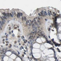


Comprehensive Human Protein Atlas IHC scoring per tissue (reliability: Enhanced — Medium consistency between antibody staining and RNA expression data. Pending external verification.). Rows are taken directly from the HPA tissue chart — click any row's HPA link to view the source.
| Tissue | Cell type | Level | Evidence | Source |
|---|---|---|---|---|
| Adipose tissue | Adipocytes | Medium | Protein (IHC) | HPA → |
| Appendix | Glandular cells | Medium | Protein (IHC) | HPA → |
| Bone marrow | Hematopoietic cells | Medium | Protein (IHC) | HPA → |
| Breast | Glandular cells | Medium | Protein (IHC) | HPA → |
| Cervix | Squamous epithelial cells | Medium | Protein (IHC) | HPA → |
| Tissue | Cell type | Level | Evidence | Source |
|---|---|---|---|---|
| Cerebral cortex | Endothelial cells | Not detected | Protein (IHC) | HPA → |
| Heart muscle | Cardiomyocytes | Not detected | Protein (IHC) | HPA → |
| Skeletal muscle | Myocytes | Not detected | Protein (IHC) | HPA → |
| Spleen | Cells in red pulp | Not detected | Protein (IHC) | HPA → |
Troubleshoot ERBB4 staining in paraffin sections by checking retrieval, controls, and the cellular compartment before comparing signal across samples.
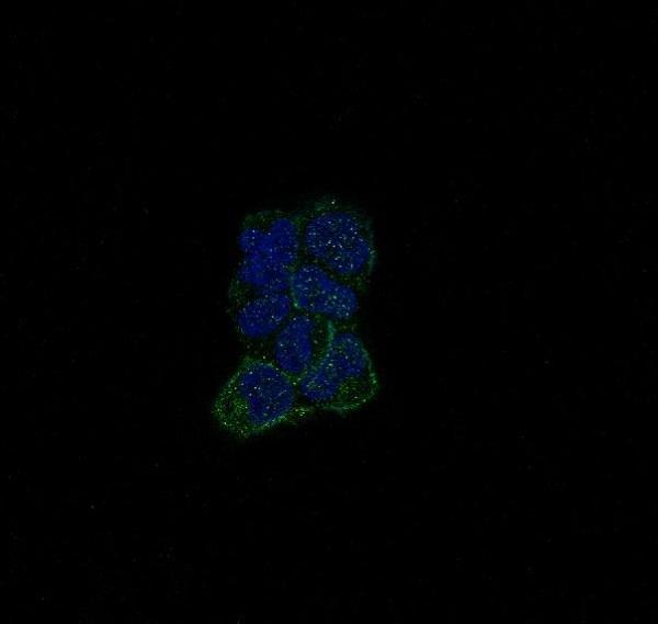
Catalog antibodies show ERBB4 staining in human paraffin tissue sections, mouse and rat brain sections, and A673 cells by IF (catalog image captions).
A00296-1 has IHC data from human colon adenocarcinoma and pancreatic ductal adenocarcinoma paraffin sections; A00296 has IHC data from human mammary and colon cancer and mouse and rat brain paraffin sections (catalog IHC captions). M00296 is listed for IF/ICC and has an IF image of A673 cells; the caption does not report the cells’ species (catalog applications and IF caption).
Which to pick: For human tissue IHC, choose A00296-1 for the EDTA pH 8.0 retrieval example or A00296 for the citrate pH 6 example; both captions describe paraffin sections but do not report the fixative (respective catalog IHC captions). For IF/ICC, choose rabbit monoclonal M00296, clone HC-5, which lists both applications and shows IF staining in A673 cells (catalog applications, clone and IF caption). For IHC across the listed species, A00296 has the strongest image support: its rabbit polyclonal antibody has paraffin-section examples from human, mouse and rat, whereas A00296-1 lists Human, Mouse and Rat reactivity but provides human IHC images only (catalog host, dilution details, reactivity and IHC captions).