This website uses cookies to ensure you get the best experience on our website.


- Table of Contents
Plan ERCC5 chromogenic IHC in paraffin sections using the nuclear and cytoplasmic tissue pattern (HPA tissue IHC). Compare high staining in colon glandular cells with undetected staining in hippocampal glial cells (HPA tissue IHC), starting the catalog antibody at 2–5 μg/ml (datasheet A01770-2).

Expected localisation, validated protocols, controls and antibodies — the at-a-glance facts below, then the full design guide.
| Expected localisation | Nuclear and cytoplasmic tissue staining (HPA tissue IHC) | |
| Staining pattern | Widespread nuclear and cytoplasmic staining (HPA tissue IHC) | |
| Antigen retrieval | EDTA pH 8.0 HIER, heat-mediated (datasheet A01770-2) | |
| Positive control | Appendix+4 more · see all | |
| Negative control | Hippocampus |
| Fixation | Matched tissue-IHC evidence does not establish the fixation claim. Validate the specimen-specific method before use. (selected-SKU IHC image A01770-2) | |
| Caveat | HPA staining may reflect proteins from multiple genes (HPA tissue IHC) | |
| Regulation | Low tissue specificity (HPA tissue RNA) | |
| Isoform / epitope | 3 isoforms; epitope coverage unknown (UniProt) |
The catalog antibody’s IHC-P protocol is paired with one published ERCC5 IHC protocol for paraffin sections of human or rat intervertebral discs (PMC12261858).
| Sample | Paraffin-embedded human colon cancer tissue; fixative not specified (datasheet A01770-2) |
| Fixation | Image fixative and duration unreported (datasheet A01770-2); verify before use. |
| Sectioning | 4–5 µm sections on charged slides (standard) |
| Deparaffinisation | Xylene, graded ethanol series to water (standard) |
| Antigen retrieval | Heat retrieval: EDTA pH 8.0 (datasheet A01770-2); 20 min, 95–100 °C (standard) |
| Peroxidase block | 3% H2O2, 10 min, room temperature (standard) |
| Blocking | 10% goat serum (datasheet A01770-2) |
| Primary antibody | Rabbit anti-ERCC5, 2-5 μg/ml (datasheet A01770-2) |
| Primary incubation | Overnight at 4 °C (datasheet A01770-2) |
| Detection | HRP-conjugated secondary, DAB chromogen (datasheet A01770-2) |
| Counterstain | Hematoxylin, blue, dehydrate and mount (standard) |
| Expected result | ERCC5-positive staining in glandular cells of appendix (HPA tissue IHC: High). HPA tissue profile: Ubiquitous nuclear and cytoplasmic expression. No signal in the no-primary control. |
ERCC5 is a nuclear DNA repair protein without a transmembrane segment (UniProt P28715: location and topology). In tissue IHC, expect staining in many cell types, including strong staining of appendix, colon, and duodenal glandular cells (HPA tissue IHC: High). HPA also reports widespread cytoplasmic staining, but rates tissue results Supported with medium consistency and cautions that the antibodies target proteins from more than one gene (HPA tissue IHC: profile and reliability).
| Glandular cells show a clear nuclear component with strong overall staining in appendix, colon, or duodenum. | This fits ERCC5's nuclear location and the reported high staining in these glandular cells (UniProt P28715: location; HPA tissue IHC: High). Score the nuclear component separately from cytoplasmic color so the result remains interpretable despite HPA's broader nuclear and cytoplasmic profile (HPA tissue IHC: profile). |
| Color is confined to cell borders, luminal material, or extracellular areas, with no convincing nuclei. | A border-only or extracellular pattern does not fit a nuclear protein without a transmembrane segment (UniProt P28715: location and topology). Treat it as a potential staining artefact and review morphology and controls (general IHC practice). Cytoplasmic color alone is less decisive because HPA reports it in tissue IHC (HPA tissue IHC: profile). |
| Hippocampal glial cells stain strongly, especially when the expected glandular cells are weak. | HPA reports hippocampal glial cells as not detected, while several glandular populations stain High (HPA tissue IHC: cell-level results). Investigate antibody cross-reactivity or endogenous chromogenic activity with appropriate controls (general IHC practice). Do not generalize this finding to all glia: caudate glial cells are reported Low (HPA tissue IHC: cell-level results). |
| Brown color spreads across nuclei, cytoplasm, and tissue spaces without a clear cell-level pattern. | Diffuse color cannot establish ERCC5 localization, even though HPA describes widespread nuclear and cytoplasmic expression (HPA tissue IHC: profile). Compare a no-primary control and inspect wash and blocking performance before scoring specific staining (general IHC practice); HPA's multi-gene antibody caution increases the need for restraint (HPA tissue IHC: reliability). |
| A high-staining glandular tissue has no convincing cellular signal. | An absent result in appendix, colon, or duodenal glandular cells conflicts with the reported High IHC staining (HPA tissue IHC: cell-level results). First check tissue preservation, retrieval, antibody and detection controls, and counterstain visibility (general IHC practice). This result alone cannot identify the failed step or prove ERCC5 is absent. |
| Choice of tissue and cell population | Appendix, colon, duodenum, gallbladder, rectum, and small intestine have High staining in glandular cells (HPA tissue IHC: cell-level results). Hippocampal glia are reported not detected, while caudate glia are Low (HPA tissue IHC: cell-level results). Evaluate the named cells within each section; a tissue label alone is insufficient. |
| Compartment and specificity | UniProt places ERCC5 in the nucleus and chromosome, whereas HPA tissue IHC describes ubiquitous nuclear and cytoplasmic staining (UniProt P28715: location; HPA tissue IHC: profile). HPA rates the tissue evidence Supported, notes medium RNA agreement, and cautions that antibodies target proteins from more than one gene (HPA tissue IHC: reliability). Cytoplasmic signal therefore warrants cautious scoring. |
| Antibody validation | HPA045845 and HPA050374 are each rated Supported for IHC (HPA antibodies: IHC validation). That rating supports using the observed tissue pattern as a comparison, while the tissue-level multi-gene caution still applies (HPA tissue IHC: reliability). It does not establish that every positive cell or compartment contains only ERCC5. |
| Isoforms and processing | UniProt lists three ERCC5 isoforms, one annotated chain spanning residues 1–1186, and no signal peptide or propeptide (UniProt P28715: isoforms and processing). The supplied record gives no antibody epitope, so isoform coverage cannot be predicted. Its lack of a transmembrane segment offers no basis to expect a cell-surface staining pattern (UniProt P28715: topology). |
| IF/ICC Q&A: where should signal appear? | In IF/ICC, look for nucleoplasmic signal; HPA lists the nucleoplasm as the supported main location and shows images for U-251MG, A-431, and U2OS (HPA subcellular ICC-IF). This observation carries HPA's caution about antibodies targeting proteins from multiple genes (HPA subcellular ICC-IF). IF/ICC conditions belong to its separate guide. |
| Situation | Likely cause | Next action |
|---|---|---|
| Expected glandular cells are unstained. | A run or specimen problem is possible; HPA reports High staining in several glandular populations (HPA tissue IHC: cell-level results). | Check section morphology and the run's positive, negative, and detection controls; review the established retrieval and antibody conditions (general IHC practice). Do not infer target-specific fixation sensitivity: none is supplied. |
| Only cytoplasm stains. | HPA reports cytoplasmic expression, but UniProt places ERCC5 in the nucleus; HPA also cautions about multi-gene antibody targeting (HPA tissue IHC: profile and reliability; UniProt P28715: location). | Record cytoplasmic and nuclear scores separately, compare controls and morphology, and avoid calling the cytoplasmic component uniquely ERCC5 on this evidence (general IHC practice; HPA tissue IHC: reliability). |
| Hippocampal glial cells show strong color. | That differs from HPA's not-detected result for those cells; nonspecific binding or endogenous detection activity are possible explanations (HPA tissue IHC: cell-level results; general IHC practice). | Examine a no-primary control and the relevant blocking controls, then compare the same run with a reported high-staining glandular tissue (general IHC practice; HPA tissue IHC: cell-level results). |
| Brown deposit follows edges or tissue spaces. | That distribution conflicts with UniProt's nuclear location and lack of a transmembrane segment; deposit or background is possible (UniProt P28715: location and topology; general IHC practice). | Inspect section integrity, reagent coverage, washes, and no-primary control before assigning a cellular score (general IHC practice). |
| Most cells are uniformly dark and nuclei cannot be distinguished. | Overdeveloped chromogen or high background can obscure compartment assessment (general IHC practice); HPA's broad tissue profile alone cannot resolve specificity (HPA tissue IHC: profile and reliability). | Review chromogen development, blocking, and the no-primary control; score only cells whose boundaries and nuclear compartment remain interpretable (general IHC practice). |
| Two IHC antibodies give different cell patterns. | Both listed antibodies are IHC Supported, while HPA warns of multi-gene targeting and only medium consistency with RNA data (HPA antibodies: IHC validation; HPA tissue IHC: reliability). | Compare their nuclear and cytoplasmic patterns in the same named cell populations and controls; report discordance rather than merging the results into one ERCC5-specific score (general IHC practice; HPA tissue IHC: reliability). |


Comprehensive Human Protein Atlas IHC scoring per tissue (reliability: Supported — Medium consistency between antibody staining and RNA expression data. Caution, targets protein from more than one gene.). Rows are taken directly from the HPA tissue chart — click any row's HPA link to view the source.
| Tissue | Cell type | Level | Evidence | Source |
|---|---|---|---|---|
| Appendix | Glandular cells | High | Protein (IHC) | HPA → |
| Colon | Glandular cells | High | Protein (IHC) | HPA → |
| Duodenum | Glandular cells | High | Protein (IHC) | HPA → |
| Fallopian tube | Ciliated cells (cell body) | High | Protein (IHC) | HPA → |
| Gallbladder | Glandular cells | High | Protein (IHC) | HPA → |
| Tissue | Cell type | Level | Evidence | Source |
|---|---|---|---|---|
| Hippocampus | Glial cells | Not detected | Protein (IHC) | HPA → |
Use the catalog antibody’s paraffin-section result as the starting point, then judge ERCC5 staining by compartment, tissue context, and controls (datasheet A01770-2; UniProt P28715; HPA tissue IHC).
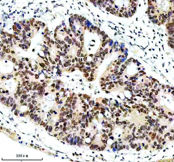
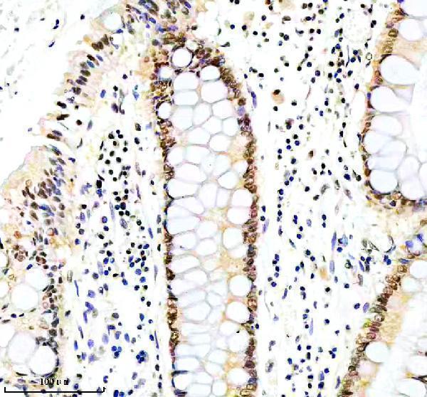
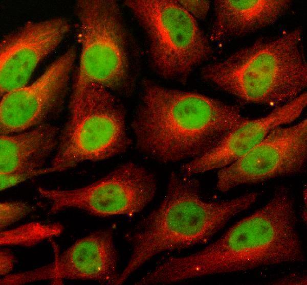
The IHC-validated anti-ERCC5 antibody has images from human paraffin-embedded colon and colon cancer sections, plus IF images from HeLa cells and a human colon cancer section (catalog image captions).
A01770-2 has IHC images from paraffin-embedded human colon and colon cancer sections (catalog IHC captions). A01770-2 also has IF images from HeLa cells and a paraffin-embedded human colon cancer section (catalog IF captions).
Which to pick: Choose A01770-2 for paraffin-section IHC; its caption documents EDTA retrieval at pH 8.0 and a primary concentration of 2 μg/ml (A01770-2 IHC caption). For IF/ICC, the same SKU lists both applications and an IF concentration of 5 μg/ml (catalog applications and dilution); its clone status is unreported (catalog clone field). The catalog lists Human, Mouse and Rat reactivity (catalog reactivity), but the supplied IHC captions show human sections only, and their fixative is unreported (A01770-2 IHC captions).