This website uses cookies to ensure you get the best experience on our website.


- Table of Contents
Plan ERG staining in paraffin sections using the nuclear and cytoplasmic tissue pattern (HPA tissue IHC). Compare positive and undetected cell populations while interpreting staining alongside the reported low consistency with RNA expression (HPA tissue IHC).

Expected localisation, validated protocols, controls and antibodies — the at-a-glance facts below, then the full design guide.
| Expected localisation | Nuclear and cytoplasmic (HPA tissue IHC) | |
| Staining pattern | Nuclear and cytoplasmic staining in several tissues (HPA tissue IHC) | |
| Antigen retrieval | Tris-EDTA pH 9.0 HIER, 95–98 °C, 20 min (rule: nuclear antigen) | |
| Positive control | Adrenal gland+4 more · see all | |
| Negative control | Cerebellum+4 more · see all |
| Fixation | Keep fixation consistent across paraffin sections (standard IHC practice; not target-specific); Matched tissue-IHC evidence does not establish the fixation claim. Validate the specimen-specific method before use. (selected-SKU IHC image M00793) | |
| Caveat | Staining has low consistency with RNA expression (HPA tissue IHC) | |
| Regulation | Blood vessel–enhanced RNA expression (HPA tissue RNA) | |
| Isoform / epitope | 6 isoforms; epitope coverage may vary (UniProt) |
The catalog antibody’s IHC-P protocol is followed by four published ERG IHC methods (PMC11106607; PMC3505676; PMC3490118; PMC3672354).
| Sample | Paraffin-embedded human kidney tissue; fixative not specified (datasheet M00793) |
| Fixation | Image fixative and duration unreported (datasheet M00793); verify before use. |
| Sectioning | 4–5 µm sections on charged slides (standard) |
| Deparaffinisation | Xylene, graded ethanol series to water (standard) |
| Antigen retrieval | Heat-induced epitope retrieval in Tris-EDTA buffer, pH 9.0, 20 min at 95–98 °C (standard rule: nuclear antigen) |
| Peroxidase block | 3% H2O2, 10 min, room temperature (standard) |
| Blocking | 10% normal serum of the secondary host, 30 min, room temperature (standard) |
| Primary antibody | Rabbit monoclonal (clone ACH-5) anti-ERG, 1:50 (datasheet M00793) |
| Primary incubation | Overnight at 4 °C (standard) |
| Detection | HRP-polymer secondary, DAB chromogen 5–10 min (standard) |
| Counterstain | Hematoxylin, blue, dehydrate and mount (standard) |
| Expected result | ERG-positive staining in glandular cells of adrenal gland (HPA tissue IHC: Medium). HPA tissue profile: Nuclear and cytoplasmic expression in several tissues. No signal in the no-primary control. |
ERG is a transcriptional regulator expected mainly in nuclei, with possible cytoplasmic staining (UniProt P11308; HPA subcellular). HPA reports medium staining in several cell populations, including adrenal glandular and bone marrow hematopoietic cells (HPA tissue IHC). Interpret those examples cautiously: the tissue profile is Approved but has low consistency with RNA expression (HPA tissue IHC). ERG has no transmembrane segment, so membrane staining is unexpected (UniProt P11308 topology).
| Distinct nuclear staining in adrenal glandular cells or bone marrow hematopoietic cells, with limited cytoplasmic signal. | This fits the reported medium tissue staining and predominant nuclear location (HPA tissue IHC; HPA subcellular). Judge the named cells and their nuclei, rather than treating every stained cell on the section as an expected positive. HPA also reports cytoplasmic expression, so limited cytoplasmic signal alone does not invalidate an otherwise convincing pattern (HPA tissue IHC). |
| Strong membrane staining, or diffuse cytoplasmic staining without a discernible nuclear pattern. | A membrane pattern conflicts with ERG's lack of a transmembrane segment (UniProt P11308 topology). Cytoplasmic ERG is possible, but an exclusively diffuse pattern warrants scrutiny because the main supported location is nucleoplasmic (HPA subcellular). Compare cellular detail and the no-primary control before assigning that staining to ERG (standard IHC practice). |
| Strong staining in an unexpected cell population, including duodenal glandular or cervical squamous epithelial cells. | Those named populations were not detected in the supplied tissue profile (HPA tissue IHC). Cross-reactivity or endogenous chromogenic activity may explain a discordant pattern (standard IHC practice). Treat the comparison as a warning, not proof of absence in every specimen: HPA labels its tissue profile Approved while reporting low consistency with RNA expression (HPA tissue IHC). |
| Diffuse color across tissue, empty spaces, or several compartments without clear cell boundaries. | That distribution does not provide a convincing cell-specific ERG readout (standard IHC practice). Background can arise from detection activity, nonspecific antibody binding, or inadequate washing (standard IHC practice). Check the no-primary control and assess whether any signal remains confined to identifiable nuclei in the expected cell population (standard IHC practice). |
| No detectable signal in adrenal glandular cells or bone marrow hematopoietic cells. | Both are reported at medium intensity, so absent staining calls for a run-level check (HPA tissue IHC). Confirm that tissue morphology and counterstain are interpretable, then check antibody application, dilution, retrieval, and detection controls against the chosen IHC protocol (standard IHC practice). HPA's low RNA–staining consistency limits any conclusion from one negative specimen (HPA tissue IHC). |
| Compartment used for scoring | Nucleus is the principal expected compartment; cytoplasm is also annotated, and supported ICC-IF localization includes cytosol (UniProt P11308; HPA subcellular). Score a clear nuclear pattern first, while recording reproducible cytoplasmic staining separately. ERG has no transmembrane segment, so a membrane-only result needs investigation (UniProt P11308 topology). |
| Choice of tissue and cell population | HPA reports medium staining in adrenal glandular cells and bone marrow hematopoietic cells, while duodenal glandular cells are not detected (HPA tissue IHC). These are cell-specific reference observations, not guarantees for every specimen. The Approved profile has low consistency between staining and RNA expression, which limits confidence in a single positive or negative comparison (HPA tissue IHC). |
| Evidence for the antibody and application | The supplied antibody records list HPA046598 and CAB018377 as IHC Approved and ICC Supported (HPA antibodies). That status supports using their reported patterns as references, but it does not establish a dilution, retrieval condition, or performance for a different antibody (HPA antibodies). Assess the antibody actually used with its own IHC controls (standard IHC practice). |
| Isoforms and antibody epitope | UniProt lists six ERG isoforms (UniProt P11308). The supplied record gives no epitope position or isoform coverage for the IHC-validated antibodies (HPA antibodies). If an observed pattern depends on a particular isoform, the available evidence cannot resolve whether the antibody recognizes it; check antibody documentation before making an isoform-specific interpretation (standard IHC practice). |
| IF/ICC Q&A: where should ERG appear? | Mainly in the nucleoplasm, with additional cytosol, according to supported ICC-IF localization (HPA subcellular). This answers the localization question for IF/ICC; the supplied records provide no IF/ICC protocol here (HPA subcellular; HPA antibodies). Interpret this page's chromogenic tissue examples as IHC observations (HPA tissue IHC). |
| Situation | Likely cause | Next action |
|---|---|---|
| Expected positive tissue is blank. | A missed primary antibody, unsuitable dilution, or failed detection step can remove visible signal (standard IHC practice). | Review the chosen antibody's IHC instructions and run controls, then verify each reagent and detection step (standard IHC practice). Use the reported medium-staining cells as a reference, while allowing for HPA's low RNA–staining consistency (HPA tissue IHC). |
| Signal is present but only diffusely cytoplasmic. | ERG can occupy cytoplasm, yet the main supported ICC-IF location is nucleoplasmic; diffuse signal may also be background (UniProt P11308; HPA subcellular; standard IHC practice). | Inspect nuclear detail in the expected cells and compare with a no-primary control (standard IHC practice). Report cytoplasmic staining separately rather than scoring it as a clear nuclear positive (HPA subcellular). |
| Unexpected cells stain strongly. | Cross-reactivity or endogenous chromogenic activity can mimic a cellular positive (standard IHC practice). | Compare the named cell population with HPA's tissue profile, then check a no-primary control and the detection system's endogenous-activity blocking (HPA tissue IHC; standard IHC practice). Do not infer that every cell type within a listed tissue shares its reported staining level (HPA tissue IHC). |
| Brown precipitate or widespread background obscures nuclei. | Nonspecific binding, residual detection activity, or insufficient washing may reduce contrast (standard IHC practice). | Examine the no-primary control; check blocking, wash steps, and detection exposure within the established IHC workflow (standard IHC practice). Reassess only where cellular boundaries and nuclei remain interpretable (standard IHC practice). |
| A presumed negative cell population shows faint color. | HPA's not-detected designation is an observation in its tissue profile, and the profile has low consistency with RNA expression (HPA tissue IHC). Weak color may also reflect background (standard IHC practice). | Identify the exact cell population and compare its staining with the no-primary control before calling it ERG positive (HPA tissue IHC; standard IHC practice). |
| A membrane-only pattern dominates the section. | Membrane localization is unsupported by the supplied ERG topology and localization records (UniProt P11308 topology; HPA subcellular). | Check morphology and detection controls, then compare a reported positive population for nuclear staining (standard IHC practice; HPA tissue IHC). Treat persistent membrane-only color as an unresolved staining discrepancy (UniProt P11308 topology). |



Comprehensive Human Protein Atlas IHC scoring per tissue (reliability: Approved — Low consistency between antibody staining and RNA expression data.). Rows are taken directly from the HPA tissue chart — click any row's HPA link to view the source.
| Tissue | Cell type | Level | Evidence | Source |
|---|---|---|---|---|
| Adrenal gland | Glandular cells | Medium | Protein (IHC) | HPA → |
| Appendix | Glandular cells | Medium | Protein (IHC) | HPA → |
| Bone marrow | Hematopoietic cells | Medium | Protein (IHC) | HPA → |
| Breast | Glandular cells | Medium | Protein (IHC) | HPA → |
| Bronchus | Respiratory epithelial cells | Medium | Protein (IHC) | HPA → |
| Tissue | Cell type | Level | Evidence | Source |
|---|---|---|---|---|
| Cerebellum | Cells in granular layer | Not detected | Protein (IHC) | HPA → |
| Cervix | Squamous epithelial cells | Not detected | Protein (IHC) | HPA → |
| Duodenum | Glandular cells | Not detected | Protein (IHC) | HPA → |
| Epididymis | Glandular cells | Not detected | Protein (IHC) | HPA → |
| Esophagus | Squamous epithelial cells | Not detected | Protein (IHC) | HPA → |
Troubleshoot ERG staining in paraffin section IHC by checking retrieval, cell compartment, controls, and scoring before interpreting chromogenic signal.
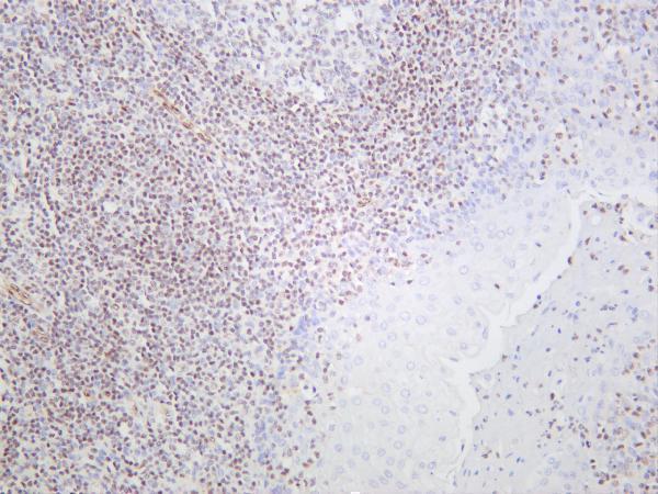
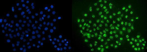
Catalog anti-ERG antibodies include IHC images of human kidney and tonsil and an IF image of A431 cells; all three list human, mouse, and rat reactivity (catalog images; catalog reactivity).
M00793 has an IHC image of paraffin-embedded human kidney, while M00793-3 has an IHC image of human tonsil with section processing unreported (M00793 image caption; M00793-3 image caption). A00793-1 has an ICC/IF image of A431 cells and lists no IHC application (A00793-1 image caption; catalog applications).
Which to pick: For tissue IHC, choose M00793 when paraffin sections match the documented kidney image; its reported dilution is 1:50, and the fixative is unreported (M00793 image caption; catalog dilution). M00793-3 is another IHC option with a human tonsil image and a 1:100–1:500 dilution range; its caption does not report section processing or fixative (M00793-3 image caption; catalog dilution). For IF/ICC, choose A00793-1 for the documented A431 example at 5 μg/ml; for mouse or rat tissue IHC, both IHC-listed SKUs claim reactivity, although their supplied IHC images show human samples (A00793-1 image caption; catalog applications and reactivity; M00793 and M00793-3 image captions).