This website uses cookies to ensure you get the best experience on our website.


- Table of Contents
Plan chromogenic IHC on paraffin sections using 2–5 μg/ml of the catalog antibody (datasheet A03621-2). Assess cytoplasmic staining in appendix glandular cells and bone marrow hematopoietic cells, with ovarian stromal cells as a low signal comparison (HPA tissue IHC).

Expected localisation, validated protocols, controls and antibodies — the at-a-glance facts below, then the full design guide.
| Expected localisation | Cytoplasmic in tissue (HPA tissue IHC); ER lumen (UniProt) | |
| Staining pattern | Broad cytoplasmic staining across many cell types (HPA tissue IHC) | |
| Antigen retrieval | EDTA pH 8.0 HIER, heat-mediated (datasheet A03621-2) | |
| Positive control | Appendix+4 more · see all | |
| Negative control | Ovary |
| Fixation | Matched tissue-IHC evidence does not establish the fixation claim. Validate the specimen-specific method before use. (selected-SKU IHC image A03621-2) | |
| Caveat | Ovarian stromal cells may be unstained (HPA tissue IHC) | |
| Regulation | Mostly expressed in secretory tissues (UniProt) | |
| Isoform / epitope | 2 isoforms; mature chain starts at residue 33; epitope dependence unknown (UniProt) |
The catalog antibody’s IHC-P protocol is accompanied by three published ERP29 IHC protocols for osteosarcoma, nasopharyngeal carcinoma and gastric cancer (PMC6444297; PMC3583588; PMC5667996).
| Sample | Paraffin-embedded human prostate cancer tissue; fixative not specified (datasheet A03621-2) |
| Fixation | Image fixative and duration unreported (datasheet A03621-2); verify before use. |
| Sectioning | 4–5 µm sections on charged slides (standard) |
| Deparaffinisation | Xylene, graded ethanol series to water (standard) |
| Antigen retrieval | Heat retrieval: EDTA pH 8.0 (datasheet A03621-2); 20 min, 95–100 °C (standard) |
| Peroxidase block | 3% H2O2, 10 min, room temperature (standard) |
| Blocking | 10% goat serum (datasheet A03621-2) |
| Primary antibody | Rabbit anti-ERP29, 2-5 μg/ml (datasheet A03621-2) |
| Primary incubation | Overnight at 4 °C (datasheet A03621-2) |
| Detection | HRP-conjugated secondary, DAB chromogen (datasheet A03621-2) |
| Counterstain | Hematoxylin, blue, dehydrate and mount (standard) |
| Expected result | ERP29-positive staining in glandular cells of appendix (HPA tissue IHC: High). HPA tissue profile: Ubiquitous cytoplasmic expression. No signal in the no-primary control. |
ERP29 is an endoplasmic reticulum lumen protein with no transmembrane segment (UniProt P30040 topology). In paraffin section IHC, expect cytoplasmic staining across many cell types, with particularly strong staining in several glandular, respiratory epithelial, hematopoietic, glial, and neuronal populations (HPA: ubiquitous cytoplasmic expression; High in the listed populations). HPA rates its tissue IHC reliability Enhanced, while reporting medium consistency between staining and RNA expression (HPA: tissue IHC reliability).
| Cytoplasmic chromogen in appendix or breast glandular cells, with a stronger signal than adjacent tissue. | This fits the reported compartment and cell types (HPA: ubiquitous cytoplasmic expression; High in appendix and breast glandular cells). Judge the identified cells, rather than treating every cell in the section as an equally strong positive. |
| Predominantly nuclear or sharply filamentous staining in an IHC section. | This is discordant with the expected tissue IHC pattern and merits an artefact or specificity check (HPA: ubiquitous cytoplasmic expression; UniProt P30040: ER lumen). HPA's separate ICC-IF observations are uncertain and do not establish that pattern for paraffin section IHC (HPA: subcellular ICC-IF). |
| Strong staining confined to ovarian stromal cells while expected positive cells remain unstained. | Consider cross-reactivity or endogenous detection activity before calling this ERP29 (HPA: ovarian stromal cells Not detected; general IHC practice). The HPA finding concerns stromal cells specifically; it does not make the whole ovary a negative tissue. |
| Even chromogen over cells, stroma, and empty areas, without a discernible cytoplasmic pattern. | This is background rather than an interpretable cell pattern (general IHC practice). Compare a no-primary control and review blocking, washing, and detection; HPA's cytoplasmic tissue pattern alone cannot identify the background source (HPA: tissue IHC profile). |
| No staining in appendix glandular cells or bronchial respiratory epithelial cells. | These are reported High populations, so an absent result calls for assay troubleshooting before a biological absence claim (HPA: High in appendix glandular and bronchial respiratory epithelial cells). Check that the relevant cells are present and that the positive control develops (general IHC practice). |
| Cell selection and intensity | HPA reports ubiquitous cytoplasmic expression but varying cell-level staining: High in breast glandular cells and bone marrow hematopoietic cells, Low in skeletal muscle myocytes and soft-tissue fibroblasts, and Not detected in ovarian stromal cells (HPA: tissue IHC). Use the named cell population when interpreting a section. |
| Compartment and processing | ERP29 is annotated in the ER lumen, lacks a transmembrane segment, and has a signal sequence at residues 1–32 with a mature chain at 33–261 (UniProt P30040: topology and processing). These annotations support a cytoplasmic ER-associated expectation; they do not identify an antibody epitope or predict paraffin-section retrieval sensitivity. |
| Antibody evidence and isoforms | Both listed antibodies have Enhanced IHC validation; HPA defines this as staining reproduced by independent antibodies or orthogonal data (HPA: HPA039363 and HPA039456 IHC validation). UniProt lists 2 isoforms (UniProt P30040: isoforms), but the supplied evidence does not establish which isoforms either antibody detects. |
| IF/ICC Q&A: should a microtubule pattern change the IHC call? | No. HPA describes microtubules as the main ICC-IF location and nucleoplasm as an additional location, both uncertain (HPA: subcellular ICC-IF). For paraffin section IHC, assess the reported cytoplasmic tissue pattern; the ICC-IF observation does not validate a nuclear or filamentous chromogenic result (HPA: tissue IHC profile). |
| Situation | Likely cause | Next action |
|---|---|---|
| Known-positive glandular or respiratory epithelial cells show no chromogen. | The run may have failed, or the expected cells may be absent from the sampled section (general IHC practice; HPA: High in appendix glandular and bronchial respiratory epithelial cells). | Confirm the cell population on the counterstained section, inspect the positive control, and review the antibody and detection steps (general IHC practice). Do not assign ERP29 absence from a failed run. |
| Signal is weak in a population reported High by HPA. | HPA levels describe its observed tissue staining, not a guaranteed intensity in every run (HPA: tissue IHC levels). Section and assay performance can affect an individual readout (general IHC practice). | Compare a concurrent known-positive section, then review the chosen antigen-retrieval and antibody conditions against the catalog IHC-P instructions (general IHC practice). No ERP29-specific retrieval effect is established here. |
| The dominant result is nuclear or filamentous staining. | The pattern conflicts with the expected cytoplasmic tissue profile; the separate microtubule and nucleoplasm ICC-IF calls are uncertain (HPA: tissue IHC profile; subcellular ICC-IF). | Check the positive-control pattern and no-primary control, then repeat with an IHC-validated antibody if needed (HPA: listed antibodies have Enhanced IHC validation; general IHC practice). |
| Ovarian stromal cells stain strongly. | That cell population is reported Not detected, so non-specific binding or endogenous detection activity is plausible (HPA: ovarian stromal cells Not detected; general IHC practice). | Verify stromal identity, compare a no-primary control, and review blocking and detection chemistry before interpreting the signal as ERP29 (general IHC practice). |
| Brown signal blankets the section or appears outside cells. | Diffuse deposition can obscure the expected cytoplasmic pattern (general IHC practice; HPA: ubiquitous cytoplasmic expression). | Compare the no-primary control, review washing and detection conditions, and score only localized cellular staining that remains distinguishable from background (general IHC practice). |
| A low-staining cell population is scored as a failed assay. | HPA reports Low staining in skeletal muscle myocytes and soft-tissue fibroblasts; faint signal there alone is an uncertain assay check (HPA: tissue IHC). | Use a reported High population as the positive comparator, such as appendix glandular cells or bone marrow hematopoietic cells, while scoring each cell type separately (HPA: tissue IHC; general IHC practice). |


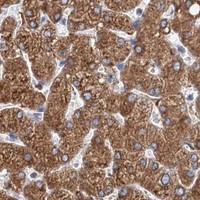


Comprehensive Human Protein Atlas IHC scoring per tissue (reliability: Enhanced — Medium consistency between antibody staining and RNA expression data.). Rows are taken directly from the HPA tissue chart — click any row's HPA link to view the source.
| Tissue | Cell type | Level | Evidence | Source |
|---|---|---|---|---|
| Appendix | Glandular cells | High | Protein (IHC) | HPA → |
| Bone marrow | Hematopoietic cells | High | Protein (IHC) | HPA → |
| Breast | Glandular cells | High | Protein (IHC) | HPA → |
| Bronchus | Respiratory epithelial cells | High | Protein (IHC) | HPA → |
| Caudate | Glial cells | High | Protein (IHC) | HPA → |
| Tissue | Cell type | Level | Evidence | Source |
|---|---|---|---|---|
| Ovary | Ovarian stroma cells | Not detected | Protein (IHC) | HPA → |
Troubleshoot ERP29 staining in paraffin sections by checking retrieval, tissue handling, cellular pattern, controls, and scoring before interpreting DAB signal.
The IHC-validated antibody has paraffin-section images from human prostate cancer, ovarian cancer, spleen and thyroid cancer, plus IF images from HepG2 cells and human colon cancer sections (A03621-2 image captions).
A03621-2 will render with its own human prostate cancer paraffin-section IHC figure; its catalog also shows IHC images from human ovarian cancer, spleen and thyroid cancer, and IF images from HepG2 cells and human colon cancer sections (A03621-2 image captions). M03621-1 is listed for IHC and ICC/IF in human, mouse and rat, but has no IHC or IF image caption in the payload and will not render as a card here (M03621-1 catalog entry; cards payload).
Which to pick: For tissue IHC, choose A03621-2 because its own caption documents paraffin-section staining with EDTA retrieval at pH 8.0 and 2 μg/ml primary antibody; the fixative is unreported (A03621-2 IHC image caption). For IF/ICC, A03621-2 has image examples in HepG2 cells and human colon cancer sections, while monoclonal M03621-1, clone 18E73, lists ICC/IF without an image example here (A03621-2 IF image captions; M03621-1 catalog entry). For cross-species planning, A03621-2 lists human, monkey, mouse and rat reactivity, while M03621-1 lists human, mouse and rat; the supplied staining images for A03621-2 cover human samples only (catalog reactivity; A03621-2 image captions).