This website uses cookies to ensure you get the best experience on our website.


- Table of Contents
ESR2 shows low nuclear staining in several tissues, including testis and ovary (HPA tissue IHC). Use this guide to plan fixation, controls, and nuclear scoring for chromogenic IHC on paraffin sections.

Expected localisation, validated protocols, controls and antibodies — the at-a-glance facts below, then the full design guide.
| Expected localisation | Nuclear staining in tissue sections (HPA tissue IHC) | |
| Staining pattern | Low nuclear staining in Leydig and ovarian stromal cells (HPA tissue IHC) | |
| Antigen retrieval | Tris-EDTA pH 9.0 HIER, 95–98 °C, 20 min (rule: nuclear antigen) | |
| Positive control | Adrenal gland | |
| Negative control | Adipose tissue+4 more · see all |
| Fixation | Keep fixation consistent across sections. (standard IHC practice; not target-specific) | |
| Caveat | Weak staining and medium antibody–RNA consistency (HPA tissue IHC) | |
| Regulation | Expression varies by tissue (UniProt) | |
| Isoform / epitope | 9 isoforms; verify epitope coverage (UniProt) |
The catalog antibody’s IHC-P protocol is paired with published ESR2 IHC methods from PMC9631985, PMC10726529, and PMC9141030.
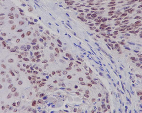
| Sample | Paraffin-embedded human cervix cancer tissue; fixative not specified (datasheet M00786-1) |
| Fixation | Image fixative and duration unreported (datasheet M00786-1); verify before use. |
| Sectioning | 4–5 µm sections on charged slides (standard) |
| Deparaffinisation | Xylene, graded ethanol series to water (standard) |
| Antigen retrieval | Heat-induced epitope retrieval in Tris-EDTA buffer, pH 9.0, 20 min at 95–98 °C (standard rule: nuclear antigen) |
| Peroxidase block | 3% H2O2, 10 min, room temperature (standard) |
| Blocking | 10% normal serum of the secondary host, 30 min, room temperature (standard) |
| Primary antibody | Rabbit monoclonal (clone IDI-5) anti-ESR2, 1:50 (datasheet M00786-1) |
| Primary incubation | Overnight at 4 °C (standard) |
| Detection | HRP-polymer secondary, DAB chromogen 5–10 min (standard) |
| Counterstain | Hematoxylin, blue, dehydrate and mount (standard) |
| Expected result | ESR2-positive staining in cells in zona glomerulosa of adrenal gland (HPA tissue IHC: Medium). HPA tissue profile: Low nuclear expression in several tissues, including testis and ovaries. No signal in the no-primary control. |
ESR2 is a nuclear receptor with no transmembrane segment (UniProt Q92731: subcellular location and topology). In paraffin-section IHC, expect nuclear staining in selected cells: HPA reports medium staining in adrenal zona glomerulosa cells and low staining in testis Leydig cells and ovarian stroma cells (HPA tissue IHC). HPA labels its tissue IHC reliability Enhanced while noting medium consistency between antibody staining and RNA expression (HPA tissue IHC).
| Distinct nuclear signal in adrenal zona glomerulosa cells. | This matches HPA’s medium staining in that cell population and ESR2’s nuclear location (HPA tissue IHC; UniProt Q92731). Judge localization and cell identity together; intensity alone does not establish specificity (general IHC practice). |
| Weak nuclear signal in testis Leydig cells or ovarian stroma cells. | Low staining is consistent with the reported pattern (HPA tissue IHC). A weak result in either population should be interpreted against a positive control and local background, since HPA’s tissue profile does not promise strong staining in every section (general IHC practice; HPA tissue IHC). |
| Predominantly cytoplasmic or membrane staining, with little nuclear signal. | That is discordant with the expected IHC compartment (UniProt Q92731: nucleus; HPA tissue IHC: low nuclear expression). Review background and antibody specificity before calling the cells ESR2-positive (general IHC practice). HPA’s uncertain vesicle observation comes from ICC-IF and does not validate cytoplasmic IHC staining (HPA subcellular ICC-IF). |
| Strong signal in a cell population HPA lists as not detected, such as breast adipocytes. | Treat the result as a specificity or detection-control concern, not proof that the whole tissue lacks ESR2 (HPA tissue IHC: breast adipocytes not detected; general IHC practice). Check cell identification, cross-reactivity and endogenous chromogenic activity (general IHC practice). |
| Diffuse colour across nuclei, cytoplasm and surrounding tissue. | The spread obscures ESR2’s expected nuclear pattern (UniProt Q92731; HPA tissue IHC). Assess nonspecific antibody binding, incomplete blocking and detection background with an appropriate negative control before scoring positive nuclei (general IHC practice). |
| Cell population and expected intensity | HPA reports medium staining in adrenal zona glomerulosa cells but low staining in Leydig and ovarian stroma cells (HPA tissue IHC). Select and score controls by the named cell population; a tissue name alone is insufficient (general IHC practice). |
| Subcellular location and protein topology | ESR2 is nuclear and has no transmembrane segment or signal peptide (UniProt Q92731). A membrane-dominant IHC pattern therefore needs investigation; these annotations do not establish how fixation or antigen retrieval affects the antibody (UniProt Q92731; source-scope limitation). |
| Isoforms and epitope coverage | UniProt lists nine ESR2 isoforms (UniProt Q92731). The payload gives no catalog antibody epitope, so isoform recognition and any resulting tissue-staining differences cannot be assigned from this record (source-scope limitation). |
| IHC evidence and antibody choice | HPA labels the overall tissue IHC reliability Enhanced, while listing CAB079300 as IHC Supported and HPA068406 only as ICC Supported (HPA tissue IHC; HPA antibodies). Apply an IHC-validated antibody to paraffin sections; do not transfer an ICC validation label to IHC (general IHC practice). |
| IF/ICC Q: Where should ESR2 fluorescence appear? | A: Mainly in the nucleoplasm; HPA also reports vesicles with uncertain support (HPA subcellular ICC-IF). That observation answers localization for IF/ICC; it does not supply a paraffin IHC protocol or validate vesicular chromogenic staining (HPA subcellular ICC-IF; source-scope limitation). |
| Situation | Likely cause | Next action |
|---|---|---|
| No nuclear signal in adrenal zona glomerulosa cells. | This misses HPA’s reported medium-positive cell population (HPA tissue IHC); the immediate cause is undetermined. | Confirm the cells and positive control, then check the catalog antibody’s IHC-P instructions for retrieval, dilution and detection conditions (general IHC practice). No ESR2-specific fixation sensitivity is reported in these payloads (source-scope limitation). |
| No signal in Leydig or ovarian stroma cells, while a positive control stains. | These are low-staining populations in HPA’s IHC profile, so a negative read can reflect limited visibility at the assay’s threshold (HPA tissue IHC; general IHC practice). | Review nuclear signal against adjacent background and assess additional sections or the HPA medium-positive adrenal control where available (general IHC practice; HPA tissue IHC). Do not infer complete tissue absence from one negative cell-level read (general IHC practice). |
| Colour is concentrated at cell membranes or throughout cytoplasm. | That distribution conflicts with UniProt’s nuclear annotation and HPA’s nuclear tissue profile (UniProt Q92731; HPA tissue IHC). | Check morphology and the negative control, then reassess antibody specificity and detection background before scoring (general IHC practice). Do not use HPA’s uncertain ICC-IF vesicle finding to justify a membrane IHC call (HPA subcellular ICC-IF). |
| Strong staining appears in breast adipocytes or bone-marrow hematopoietic cells. | HPA lists those particular cell populations as not detected; this discrepancy can reflect cell misidentification, cross-reactivity or detection background (HPA tissue IHC; general IHC practice). | Verify the stained cell type and compare with a no-primary control and a known-positive cell population (general IHC practice). Keep the conclusion limited to the examined cells, since HPA’s negative labels are cell-specific (HPA tissue IHC). |
| Diffuse brown background obscures nuclear boundaries. | Nonspecific binding or endogenous chromogenic activity can make nuclear scoring unreliable (general IHC practice). | Inspect no-primary and detection controls, then review blocking, wash and chromogen steps under the assay’s instructions (general IHC practice). Score ESR2 only where discrete nuclear staining remains distinguishable (UniProt Q92731; general IHC practice). |
| ICC-IF images show vesicular signal, but the paraffin IHC slide shows cytoplasmic colour. | HPA calls the vesicle location uncertain in ICC-IF and reports mainly nucleoplasmic localization; the two assay observations are not interchangeable (HPA subcellular ICC-IF). | Interpret the paraffin slide against the nuclear IHC pattern and its controls (HPA tissue IHC; general IHC practice). Use the separate IF/ICC guide for fluorescence assay design; this IHC result alone cannot resolve the uncertain vesicle annotation (HPA subcellular ICC-IF). |


Comprehensive Human Protein Atlas IHC scoring per tissue (reliability: Enhanced — Medium consistency between antibody staining and RNA expression data.). Rows are taken directly from the HPA tissue chart — click any row's HPA link to view the source.
| Tissue | Cell type | Level | Evidence | Source |
|---|---|---|---|---|
| Adrenal gland | Cells in zona glomerulosa | Medium | Protein (IHC) | HPA → |
| Tissue | Cell type | Level | Evidence | Source |
|---|---|---|---|---|
| Adipose tissue | Adipocytes | Not detected | Protein (IHC) | HPA → |
| Appendix | Glandular cells | Not detected | Protein (IHC) | HPA → |
| Bone marrow | Hematopoietic cells | Not detected | Protein (IHC) | HPA → |
| Breast | Adipocytes | Not detected | Protein (IHC) | HPA → |
| Bronchus | Respiratory epithelial cells | Not detected | Protein (IHC) | HPA → |
Troubleshoot ESR2 staining by checking nuclear signal, cell identity, epitope coverage and matched controls before interpreting intensity.
An anti-ESR2 antibody has IHC data from paraffin-embedded human cervix cancer (M00786-1 IHC image caption). Human is the only listed species, and no IF image is supplied (catalog reactivity; IF images).
M00786-1 is listed for human IHC (catalog applications; reactivity). Its IHC image shows paraffin-embedded human cervix cancer; no IF image is supplied (M00786-1 IHC image caption; IF images).
Which to pick: Choose M00786-1 for human tissue IHC: it is rabbit monoclonal clone IDI-5, and its own image shows paraffin-embedded human cervix cancer (catalog host and clone; M00786-1 IHC image caption). No listed SKU supports an IF/ICC or cross-species recommendation (catalog applications: IHC, WB; reactivity: Human; IF images: none). The caption does not report the fixative (M00786-1 IHC image caption).