EXOSC8 / Exosome complex component RRP43 · IHC design guide

Design Immunohistochemistry for EXOSC8

Plan chromogenic EXOSC8 IHC on paraffin sections with the IHC-validated antibody at 2–5 μg/ml (datasheet A09191-1). Assess cytoplasmic and nuclear staining, and interpret intensity cautiously because tissue staining reliability is uncertain (HPA tissue IHC).

Evidence assembled Oct 2026 · For research use; verify linked source records and product datasheet before use
Immunohistochemistry protocol sheet for EXOSC8 (IHC for EXOSC8): expected localisation Observed cytoplasmic and nuclear staining (HPA tissue IHC), antibody A09191-1, validated IHC image, and IHC protocol steps
Printable EXOSC8 IHC protocol sheet — expected localisation Observed cytoplasmic and nuclear staining (HPA tissue IHC), antibody A09191-1, controls and protocol steps. Open the full EXOSC8 IHC guide →

EXOSC8 Immunohistochemistry Experimental Design Guide

Expected localisation, validated protocols, controls and antibodies — the at-a-glance facts below, then the full design guide.

Must know before staining
Expected localisation Observed cytoplasmic and nuclear staining (HPA tissue IHC)
Staining pattern Broad cytoplasmic and nuclear staining across cell types (HPA tissue IHC)
Antigen retrieval EDTA pH 8.0 HIER, heat-mediated (datasheet A09191-1)
Positive control ⓘ Colon+4 more · see all
Negative control ⓘ No source-confirmed negative tissue/cell row; use no-primary + isotype controls
Important caveats
Reasons your staining may differ from the expected pattern.
Fixation Keep paraffin-section fixation consistent across samples (standard IHC practice; not target-specific)
Caveat Antibody staining has low concordance with RNA data (HPA tissue IHC)
Regulation Broad expression; cell intensity varies (HPA tissue IHC)
Isoform / epitope No isoforms annotated; chain spans aa 2–276 (UniProt)
Section 1

Recommended EXOSC8 IHC & IF Protocols

Compare the catalog antibody’s IHC-P protocol (datasheet: A09191-1) with the published patient-derived tissue workflow (PMC12776135).

Recommended immunohistochemistry (IHC-P) protocol parameters
SampleParaffin-embedded human gall bladder adenosquamous carcinoma tissue; fixative not specified (datasheet A09191-1)
FixationImage fixative and duration unreported (datasheet A09191-1); verify before use.
Sectioning4–5 µm sections on charged slides (standard)
DeparaffinisationXylene, graded ethanol series to water (standard)
Antigen retrievalHeat retrieval: EDTA pH 8.0 (datasheet A09191-1); 20 min, 95–100 °C (standard)
Peroxidase block3% H2O2, 10 min, room temperature (standard)
Blocking10% goat serum (datasheet A09191-1)
Primary antibodyRabbit anti-EXOSC8, 2-5 μg/ml (datasheet A09191-1)
Primary incubationOvernight at 4 °C (datasheet A09191-1)
DetectionStreptavidin-biotin complex (SABC), DAB chromogen (datasheet A09191-1)
CounterstainHematoxylin, blue, dehydrate and mount (standard)
Expected resultEXOSC8-positive staining in endothelial cells of colon (HPA tissue IHC: High). HPA tissue profile: Ubiquitous cytoplasmic and nuclear expression. No signal in the no-primary control.
💡Decision noteStart with EDTA pH 8.0 heat retrieval (datasheet: A09191-1); the published workflow does not specify retrieval (PMC12776135).
Section 2

What Is the Expected EXOSC8 Staining Pattern?

EXOSC8 is a soluble protein localized to the cytoplasm, nucleus and nucleolus (UniProt Q96B26: localization; no transmembrane segment). In tissue IHC, expect cytoplasmic and nuclear staining across many cell types, with strong staining reported in selected glandular, endothelial and decidual cells (HPA: ubiquitous profile; High examples). Treat intensity as provisional: HPA rates tissue IHC reliability Uncertain because staining and RNA expression have low consistency (HPA: reliability).

What am I looking at on my slide?
Nuclear and cytoplasmic staining in glandular cells of duodenum or rectum.This fits the reported High staining in those cells (HPA: duodenum and rectum). Nuclear and cytoplasmic distribution also fits EXOSC8 localization (UniProt Q96B26). Neither observation alone establishes antibody specificity because tissue IHC reliability is Uncertain (HPA: reliability).
Staining is confined to the cell surface or extracellular space, with little intracellular signal.That distribution conflicts with the reported cytoplasmic and nuclear tissue pattern (HPA: profile) and the absence of a transmembrane segment (UniProt Q96B26: topology). Consider nonspecific staining or a section and detection artifact before interpreting it as EXOSC8.
The strongest signal appears in cells outside the reported High populations.Compare identifiable cell types within the section: HPA reports High staining in colonic endothelial cells but Low staining in adipocytes and myocytes (HPA: tissue IHC). A reversed pattern can indicate cross-reactivity or endogenous detection activity; HPA does not define any tissue as definitively negative.
Brown signal spreads across cells and surrounding tissue without clear intracellular boundaries.Diffuse background cannot establish the nuclear and cytoplasmic pattern reported for EXOSC8 (HPA: profile). In chromogenic IHC, inadequate blocking, residual detection activity or excessive detection can produce background (general IHC practice). Compare the primary-antibody omission control and intact tissue morphology.
No discernible signal appears in a reported High cell population.Loss of staining in duodenal glandular cells or pancreatic exocrine glandular cells warrants a technical check (HPA: High in both). It is not, by itself, evidence that EXOSC8 is absent: HPA assigns tissue IHC an Uncertain reliability rating (HPA: reliability).
💡Expected EXOSC8 appearanceA plausible positive IHC result is intracellular nuclear and cytoplasmic signal, potentially High in duodenal glandular or colonic endothelial cells (HPA: profile and tissue IHC); isolated surface or extracellular staining conflicts with EXOSC8 localization (UniProt Q96B26: localization and topology).
How each factor affects the staining
Compartment and topologyEXOSC8 is reported in cytoplasm, nucleus and nucleolus and has no transmembrane segment (UniProt Q96B26). Score intracellular compartments separately; a membrane-only pattern needs independent verification.
Tissue and cell-type referenceHPA reports High staining in several glandular populations and colonic endothelial cells, versus Low in adipocytes and muscle cells (HPA: tissue IHC). Low is a relative comparison, not a negative control.
Strength of tissue evidenceHPA calls the tissue profile ubiquitous but rates IHC reliability Uncertain owing to low agreement with RNA expression (HPA: profile and reliability). Its listed intensities guide comparison; they do not validate every stained cell.
Antibody agreementThe listed IHC antibody assessments are Uncertain; no supplied IHC assessment is Enhanced (HPA: HPA039702, HPA043942, CAB034240). Interpret a matching pattern with controls rather than treating one antibody's signal as independent confirmation.
Does IF show the same pattern?ICC-IF supports nucleoli fibrillar center, nucleoplasm and cytosol; mitotic chromosome and vesicle assignments are Uncertain (HPA: subcellular). These observations can inform compartment interpretation, but they are ICC-IF evidence, not tissue IHC validation or an IF protocol.
Why is my staining missing, weak or wrong?
SituationLikely causeNext action
No staining in a reported High population.The assay may have insufficient detectable signal; the reference itself has Uncertain IHC reliability (HPA: tissue IHC and reliability).Check section integrity, primary-antibody use, detection reagents and a concurrently processed reference section (general IHC practice). Review antigen retrieval as a general assay variable without assuming EXOSC8 has known fixation sensitivity.
Only cytoplasm stains, or only nuclei stain.EXOSC8 has both reported compartments, but their visibility may differ by cell and assay (UniProt Q96B26: localization; HPA: profile).Score nuclear and cytoplasmic signal separately across intact cells. Compare a reported High population and controls before calling either compartment absent; HPA's IHC profile remains Uncertain (HPA: reliability).
Strong staining lies mainly at cell borders or outside cells.This conflicts with EXOSC8's intracellular localization and lack of a transmembrane segment (UniProt Q96B26).Inspect morphology and the primary-antibody omission control; repeat detection or blocking checks if the same distribution appears without primary antibody (general IHC practice).
A Low reference stains as strongly as a High reference.HPA reports relative differences, including Low muscle or adipose staining and High glandular staining, with Uncertain IHC reliability (HPA: tissue IHC).Compare equivalent cell types and staining conditions, then check background controls. Do not call the Low population EXOSC8-negative or use this contrast alone to validate specificity.
The whole section has diffuse brown background.Nonspecific binding or endogenous chromogenic detection activity can obscure compartment scoring (general IHC practice).Inspect the primary-antibody omission control, blocking and detection steps; assess specific intracellular signal only after background is controlled (general IHC practice).
IHC appears different from ICC-IF images.ICC-IF supports nucleoli fibrillar center, nucleoplasm and cytosol, whereas tissue IHC is summarized broadly as nuclear and cytoplasmic (HPA: subcellular and tissue profile).Compare only shared compartments and keep the applications distinct. Do not infer tissue IHC validation from ICC-IF localization; HPA rates tissue IHC Uncertain (HPA: reliability).

Sample controls for EXOSC8 IHC & IF

🧪Run duodenum first and assess its glandular cells for EXOSC8 staining (High; HPA: duodenum glandular cells). No source-confirmed negative tissue or internal negative cell type is listed among the selected HPA candidates, so use no-primary and isotype controls to define background; cells without specific staining on the positive slide should show only that background (HPA: selected negative rows empty).
Positive control tissue: Colon (Endothelial cells, HPA High)
Negative control tissue: No source-confirmed negative tissue/cell row among selected HPA candidates. Use a no-primary (secondary-only) and an isotype control instead.
ICC-IF cell lines (HPA subcellular resource): HPA ICC-IF images show EXOSC8 in HEL, Rh30, U2OS, with annotated localisation: Nucleoli fibrillar center (supported), Mitotic chromosome (uncertain) (HPA subcellular).
Technical controls: Include a no-primary, secondary-only control and a concentration-matched rabbit IgG isotype control, then confirm specificity with an EXOSC8 knockout sample or a peptide-block control if the immunizing peptide is available (hero IHC caption: rabbit primary antibody; standard IHC practice). For the caption’s biotin-based DAB detection, check endogenous peroxidase and biotin background in duodenum (hero IHC caption: biotinylated secondary and SABC–DAB; standard IHC practice).
⚠️Feasibility: The supplied evidence reports no EXOSC8-specific fixation window or fixation effect, and the paraffin-section caption does not state a fixative (hero IHC caption: fixative unreported). Heat retrieval in EDTA at pH 8.0 was used for the catalog antibody’s paraffin-section image; that example does not establish that retrieval is required under all conditions (hero IHC caption: EDTA retrieval). The evidence does not establish whether frozen sections or IF are easier for this antibody; with biotin-based DAB detection in duodenum, check for endogenous biotin background (hero IHC caption: SABC–DAB; standard IHC practice).

HPA tissue IHC evidence for EXOSC8

Comprehensive Human Protein Atlas IHC scoring per tissue (reliability: Uncertain — Low consistency between antibody staining and RNA expression data. Pending external verification.). Rows are taken directly from the HPA tissue chart — click any row's HPA link to view the source.

Positive expression · recommended positive controls

TissueCell typeLevelEvidenceSource
Colon Endothelial cells High Protein (IHC) HPA →
Duodenum Glandular cells High Protein (IHC) HPA →
Endometrium Cells in endometrial stroma Medium Protein (IHC) HPA →
Epididymis Glandular cells High Protein (IHC) HPA →
Pancreas Exocrine glandular cells High Protein (IHC) HPA →

Undetected expression · recommended negative controls

TissueCell typeLevelEvidenceSource
No source-confirmed negative tissue/cell row among selected HPA candidates. Use a no-primary (secondary-only) and an isotype control instead.
Section 3

Advanced EXOSC8 IHC Tips

Troubleshoot EXOSC8 staining in paraffin sections by checking retrieval, intracellular localisation, assay controls and cell-level scoring before interpreting differences between tissues.

Which retrieval conditions should I try when EXOSC8 staining is weak in paraffin sections?
Start with heat-mediated retrieval in EDTA at pH 8.0 (datasheet A09191-1). The selected paraffin-section example used that retrieval before overnight incubation at 4°C with 2 μg/ml catalog antibody and DAB detection (datasheet A09191-1). If staining remains weak, vary heating time in small increments on matched sections while keeping antibody concentration and detection conditions fixed (standard IHC practice). Compare signal in nuclei and cytoplasm with background in the same cell populations; both compartments are plausible for EXOSC8, so a brighter section alone does not establish better retrieval (UniProt Q96B26 localisation; HPA tissue IHC, reliability uncertain).
How should I troubleshoot suspected fixation-related loss of EXOSC8 staining?
Target-specific fixation sensitivity is unknown because the selected paraffin-section caption does not state its fixative (datasheet A09191-1). Record the actual fixative, fixation duration and section age for each specimen, then compare similarly processed sections using the same EDTA pH 8.0 retrieval and detection conditions (standard IHC practice; datasheet A09191-1). If a control section also weakens, test a modest retrieval adjustment before increasing primary antibody, which can raise background (standard IHC practice). Do not infer a preferred fixative from EXOSC8 localisation, absence of a transmembrane segment, or its annotated N-terminal acetylation; those annotations do not measure fixation sensitivity (UniProt Q96B26).
Which staining compartments are plausible for EXOSC8, and which patterns need closer inspection?
Assess nuclear and cytoplasmic staining separately because both locations are annotated for EXOSC8, including the nucleolus (UniProt Q96B26 localisation). The HPA subcellular record supports nucleolar fibrillar-center, nucleoplasmic and cytosolic localisation, while its mitotic-chromosome and vesicle calls are uncertain (HPA subcellular). In chromogenic sections, compare cells with intact nuclear outlines and tissue morphology; apparent nucleolar signal should be evaluated within identifiable nuclei rather than from isolated dark deposits (standard IHC practice). EXOSC8 has no annotated transmembrane segment, so a dominant crisp cell-membrane rim merits review of background, detection and section quality before biological interpretation (UniProt Q96B26 topology).
Can an EXOSC8 isoform or epitope explain discordant staining between sections?
The supplied UniProt record lists 0 isoforms and no annotated domain, so it provides no basis for assigning different IHC patterns to particular isoforms (UniProt Q96B26). The selected caption establishes paraffin-section staining with A09191-1 but does not identify the antibody epitope, preventing an epitope-specific prediction about retrieval sensitivity (datasheet A09191-1). Keep section preparation, EDTA pH 8.0 retrieval and primary-antibody conditions matched when comparing specimens, then check whether changes track tissue handling (datasheet A09191-1; standard IHC practice). If an independent antibody is available, agreement in cell type and intracellular compartment provides stronger support than matching overall DAB intensity alone (standard IHC validation practice).
How can I use IF to investigate an ambiguous EXOSC8 IHC pattern?
Use IF as a separate check of the nuclear and cytoplasmic pattern seen by chromogenic IHC, not as a direct intensity comparison (UniProt Q96B26 localisation; standard microscopy practice). Multiplex EXOSC8 with a marker for the cell population being evaluated and a nuclear counterstain, then assess overlap cell by cell (standard IF practice). Choose a fluorophore channel with low measured tissue autofluorescence and include single-stain and no-primary controls to check bleed-through and background (standard IF practice). Because EXOSC8 is intracellular and has no annotated transmembrane segment, permeabilise sufficiently to access nuclear and cytoplasmic antigen; the antibody epitope itself is unspecified, so optimise this empirically (UniProt Q96B26 topology and localisation; datasheet A09191-1).
What should I check if EXOSC8 DAB staining is diffuse or unexpectedly widespread?
Run a no-primary control and inspect tissue edges, folds and damaged areas before assigning diffuse brown colour to EXOSC8 (standard IHC practice). For DAB detection, check endogenous peroxidase blocking; the selected caption specifies biotinylated secondary antibody and streptavidin-biotin detection, so evaluate background from that detection workflow as well (datasheet A09191-1; standard IHC practice). Titrate primary antibody around the documented 2 μg/ml condition while keeping EDTA pH 8.0 retrieval constant, and compare signal with its matched control (datasheet A09191-1; standard IHC practice). Widespread intracellular staining is plausible, but the HPA tissue profile has uncertain reliability and cannot by itself validate diffuse signal (HPA tissue IHC).
How should I score EXOSC8 staining across different cell populations and sections? ⚠ ANSWER MARKED FOR VERIFICATION
Define the cell population and compartment before scoring, since EXOSC8 can appear in nuclei and cytoplasm (UniProt Q96B26 localisation). Report an H-score from intensity and percentage of positive cells for each compartment, or report percentage positive at a prespecified threshold; keep the scoring rule identical across sections (standard IHC quantification practice). Normalise counts or positive-cell density per mm² to the analysed, viable tissue area and report the number of evaluable cells, excluding folds and necrosis (standard digital pathology practice). Include matched control sections and document retrieval and staining batches, because the HPA tissue-IHC profile has uncertain reliability and does not establish a universal positivity cutoff (HPA tissue IHC; standard IHC practice).
When is an EXOSC8-positive cell convincing rather than a staining artefact?
A convincing result places signal in morphologically intact cells with nuclear, nucleolar or cytoplasmic staining compatible with EXOSC8, and exceeds the matched no-primary background (UniProt Q96B26 localisation; standard IHC practice). Check cell identity before drawing tissue-level conclusions: HPA reports high staining in colon endothelial cells and duodenal glandular cells, but rates its tissue-IHC reliability uncertain (HPA tissue IHC). Treat staining confined to section edges, necrosis or tissue folds as suspect, and investigate residual endogenous peroxidase when DAB appears in the control (standard IHC practice). A strong membrane-only pattern requires particular scrutiny because EXOSC8 has no annotated transmembrane segment (UniProt Q96B26 topology).
Boster reagents

Best EXOSC8 / Exosome complex component RRP43 IHC Antibodies

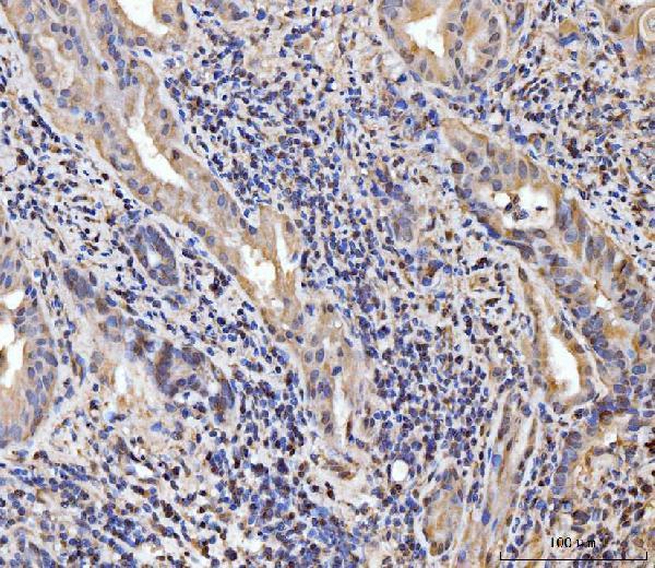
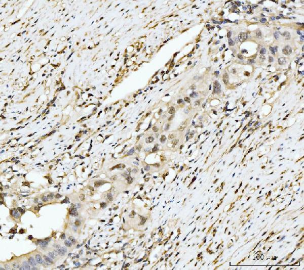
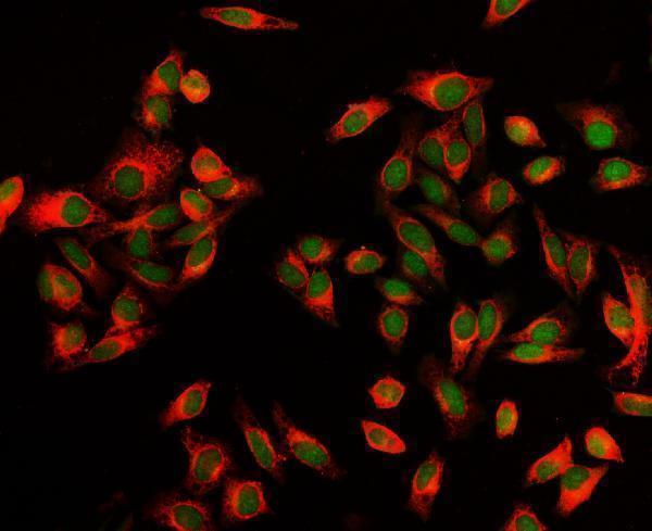
A09191-1 has IHC images from human paraffin sections and an IF image from HEP3B cells (catalog image captions); the listed reactivity is human (catalog).

Real IHC data IHC analysis of EXOSC8 using anti-EXOSC8 antibody (A09191-1). EXOSC8 was detected in a paraffin-embedded section of human gall bladder adenosquamous carcinoma tissue. Heat mediated antigen retrieval was performed in EDTA buffer (pH 8.0, epitope retrieval solution). The tissue section was blocked with 10% goat serum. The tissue section was then incubated with 2 μg/ml rabbit anti-EXOSC8 Antibody (A09191-1) overnight at 4°C. Biotinylated goat anti-rabbit IgG was used as secondary antibody and incubated for 30 minutes at 37°C. The tissue section was developed using Strepavidin-Biotin-Complex (SABC) (Catalog # SA1022) with DAB as the chromogen.
Anti-EXOSC8 Antibody ®
Cat # A09191-1

A09191-1 is listed for human IHC and has images from paraffin sections of gall bladder adenosquamous carcinoma, appendiceal adenocarcinoma, breast cancer and gastric adenocarcinoma (catalog applications, reactivity and IHC captions). The same SKU is listed for IF/ICC and has an IF image from HEP3B cells (catalog applications and IF caption).

Which to pick: Choose A09191-1 for human tissue IHC: its images document paraffin sections, with 2 μg/ml antibody used in the pictured experiments; the fixative is unreported (catalog IHC captions). Choose A09191-1 for IF/ICC: both applications are listed, and its HEP3B IF image used 5 μg/ml antibody (catalog applications and IF caption). Cross-species use has no listed validation because the catalog reports human reactivity only; clonality is unreported (catalog reactivity and clone field).

Each figure is that product's own IHC / IF validation image from its datasheet.

References

  1. UniProt Consortium. UniProt entry Q96B26 (EXOS8_HUMAN, Exosome complex component RRP43).
  2. Human Protein Atlas. EXOSC8 tissue IHC expression (reliability: Uncertain).
  3. Human Protein Atlas. EXOSC8 subcellular location (ICC-IF): Mainly localized to the nucleoli fibrillar center and mitotic chromosome. In addition localized to the nucleoplasm, vesicles and cytosol..
  4. Human Protein Atlas. EXOSC8 antibody validation summary (4 antibodies).
  5. Molecular characterization and prognostic modeling of liquid-liquid phase separation-related genes in osteosarcoma based on single-cell sequencing and weighted gene co-expression network analysis. Translational cancer research 2025 — PMC12776135.
  6. Comprehensive bioinformatics analysis of EXOSC family genes in head and neck squamous cell carcinoma. Scientific reports 2025 — PMC12365086.
  7. PubMed PMID:15057823 — UniProt-cited evidence.
  8. PubMed PMID:15489334 — UniProt-cited evidence.
  9. PubMed PMID:9466265 — UniProt-cited evidence.