F2R / Proteinase-activated receptor 1 · IHC design guide

Design Immunohistochemistry for F2R

Plan paraffin section IHC for F2R using its membrane topology and the reported endothelial staining in blood vessels (UniProt; HPA tissue IHC). Compare vessel staining with the surrounding cells and keep fixation consistent across sections (HPA tissue IHC; standard IHC practice).

Evidence assembled Oct 2026 · For research use; verify linked source records and product datasheet before use
Immunohistochemistry protocol sheet for F2R (IHC for F2R): expected localisation Cell membrane expected; endothelial staining observed (UniProt; HPA tissue IHC), antibody A03352-1, validated IHC image, and IHC protocol steps
Printable F2R IHC protocol sheet — expected localisation Cell membrane expected; endothelial staining observed (UniProt; HPA tissue IHC), antibody A03352-1, controls and protocol steps. Open the full F2R IHC guide →

F2R Immunohistochemistry Experimental Design Guide

Expected localisation, validated protocols, controls and antibodies — the at-a-glance facts below, then the full design guide.

Must know before staining
Expected localisation Cell membrane expected; endothelial staining observed (UniProt; HPA tissue IHC)
Staining pattern Endothelial cells stain in blood vessels; subcellular pattern unspecified (HPA tissue IHC)
Antigen retrieval EDTA pH 8.0 HIER, heat-mediated (datasheet A03352-1)
Positive control ⓘ Bone marrow+4 more · see all
Negative control ⓘ Adipose tissue+4 more · see all
Important caveats
Reasons your staining may differ from the expected pattern.
Fixation Keep fixation consistent across sections (standard IHC practice; not target-specific)
Caveat Vessels may stain where the surrounding cells do not (HPA tissue IHC)
Regulation Expression regulation is not specified (UniProt)
Isoform / epitope No isoforms annotated; extracellular vs cytoplasmic epitope matters (UniProt)
Section 1

Recommended F2R IHC & IF Protocols

Compare the catalog antibody’s IHC-P protocol (datasheet: A03352-1) with published F2R staining in intestinal sections (PMC11681937) and gastric tissues (PMC10827804).

Recommended immunohistochemistry (IHC-P) protocol parameters
SampleParaffin-embedded human breast cancer tissue; fixative not specified (datasheet A03352-1)
FixationImage fixative and duration unreported (datasheet A03352-1); verify before use.
Sectioning4–5 µm sections on charged slides (standard)
DeparaffinisationXylene, graded ethanol series to water (standard)
Antigen retrievalHeat retrieval: EDTA pH 8.0 (datasheet A03352-1); 20 min, 95–100 °C (standard)
Peroxidase block3% H2O2, 10 min, room temperature (standard)
Blocking10% goat serum (datasheet A03352-1)
Primary antibodyRabbit anti-F2R, 0.5-1μg/ml (datasheet A03352-1)
Primary incubationOvernight at 4 °C (datasheet A03352-1)
DetectionStreptavidin-biotin complex (SABC), DAB chromogen (datasheet A03352-1)
CounterstainHematoxylin, blue, dehydrate and mount (standard)
Expected resultF2R-positive staining in hematopoietic cells of bone marrow (HPA tissue IHC: Medium). HPA tissue profile: Expression in several tissues and in blood vessles. No signal in the no-primary control.
💡Decision noteStart with heat-mediated EDTA pH 8.0 retrieval for the catalog antibody (datasheet: A03352-1). The intestinal study used citrate retrieval (PMC11681937); the gastric study did not specify retrieval (PMC10827804).
Section 2

What Is the Expected F2R Staining Pattern?

F2R is a seven-pass cell-membrane receptor (UniProt P25116 topology). In paraffin-section IHC, look for staining along cell borders, especially in vascular endothelial cells, where HPA reports an Approved staining pattern (HPA: tissue IHC, Approved). Other documented sites include hematopoietic, stromal, glomerular and germinal-center cells (HPA: tissue IHC, Medium). Assess the named cell population within each tissue; a whole-section score can obscure its pattern.

What am I looking at on my slide?
Cell borders stain in vessel-lining cells, with restrained staining elsewhere.This fits the membrane assignment (UniProt P25116: cell membrane) and HPA's Approved endothelial-cell staining (HPA: tissue IHC). Judge the vessel-lining cells individually; HPA does not provide a universal endothelial intensity threshold.
Relevant nonvascular cells stain at moderate intensity.Medium staining is documented for hematopoietic cells in bone marrow, glomerular cells in kidney, and germinal-center cells in lymph node (HPA: tissue IHC). Match the observed cells to the named population before calling a tissue positive.
Signal is confined to nuclei, with little or no cell-border staining.A predominantly nuclear pattern conflicts with F2R's membrane assignment (UniProt P25116: cell membrane; HPA: subcellular summary, Membrane). Treat it as suspect and check morphology, counterstain, detection controls and antibody conditions before assigning F2R positivity.
Unlisted cell types stain as strongly as the documented positive populations.That distribution warrants a specificity check; it does not establish cross-reactivity by itself. For context, HPA reports no detected staining in adipocytes of adipose tissue and glandular cells of adrenal gland (HPA: tissue IHC). Compare like-for-like cell populations and run detection controls.
The section has uniform haze, or a documented positive population has no signal.Uniform haze limits localization and may reflect background in the general IHC workflow. An absent signal needs assessment in the specific population: HPA records Medium staining in several named populations, while its examples do not guarantee every specimen will be positive (HPA: tissue IHC).
💡Expected F2R appearanceCall a positive result when cell-border staining is discernible in vessel-lining cells (HPA: Approved endothelial staining; UniProt P25116: cell membrane), or in a matching HPA-listed Medium cell population; predominantly nuclear staining or uniform color across unrelated cells is a suspect result, not a supported F2R pattern.
How each factor affects the staining
Subcellular assignment and epitope positionF2R has seven transmembrane segments, extracellular loops and cytoplasmic regions (UniProt P25116 topology). Interpret a cell-border pattern as consistent with the receptor. Without the antibody's epitope, topology alone cannot predict which retrieval or permeabilisation condition will work.
Receptor processing and glycosylationUniProt annotates a signal peptide at residues 1–21, a propeptide at 22–41, the mature chain at 42–425, and glycosylation sites (UniProt P25116). These annotations make epitope identity relevant to interpretation; they do not establish an IHC staining change or fixation sensitivity.
Cell population and tissue contextHPA lists Medium staining in endometrial stromal cells, placental decidual cells, skin keratinocytes, splenic red-pulp cells and nasopharyngeal respiratory epithelium (HPA: tissue IHC). Its Not detected calls apply to specified cells, such as bronchial respiratory epithelium, rather than every cell in that tissue.
Strength of antibody evidenceHPA calls the tissue staining reliability Approved and lists CAB008973 and CAB012962 as IHC Approved (HPA: tissue IHC; HPA: antibodies). Neither entry is listed as IHC Enhanced in the supplied record. These labels support comparison with the shown pattern, not a claim of identical staining across antibodies or specimens.
IF/ICC interpretation?For IF/ICC, HPA summarizes F2R as Membrane but supplies no cell-line images, and neither listed antibody has an ICC status in this payload (HPA: subcellular; HPA: antibodies). Use that as a localization clue only; this IHC-P section provides no IF/ICC protocol recommendation.
Why is my staining missing, weak or wrong?
SituationLikely causeNext action
No staining in an expected positive areaThe examined cells may differ from the documented population, or the general IHC detection workflow may have failed; HPA's Medium calls are cell-specific (HPA: tissue IHC).Identify the cells on the section, compare a documented HPA-positive population, and verify that the IHC-validated antibody and detection controls produce interpretable staining. Optimize retrieval using the antibody's IHC-P instructions; F2R-specific retrieval sensitivity is unreported here.
Predominantly nuclear stainingA nuclear-only pattern is inconsistent with F2R's membrane localization (UniProt P25116: cell membrane; HPA: subcellular summary, Membrane).Check the counterstain and cell borders, then compare the same run with a documented positive population and appropriate detection controls. Do not score nuclear color alone as F2R positivity.
Diffuse color across vessels, stroma and empty areasThe distribution lacks the cellular boundaries needed for a membrane call (UniProt P25116: cell membrane); nonspecific background or detection activity is possible under general IHC practice.Inspect a no-primary control and reagent-only areas, and adjust blocking, washes or detection conditions as indicated by those controls. Score only localized cellular staining above the background.
Strong signal in a population HPA calls Not detectedThe result differs from the reference for that named population; for example, HPA calls adipocytes in adipose tissue Not detected (HPA: tissue IHC). Tissue labels alone cannot resolve mixed-cell staining.Confirm the stained cell type and compare with a documented positive population on the same run. If the discrepancy persists, review antibody specificity and detection controls before interpreting it as biological F2R expression.
Patchy or weak color in a nominally positive tissueThe target population may occupy only part of the section; HPA reports Medium staining for named cells, not a uniform whole-tissue signal (HPA: tissue IHC).Locate the relevant cells using morphology and counterstain, then score their membrane-associated signal separately from adjacent cells. Compare sections processed in the same run before changing retrieval or dilution.
Membrane signal cannot be separated from cytoplasmic colorF2R's documented localization favors a cell-border readout, while the supplied sources give no antibody epitope or specimen-specific trafficking pattern (UniProt P25116: cell membrane; HPA: subcellular summary, Membrane).Review higher-magnification morphology and the no-primary control; use a documented endothelial or other HPA-positive population as a reference (HPA: tissue IHC). Record uncertain localization instead of assigning a membrane-positive score.

Sample controls for F2R IHC & IF

🧪Run kidney first and look for staining in glomerular cells (HPA: Kidney, cells in glomeruli, Medium). Use adrenal gland glandular cells as a negative tissue (HPA: Adrenal gland, glandular cells, Not detected); on the kidney slide, nonglomerular cells can serve as internal negatives only where they show counterstain without specific DAB signal and their negative status is independently established.
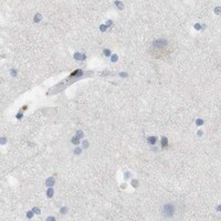
Positive control tissue: Bone marrow (Hematopoietic cells, HPA Medium)
Negative control tissue: Adipose tissue (HPA Not detected)
ICC-IF cell lines (HPA subcellular resource): HPA carries no ICC-IF cell line for F2R; derive a cell-line control from the positive tissue's cell type (Hematopoietic cells) and confirm it by RNA or western blot first.
Technical controls: Include a no-primary, secondary-only control and a concentration-matched rabbit IgG isotype control for the rabbit primary (selected-SKU caption: rabbit anti-F2R), plus F2R-knockout tissue as a biological negative if available. Quench endogenous peroxidase and assess endogenous biotin background in kidney, particularly with the biotin-based DAB detection used in the example (selected-SKU caption: biotinylated secondary, SABC and DAB; standard IHC practice).
⚠️Feasibility: No target-specific fixation window or fixation effect is reported in the supplied evidence; the selected-SKU paraffin-section caption does not state the fixative (selected-SKU caption: fixative unreported). That IHC example uses heat retrieval in EDTA at pH 8.0 and 1 μg/mL primary overnight at 4°C, so retrieval is part of the documented workflow, but its necessity has not been established (selected-SKU caption). The supplied evidence gives no basis to call frozen sections or IF easier; for kidney IHC, check tubular biotin background when using SABC detection (selected-SKU caption: SABC; standard IHC practice).

HPA tissue IHC evidence for F2R

Comprehensive Human Protein Atlas IHC scoring per tissue (reliability: Approved — Antibody staining in endothelial cells, view images.). Rows are taken directly from the HPA tissue chart — click any row's HPA link to view the source.

Positive expression · recommended positive controls

TissueCell typeLevelEvidenceSource
Bone marrow Hematopoietic cells Medium Protein (IHC) HPA →
Endometrium Cells in endometrial stroma Medium Protein (IHC) HPA →
Kidney Cells in glomeruli Medium Protein (IHC) HPA →
Lymph node Germinal center cells Medium Protein (IHC) HPA →
Nasopharynx Respiratory epithelial cells Medium Protein (IHC) HPA →

Undetected expression · recommended negative controls

TissueCell typeLevelEvidenceSource
Adipose tissue Adipocytes Not detected Protein (IHC) HPA →
Adrenal gland Glandular cells Not detected Protein (IHC) HPA →
Appendix Glandular cells Not detected Protein (IHC) HPA →
Breast Adipocytes Not detected Protein (IHC) HPA →
Bronchus Respiratory epithelial cells Not detected Protein (IHC) HPA →
Section 3

Advanced F2R IHC Tips

Troubleshoot F2R staining in paraffin sections by checking retrieval, epitope access, membrane localisation, cell identity, and assay controls.

How should I retrieve F2R antigen when staining paraffin sections?
Start with heat-mediated antigen retrieval in EDTA at pH 8.0 (datasheet A03352-1). The selected paraffin-section image used that retrieval before staining human breast cancer tissue with 1 μg/mL antibody overnight at 4°C (datasheet A03352-1). If staining is weak, compare a longer or shorter heating interval on adjacent sections while keeping the EDTA formulation constant; record the heating and cooling times for each run (standard IHC practice). Judge improvement by clearer cell-associated staining and preserved morphology, using a no-primary control to identify retrieval-related background (standard IHC practice). Avoid interpreting a stronger diffuse signal alone as improved F2R detection, because the expected location is the cell membrane (UniProt P25116 localisation).
Could fixation explain weak or uneven F2R staining?
The selected paraffin-section caption does not state a fixative, so target-specific F2R sensitivity to fixation is unknown (datasheet A03352-1). Record the fixative, duration, tissue thickness, and processing history for each specimen before comparing staining intensity across cases (standard IHC practice). If sections differ in fixation history, test adjacent sections with the same EDTA pH 8.0 retrieval and staining run, then assess morphology alongside signal (datasheet A03352-1; standard IHC practice). Excessive or uneven fixation can alter antigen access in IHC generally, but neither the tissue staining pattern nor F2R topology establishes such an effect for this antibody (standard IHC practice). Use matched processing and an on-slide reference when comparing samples quantitatively (standard IHC practice).
Where should convincing F2R staining appear in a tissue section?
Prioritise cell-boundary staining, since F2R is annotated at the cell membrane and has 7 transmembrane segments (UniProt P25116 localisation and topology). Examine vascular endothelial cells as an expected context; the tissue atlas reports approved endothelial staining and expression in blood vessels (HPA: tissue IHC). Platelets are also listed in the tissue-specificity record, although their small size makes cell-level localisation difficult in routine sections (UniProt P25116 tissue specificity; standard IHC practice). Compare the suspected signal with a counterstain and tissue architecture, especially around vessel lumens, before assigning it to a cell type (standard IHC practice). Predominantly nuclear or structureless extracellular DAB signal needs further validation against no-primary and tissue controls (UniProt P25116 localisation; standard IHC practice).
How do epitope position and F2R processing affect staining interpretation?
The supplied record lists 0 isoforms, so an isoform-specific staining claim is unsupported here (UniProt P25116 isoforms). F2R is processed from a precursor with signal peptide residues 1–21 and propeptide residues 22–41; the listed receptor chain begins at residue 42 (UniProt P25116 processing). Its extracellular and cytoplasmic regions occupy different sides of a 7-pass membrane, making antibody epitope mapping relevant to accessibility (UniProt P25116 topology; standard IHC practice). The epitope for A03352-1 is not supplied, so avoid attributing a staining difference to a particular domain, processing event, or glycosylation site (datasheet A03352-1; UniProt P25116 glycosylation). Request an epitope map before making those mechanistic interpretations (standard IHC practice).
How should I adapt F2R localisation checks to multiplex IF?
Treat IF as a separately optimised assay: the supplied antibody evidence describes chromogenic staining of a paraffin section, with no IF conditions given (datasheet A03352-1). Multiplex F2R with a validated endothelial-cell marker and inspect whether the F2R signal follows cell boundaries in the same cells (HPA: endothelial staining; UniProt P25116 localisation; standard IF practice). Choose a fluorophore channel with low tissue autofluorescence after viewing an unstained section, and include single-stain controls to check spectral bleed-through (standard IF practice). Permeabilise cautiously if the mapped epitope is cytoplasmic; an extracellular epitope may be assessed without permeabilisation when the preparation permits access (UniProt P25116 topology; standard IF practice). Because the epitope is unspecified, compare permeabilised and nonpermeabilised conditions before inferring which membrane face is recognised (datasheet A03352-1; standard IF practice).
What should I check when F2R DAB staining looks diffuse?
Run a no-primary control alongside the sample to reveal signal from the detection system or tissue rather than the primary antibody (standard IHC practice). The selected image used a biotinylated secondary, streptavidin–biotin complex, and DAB, so assess background from that detection chain when reproducing its method (datasheet A03352-1; standard IHC practice). Apply an endogenous peroxidase block and appropriate protein block, then compare staining with the reported 10% goat-serum block and 1 μg/mL primary concentration (datasheet A03352-1; standard IHC practice). Check whether pigment, necrosis, or section edges retain color in controls before calling diffuse DAB F2R-positive (standard IHC practice). A plausible result should retain cell-associated membrane localisation (UniProt P25116 localisation).
How can I score F2R staining across tissue samples? ⚠ ANSWER MARKED FOR VERIFICATION
Define the cell population and compartment before scoring; F2R is membrane annotated, and endothelial staining has tissue-IHC support (UniProt P25116 localisation; HPA: tissue IHC). For a defined population, report the percentage of positive cells and staining intensity, or calculate an H-score as the sum of percentage at each intensity multiplied by its score (standard IHC practice). If counting vessel-associated positive cells, report density per mm² of viable tissue or per measured vessel area, stating which denominator was used (standard IHC practice). Keep section thickness, retrieval, DAB development, imaging, and thresholds consistent across the comparison set (standard IHC practice). Exclude artefactual edges and necrosis using prespecified rules, and score a reference section with each run (standard IHC practice).
How can I distinguish genuine F2R staining from artefact?
Look first for cell-associated staining compatible with a membrane receptor, especially in vascular endothelium where tissue-IHC staining is reported (UniProt P25116 localisation; HPA: endothelial staining). Confirm cell identity from morphology and, when needed, an independently validated cell marker on an adjacent section (standard IHC practice). Interpret staining in other cells cautiously: the atlas reports medium staining in several cell populations, including glomerular cells and hematopoietic cells, so endothelial restriction is not an absolute rule (HPA: tissue IHC). Strong nuclear staining, signal confined to section edges or necrosis, and color persisting in a no-primary control each warrant an artefact check (UniProt P25116 localisation; standard IHC practice). Check endogenous peroxidase activity before assigning DAB deposits to F2R (standard IHC practice).
Boster reagents

Best F2R / Proteinase-activated receptor 1 IHC Antibodies

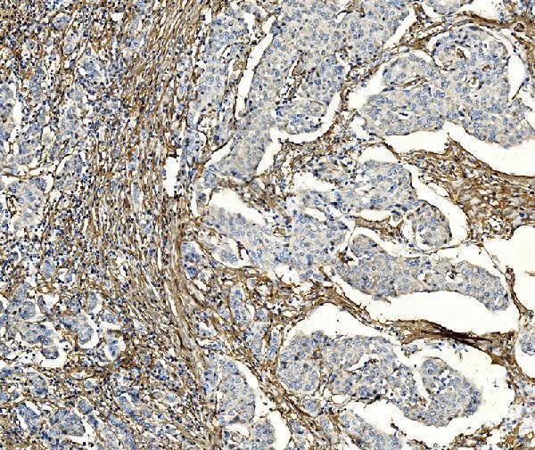
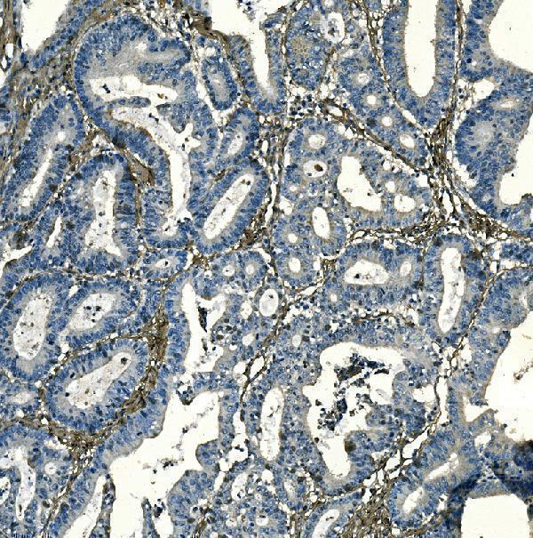
Catalog antibodies A03352-1 and RP1092 have paraffin-section IHC images from human breast and rectal cancer tissue and human placenta, respectively; neither has IF image data (catalog image captions).

Real IHC data IHC analysis of F2R using anti-F2R antibody (A03352-1). F2R was detected in paraffin-embedded section of human breast cancer tissue. Heat mediated antigen retrieval was performed in EDTA buffer (pH8.0, epitope retrieval solution). The tissue section was blocked with 10% goat serum. The tissue section was then incubated with 1μg/ml rabbit anti-F2R Antibody (A03352-1) overnight at 4°C. Biotinylated goat anti-rabbit IgG was used as secondary antibody and incubated for 30 minutes at 37°C. The tissue section was developed using Strepavidin-Biotin-Complex (SABC) (Catalog # SA1022) with DAB as the chromogen.
Anti-PAR1/Thrombin Receptor/F2R Antibody ®
Cat # A03352-1
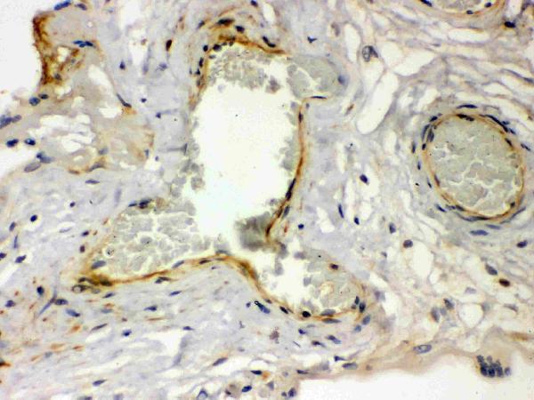
Real IHC data Anti-Thrombin Receptor antibody, RP1092, IHC(P) IHC(P): Human Placenta Tissue
Anti-Thrombin Receptor/F2R Antibody ®
Cat # RP1092

A03352-1 has IHC images from paraffin sections of human breast and rectal cancer tissue and lists Human, Mouse, and Rat reactivity (A03352-1 image captions; catalog applications/reactivity). RP1092 has an IHC-P image from human placenta and lists Human reactivity (RP1092 image caption; catalog applications/reactivity).

Which to pick: For tissue IHC, choose A03352-1 for the documented human breast or rectal cancer paraffin-section examples, or RP1092 for the documented human placenta IHC-P example (respective image captions). For cross-species work, A03352-1 lists Mouse and Rat reactivity, although its supplied IHC images show only human tissue (catalog reactivity; A03352-1 image captions). Neither SKU lists IF/ICC validation, and neither supplied IHC caption reports a fixative; clonality is unreported, so these data do not support an IF/ICC pick or a clone-based preference (catalog applications; image captions; clone fields).

Each figure is that product's own IHC / IF validation image from its datasheet.

References

  1. UniProt Consortium. UniProt entry P25116 (PAR1_HUMAN, Proteinase-activated receptor 1).
  2. Human Protein Atlas. F2R tissue IHC expression (reliability: Approved).
  3. Human Protein Atlas. F2R subcellular location (ICC-IF): Membrane.
  4. Human Protein Atlas. F2R antibody validation summary (2 antibodies).
  5. Ameloblast transcriptome changes from secretory to maturation stages. Connective tissue research 2014 — PMC4252044.
  6. Uncovering novel therapeutic clues for hypercoagulable active ulcerative colitis: novel findings from old data. Gastroenterology report 2024 — PMC11681937.
  7. Bioinformatics analysis identifies coagulation factor II receptor as a potential biomarker in stomach adenocarcinoma. Scientific reports 2024 — PMC10827804.
  8. Heterogeneity induced GZMA-F2R communication inefficient impairs antitumor immunotherapy of PD-1 mAb through JAK2/STAT1 signal suppression in hepatocellular carcinoma. Cell death & disease 2022 — PMC8901912.
  9. PubMed PMID:1672265 — UniProt-cited evidence.
  10. PubMed PMID:15489334 — UniProt-cited evidence.
  11. PubMed PMID:7744748 — UniProt-cited evidence.