This website uses cookies to ensure you get the best experience on our website.


- Table of Contents
Plan FABP6 IHC in paraffin sections using small-intestinal endocrine cells as a positive tissue reference (HPA tissue IHC). This guide covers fixation, chromogenic staining, controls, and interpretation of cytoplasmic and membranous staining (HPA tissue IHC).

Expected localisation, validated protocols, controls and antibodies — the at-a-glance facts below, then the full design guide.
| Expected localisation | Cytoplasmic and membranous staining (HPA tissue IHC) | |
| Staining pattern | High in small-intestinal endocrine cells; cytoplasmic/membranous (HPA tissue IHC) | |
| Antigen retrieval | EDTA pH 8.0 HIER, heat-mediated (datasheet A07132) | |
| Positive control | Small intestine | |
| Negative control | Adipose tissue+4 more · see all |
| Fixation | Keep fixation consistent across sections (standard IHC practice; not target-specific). Selected-image fixative and duration unreported (datasheet A07132); verify before use. | |
| Caveat | Appendix endocrine cells are undetected (HPA tissue IHC) | |
| Regulation | Intestine-enriched expression (HPA RNA) | |
| Isoform / epitope | 2 isoforms differ in gut distribution; epitope coverage unknown (UniProt) |
The catalog antibody’s IHC-P protocol (datasheet A07132) is accompanied by published FABP6 protocols for human colorectal tissue (PMC6814121), mouse ovary (PMC4498371), and human prostate tissue (PMC5347732).
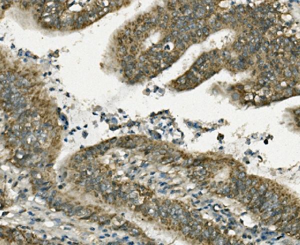
| Sample | Paraffin-embedded human rectal cancer tissue; fixative not specified (datasheet A07132) |
| Fixation | Image fixative and duration unreported (datasheet A07132); verify before use. |
| Sectioning | 4–5 µm sections on charged slides (standard) |
| Deparaffinisation | Xylene, graded ethanol series to water (standard) |
| Antigen retrieval | Heat retrieval: EDTA pH 8.0 (datasheet A07132); 20 min, 95–100 °C (standard) |
| Peroxidase block | 3% H2O2, 10 min, room temperature (standard) |
| Blocking | 10% goat serum (datasheet A07132) |
| Primary antibody | Rabbit anti-FABP6, 0.5-1μg/ml (datasheet A07132) |
| Primary incubation | Overnight at 4 °C (datasheet A07132) |
| Detection | Streptavidin-biotin complex (SABC), DAB chromogen (datasheet A07132) |
| Counterstain | Hematoxylin, blue, dehydrate and mount (standard) |
| Expected result | FABP6-positive staining in endocrine cells of small intestine (HPA tissue IHC: High). HPA tissue profile: Distinct cytoplasmic and membranous expression in small intestine. No signal in the no-primary control. |
FABP6 staining is expected in the cytoplasm and along cell membranes of the small intestine, with high staining reported in endocrine cells (HPA: tissue IHC, Enhanced reliability). UniProt also places FABP6 near the nucleus on the apical side of normal and neoplastic cells; it has no transmembrane segment (UniProt P51161: subcellular location and topology).
| Distinct cytoplasmic staining, with membrane-associated signal, in small-intestinal endocrine cells. | This matches the reported compartment pattern and high cell-level staining (HPA: small-intestine IHC). Compare cell identity and distribution with the counterstained tissue architecture (general IHC practice). |
| Signal is predominantly nuclear, with little cytoplasmic or membrane-associated staining. | A predominantly nuclear pattern does not match the reported locations (HPA: tissue IHC; UniProt P51161: subcellular location). Treat it as suspect; inspect morphology and control sections before assigning it to FABP6 (general IHC practice). |
| Strong staining appears in adipocytes or adrenal glandular cells. | Those specific cell populations were not detected by HPA tissue IHC (HPA: adipose tissue and adrenal gland). Check for nonspecific antibody binding or endogenous chromogenic activity; a positive cell elsewhere in either tissue cannot be judged from these entries alone (general IHC practice). |
| Color spreads across the section without a clear cellular pattern. | Diffuse deposition does not reproduce the distinct cytoplasmic and membranous pattern (HPA: small-intestine IHC). Check background on the no-primary control, then review blocking, washes and detection conditions (general IHC practice). |
| No convincing staining appears in a small-intestine section with identifiable endocrine cells. | This conflicts with the reported high staining in those cells (HPA: small-intestine IHC). First confirm tissue and cell identification, then assess the antibody and detection run with appropriate controls (general IHC practice). |
| Compartment and topology | FABP6 is annotated in cytoplasm and at membranes, with no transmembrane segment (UniProt P51161). Interpret membrane-associated color alongside the cytoplasmic pattern; it does not establish a membrane-spanning protein (UniProt P51161: topology). |
| Tissue and cell selection | HPA reports high staining in small-intestinal endocrine cells and no detection in specified adipocytes or adrenal glandular cells (HPA: tissue IHC). These observations guide positive and negative comparisons, without defining every cell in those tissues. |
| Isoform coverage | UniProt lists 2 isoforms with different intestinal distributions, and reports isoform 2 in colorectal adenocarcinomas and adjacent normal mucosa (UniProt P51161: tissue specificity). No supplied antibody epitope establishes which isoform the IHC stain detects. |
| Antibody validation | HPA labels the tissue pattern Enhanced and lists 2 antibodies with Enhanced IHC status, HPA012601 and CAB024713 (HPA: tissue IHC and antibodies). This supports the reported pattern; it does not establish performance under every local staining condition. |
| Processing and retrieval evidence | UniProt lists no signal peptide or propeptide and annotates a Gastrotropin chain spanning residues 2–128 (UniProt P51161: processing). The supplied sources give no FABP6-specific retrieval response or fixation sensitivity, so assess those conditions with controls (general IHC practice). |
| Situation | Likely cause | Next action |
|---|---|---|
| The expected small-intestine signal is absent or weak. | Possible causes include incorrect tissue or cell identification, an ineffective detection run, or unsuitable local IHC conditions (general IHC practice). The expected endocrine-cell level is high (HPA: small-intestine IHC). | Verify the section and endocrine-cell morphology; inspect a run control, then optimize retrieval and antibody conditions using the IHC-validated antibody's instructions (general IHC practice). Do not infer a FABP6-specific fixation effect from this result. |
| Adipocytes or adrenal glandular cells stain strongly. | HPA reports those cell populations as not detected (HPA: tissue IHC). Nonspecific binding or endogenous detection activity can produce misleading chromogenic color (general IHC practice). | Compare no-primary and detection controls; review blocking and chromogen development, then repeat with a validated antibody if needed (general IHC practice). Score the named cell population rather than treating the whole tissue as negative (HPA: tissue IHC). |
| Color appears mainly in nuclei. | Nuclear dominance conflicts with the cytoplasmic and membrane-associated locations (HPA: tissue IHC; UniProt P51161: subcellular location). Counterstain or precipitate can also complicate compartment calls (general IHC practice). | Inspect cellular boundaries and the no-primary control; adjust detection or counterstain conditions if they obscure the cytoplasm (general IHC practice). Require a reproducible expected pattern before scoring FABP6 positive (HPA: tissue IHC). |
| Background obscures the distinct cellular pattern. | Diffuse color may arise from nonspecific binding, incomplete washes or detection background (general IHC practice); it prevents assessment of the reported pattern (HPA: small-intestine IHC). | Inspect no-primary and negative-cell controls; review blocking, wash steps and development time (general IHC practice). Reassess only where cytoplasmic and membrane-associated staining is separable from background (HPA: tissue IHC). |
| Staining varies between intestinal regions or a tumor and adjacent mucosa. | UniProt reports different distributions for isoforms 1 and 2, including isoform 2 in colorectal adenocarcinomas and adjacent mucosa (UniProt P51161: tissue specificity). The supplied IHC data do not resolve isoforms. | Record region, morphology and compartment for each area; compare like cell populations before interpreting differences (general IHC practice). Do not assign an isoform from this IHC pattern without isoform-specific antibody evidence. |
| Can this pattern be used as an IF/ICC readout? | HPA supplies no main ICC-IF location and no cell-line ICC-IF images for FABP6 (HPA: subcellular record). Its Enhanced validation entries concern IHC (HPA: antibodies). | Use the cytoplasmic and membrane-associated locations as a hypothesis (UniProt P51161; HPA: tissue IHC). Establish an IF/ICC pattern with appropriate controls in the separate IF/ICC guide (general IF practice). |


Comprehensive Human Protein Atlas IHC scoring per tissue (reliability: Enhanced — High consistency between antibody staining and RNA expression data.). Rows are taken directly from the HPA tissue chart — click any row's HPA link to view the source.
| Tissue | Cell type | Level | Evidence | Source |
|---|---|---|---|---|
| Small intestine | Endocrine cells | High | Protein (IHC) | HPA → |
| Tissue | Cell type | Level | Evidence | Source |
|---|---|---|---|---|
| Adipose tissue | Adipocytes | Not detected | Protein (IHC) | HPA → |
| Adrenal gland | Glandular cells | Not detected | Protein (IHC) | HPA → |
| Appendix | Endocrine cells | Not detected | Protein (IHC) | HPA → |
| Bone marrow | Hematopoietic cells | Not detected | Protein (IHC) | HPA → |
| Breast | Adipocytes | Not detected | Protein (IHC) | HPA → |
Troubleshoot FABP6 staining in paraffin sections by checking retrieval, cellular pattern, controls and scoring against the available tissue evidence.
Two anti-FABP6 antibodies have IHC images: A07132 in paraffin sections of human rectal cancer and rat intestine, and PA2158 in human lung cancer tissue (catalog image captions).
A07132 lists IHC and human, mouse, and rat reactivity; its images show paraffin-section staining in human rectal cancer and rat intestine (catalog applications, reactivity, and image captions). PA2158 lists human IHC and has an IHC(P) image of human lung cancer tissue (catalog applications, reactivity, and image caption).
Which to pick: For human tissue IHC, A07132 has a documented paraffin-section protocol, while PA2158 has a human lung cancer IHC(P) image (A07132 and PA2158 image captions). For work across species, choose A07132: it lists human, mouse, and rat reactivity, with IHC images for human and rat tissue (A07132 catalog reactivity and image captions). Neither SKU lists IF/ICC validation or clonality, so neither has a supported IF/ICC recommendation; the IHC captions do not report the fixative (catalog applications, clone fields, and image captions).