This website uses cookies to ensure you get the best experience on our website.


- Table of Contents
Plan paraffin-section FADS1 IHC using a 1:50 starting dilution for M02535 (datasheet). Score cytoplasmic staining in adipocytes and glandular cells (HPA tissue IHC), while accounting for low agreement between antibody staining and RNA expression (HPA tissue IHC).

Expected localisation, validated protocols, controls and antibodies — the at-a-glance facts below, then the full design guide.
| Expected localisation | Cytoplasmic tissue staining (HPA tissue IHC); ER and mitochondrion (UniProt) | |
| Staining pattern | Cytoplasmic staining in adipocytes and glandular cells (HPA tissue IHC) | |
| Antigen retrieval | EDTA pH 8.0 HIER, heat-mediated (datasheet M02535) | |
| Positive control | Adipose tissue+4 more · see all | |
| Negative control | Soft tissue |
| Fixation | Keep fixation conditions consistent across sections. (standard IHC practice; not target-specific) | |
| Caveat | Antibody staining has low agreement with RNA expression (HPA tissue IHC) | |
| Regulation | Expression regulation not specified (UniProt) | |
| Isoform / epitope | Two isoforms; epitope coverage is unspecified (UniProt) |
The catalog antibody’s IHC-P protocol is accompanied by three published FADS1 staining protocols for mouse eye sections and tumor specimens (datasheet M02535; PMC12136103; PMC4934030; PMC12905685).
| Sample | Paraffin-embedded human liver cancer tissue; fixative not specified (datasheet M02535) |
| Fixation | Image fixative and duration unreported (datasheet M02535); verify before use. |
| Sectioning | 4–5 µm sections on charged slides (standard) |
| Deparaffinisation | Xylene, graded ethanol series to water (standard) |
| Antigen retrieval | Heat retrieval: EDTA pH 8.0 (datasheet M02535); 20 min, 95–100 °C (standard) |
| Peroxidase block | 3% H2O2, 10 min, room temperature (standard) |
| Blocking | 10% goat serum (datasheet M02535) |
| Primary antibody | Rabbit monoclonal (clone 26F55) anti-FADS1, 1:50 (datasheet M02535) |
| Primary incubation | Overnight at 4 °C (datasheet M02535) |
| Detection | HRP-conjugated secondary, DAB chromogen (datasheet M02535) |
| Counterstain | Hematoxylin, blue, dehydrate and mount (standard) |
| Expected result | FADS1-positive staining in adipocytes of adipose tissue (HPA tissue IHC: Medium). HPA tissue profile: General cytoplasmic expression. No signal in the no-primary control. |
FADS1 is an endoplasmic reticulum membrane protein also annotated in mitochondria, with four transmembrane segments (UniProt O60427). In paraffin sections, expect predominantly cytoplasmic staining in relevant cells: HPA reports general cytoplasmic expression and medium staining in adipocytes, adrenal glandular cells, respiratory epithelial cells and cortical glia (HPA tissue IHC). Its tissue IHC status is Approved, with low consistency between antibody staining and RNA expression (HPA tissue IHC).
| Cytoplasmic signal in adipocytes or adrenal glandular cells, with nuclei largely clear. | This fits the reported general cytoplasmic pattern and medium staining in those cells (HPA tissue IHC). Assess the specific cells stained and their intensity, rather than calling an entire section uniformly positive. |
| Signal is confined to nuclei, with little or no cytoplasmic staining. | A nuclear-only pattern conflicts with the reported cytoplasmic IHC profile (HPA tissue IHC) and membrane-associated locations (UniProt O60427). Review staining specificity and section quality before interpreting it as FADS1. |
| Strong staining appears in a cell population where the reference reports no detection. | Peripheral nerve in soft tissue is reported as not detected (HPA tissue IHC). Unexpected staining there warrants checks for cross-reactivity or endogenous detection activity; that reference does not make all soft tissue a negative control. |
| Chromogen spreads across stroma, empty spaces or many cell types without clear cell boundaries. | Diffuse, poorly localised colour is less consistent with the general cytoplasmic profile (HPA tissue IHC). Consider nonspecific antibody binding, inadequate washing or detection background (general IHC practice). |
| No signal appears in adipocytes and adrenal glandular cells in an otherwise interpretable run. | Both are reported at medium intensity (HPA tissue IHC), so an absent result prompts a technical check. Because antibody staining and RNA data have low consistency, do not infer biological absence from one failed section (HPA tissue IHC). |
| Compartment and epitope location | Four membrane-spanning segments and cytoplasmic and lumenal regions are annotated (UniProt O60427 topology). The supplied record gives no antibody epitope, so topology cannot predict whether a particular retrieval condition exposes it. |
| Choice of reference cells | Medium staining is reported in several cell types, including adipocytes, adrenal glandular cells and cortical glia (HPA tissue IHC). Peripheral nerve in soft tissue is reported as not detected (HPA tissue IHC); compare the named cells, not whole tissues. |
| Strength of tissue-pattern evidence | The tissue IHC profile is Approved but has low consistency with RNA expression (HPA tissue IHC). HPA042705 is listed as IHC Approved, without an Enhanced designation in the supplied antibody record (HPA antibodies). Treat unexpected cell patterns cautiously. |
| Isoforms and processing | Two isoforms are listed, with no signal peptide, propeptide or cleaved chain annotated (UniProt O60427). The supplied evidence does not locate the antibody epitope or establish isoform-specific staining; avoid assigning a colour pattern to one isoform. |
| IF/ICC: should its pattern match IHC? | HPA summarises the subcellular result as membrane, but provides no main location or cell-line ICC-IF images (HPA subcellular). The supplied antibody record lists IHC Approved and no ICC status (HPA antibodies), so this IHC pattern is not an IF/ICC validation. |
| Retrieval and fixation evidence | Target-specific fixation effects are not established by the supplied assay evidence. Verify with a matched IHC source before attributing a result to fixation. |
| Situation | Likely cause | Next action |
|---|---|---|
| A known-positive cell population is unstained. | The assay may have failed, or the tested section may differ from the HPA example; medium staining is reported in adipocytes and adrenal glandular cells (HPA tissue IHC). | Run a listed positive tissue alongside the test section; check antibody application, detection reagents and counterstain, then compare retrieval conditions with matched controls (general IHC practice). |
| Colour appears mainly in nuclei. | This disagrees with HPA's general cytoplasmic profile (HPA tissue IHC) and UniProt's membrane-associated locations (UniProt O60427). Nonspecific staining or misidentified pigment is possible (general IHC interpretation). | Inspect a counterstained section and a no-primary control; interpret FADS1 only where cell boundaries and cytoplasmic signal are convincing (general IHC practice). |
| Peripheral nerve shows conspicuous signal. | Peripheral nerve in soft tissue is reported as not detected (HPA tissue IHC). Cross-reactivity or endogenous detection activity is possible, but a single discordant section cannot identify the cause. | Compare a matched no-primary control and another HPA-listed cell population; if the signal persists only with primary antibody, assess antibody specificity (general IHC practice). |
| Diffuse chromogen obscures cell-level scoring. | Background may arise from nonspecific binding, insufficient washing or endogenous detection activity (general IHC practice); it does not match the reported general cytoplasmic profile (HPA tissue IHC). | Use a no-primary control to localise background, then review blocking, washes and chromogen development before rescoring identifiable cells (general IHC practice). |
| A tissue looks weaker than expected from UniProt's expression summary. | UniProt reports highest expression in liver, brain, adrenal gland and heart (UniProt O60427), while HPA's IHC antibody staining has low consistency with RNA data (HPA tissue IHC). | Score the observed cell type against HPA's tissue IHC examples and record intensity separately; do not convert a tissue-level expression statement into a required chromogenic score. |
| A retested section changes after antigen retrieval is adjusted. | Retrieval can alter a general IHC assay's staining, but the supplied sources report no FADS1-specific fixation or retrieval effect (general IHC practice; UniProt O60427; HPA tissue IHC). | Compare conditions on matched sections with positive and no-primary controls; choose a condition that preserves interpretable cellular staining (general IHC practice). |
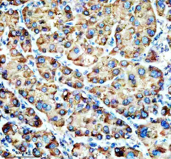
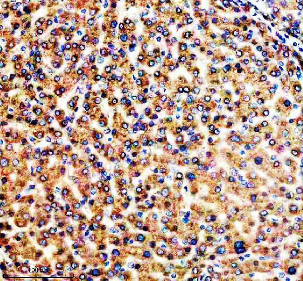


Comprehensive Human Protein Atlas IHC scoring per tissue (reliability: Approved — Low consistency between antibody staining and RNA expression data.). Rows are taken directly from the HPA tissue chart — click any row's HPA link to view the source.
| Tissue | Cell type | Level | Evidence | Source |
|---|---|---|---|---|
| Adipose tissue | Adipocytes | Medium | Protein (IHC) | HPA → |
| Adrenal gland | Glandular cells | Medium | Protein (IHC) | HPA → |
| Appendix | Glandular cells | Medium | Protein (IHC) | HPA → |
| Bone marrow | Hematopoietic cells | Medium | Protein (IHC) | HPA → |
| Breast | Adipocytes | Medium | Protein (IHC) | HPA → |
| Tissue | Cell type | Level | Evidence | Source |
|---|---|---|---|---|
| Soft tissue | Peripheral nerve | Not detected | Protein (IHC) | HPA → |
Troubleshoot FADS1 staining in paraffin sections using the documented IHC conditions, protein topology, and tissue staining evidence.
Anti-FADS1 IHC images cover human paraffin sections (M02535 and A02535-1 IHC captions); IF images cover human paraffin sections (A02535-1 IF captions).
M02535 has IHC images from human liver, liver cancer and thyroid cancer paraffin sections; its listed reactivity includes human, mouse and rat (M02535 IHC captions; catalog reactivity). A02535-1 has IHC images from human liver, lung and ovarian cancer paraffin sections and IF images from human liver and ovarian cancer paraffin sections (A02535-1 IHC and IF captions).
Which to pick: For tissue IHC, M02535 is a rabbit monoclonal used at 1:50, while A02535-1 was used at 2 μg/ml; each SKU’s human paraffin-section IHC caption documents EDTA retrieval at pH 8.0 (catalog; respective IHC captions). For IF, choose A02535-1 if an IF tissue image is useful; for ICC, M02535 lists that application, though its supplied images are IHC only (A02535-1 IF captions; M02535 applications and image list). For mouse or rat work, M02535 lists those species, but its supplied IHC images show human sections only; neither SKU’s IHC caption reports the fixative (M02535 reactivity and IHC captions; A02535-1 IHC captions).