This website uses cookies to ensure you get the best experience on our website.


- Table of Contents
Plan FANCC IHC-P around nuclear and cytoplasmic tissue staining, with high staining in testis spermatogonia (HPA tissue IHC). The IHC-validated antibody has a 1:50–1:100 dilution range (datasheet: A02387-1); this guide covers fixation, chromogenic detection, and interpretation.

Expected localisation, validated protocols, controls and antibodies — the at-a-glance facts below, then the full design guide.
| Expected localisation | Nuclear and cytoplasmic staining in most tissues (HPA tissue IHC) | |
| Staining pattern | High in spermatogonia; nuclear and cytoplasmic in most tissues (HPA tissue IHC) | |
| Antigen retrieval | Tris-EDTA pH 9.0 HIER, 95–98 °C, 20 min (rule: nuclear antigen) | |
| Positive control | Testis+4 more · see all | |
| Negative control | Adipose tissue+2 more · see all |
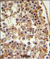
| Fixation | Formalin-fixed paraffin testis is shown (selected-SKU IHC image A02387-1); Matched tissue-IHC evidence does not establish the fixation claim. Validate the specimen-specific method before use. (selected-SKU IHC image A02387-1) | |
| Caveat | Staining and RNA levels show medium consistency (HPA tissue IHC) | |
| Regulation | Liver-enhanced RNA expression (HPA tissue RNA) | |
| Isoform / epitope | 0 annotated isoforms; no processing or mapped C-term epitope (UniProt; datasheet) |
Compare the catalog antibody's IHC-P protocol with 2 published breast carcinoma IHC protocols (PMC2633285; PMC6439399).
| Sample | FFPE tissue sections (datasheet A02387-1; sample unspecified) |
| Fixation | Image formalin-fixed; duration unreported (datasheet A02387-1); verify before use. |
| Sectioning | 4–5 µm sections on charged slides (standard) |
| Deparaffinisation | Xylene, graded ethanol series to water (standard) |
| Antigen retrieval | Heat-induced epitope retrieval in Tris-EDTA buffer, pH 9.0, 20 min at 95–98 °C (standard rule: nuclear antigen) |
| Peroxidase block | 3% H2O2, 10 min, room temperature (standard) |
| Blocking | 10% normal serum of the secondary host, 30 min, room temperature (standard) |
| Primary antibody | Rabbit anti-FANCC, 1:50-1:100 (datasheet A02387-1) |
| Primary incubation | Overnight at 4 °C (standard) |
| Detection | HRP-polymer secondary, DAB chromogen 5–10 min (standard) |
| Counterstain | Hematoxylin, blue, dehydrate and mount (standard) |
| Expected result | FANCC-positive staining in spermatogonia cells of testis (HPA tissue IHC: High). HPA tissue profile: Nuclear and cytoplasmic expression in most tissues. No signal in the no-primary control. |
FANCC is predominantly nuclear, with a smaller cytoplasmic pool (UniProt Q00597). IHC shows nuclear and cytoplasmic staining in most tissues (HPA tissue IHC: Approved; medium consistency with RNA). Expect the strongest listed signal in testis spermatogonia; several glandular, hematopoietic, epithelial, and neural cell populations show medium signal (HPA tissue IHC). FANCC has no transmembrane segment (UniProt Q00597 topology).
| Nuclear staining with some cytoplasmic staining in testis spermatogonia. | This fits the expected compartments and the highest listed cell specific signal (UniProt Q00597; HPA tissue IHC: High in spermatogonia). Score the cells and compartments separately; neighboring cells need not match their intensity (general IHC practice). |
| Predominantly nuclear staining in a medium signal reference, such as adrenal glandular cells or bone marrow hematopoietic cells. | A moderate result can support the expected pattern; FANCC need not appear uniformly strong across tissues (HPA tissue IHC: Medium in these cells; UniProt Q00597: major form nuclear). Compare like cell populations rather than whole section color (general IHC practice). |
| Strong membrane outlining or an exclusively cytoplasmic pattern across the positive cells. | Question the localization before calling it FANCC: the major form is nuclear, and FANCC lacks a transmembrane segment (UniProt Q00597). Check compartment assignment against the counterstain and assess antibody and detection controls (general IHC practice). |
| Prominent color in an HPA undetected cell population, such as adipocytes or lymph node germinal center cells. | This conflicts with the listed cell specific observations (HPA tissue IHC: Not detected in these cells). Cross reactivity or endogenous detection activity is possible; an HPA undetected call is a comparator, not proof that every cell in that tissue must be blank (general IHC practice). |
| Diffuse color across many compartments, or no staining in testis spermatogonia. | Diffuse color limits compartment scoring; testis spermatogonia are listed as High (HPA tissue IHC). Check background controls for the former and a known positive section for the latter before treating either result as FANCC biology (general IHC practice). |
| Biological compartment | The major FANCC form is nuclear and the minor form cytoplasmic (UniProt Q00597); HPA reports both compartments in most tissues (HPA tissue IHC). Use a nuclear counterstain to judge whether chromogen overlaps nuclei (general IHC practice). |
| Choice of positive and comparison cells | Testis spermatogonia are High; adrenal glandular cells and bone marrow hematopoietic cells are Medium (HPA tissue IHC). Adipocytes, lymph node germinal center cells, and spleen red pulp cells are listed as Not detected (HPA tissue IHC). |
| Strength of the reference evidence | HPA rates the tissue IHC profile Approved, with medium consistency between staining and RNA (HPA tissue IHC). Its antibody list marks CAB017793 IHC Approved; it does not assign an IHC rating to HPA030771 (HPA antibodies). |
| Protein architecture | FANCC has no transmembrane segment, signal peptide, propeptide, annotated isoforms, or glycosylation sites (UniProt Q00597). These annotations support checking a membrane dominant result, but do not establish an epitope retrieval requirement or fixation sensitivity. |
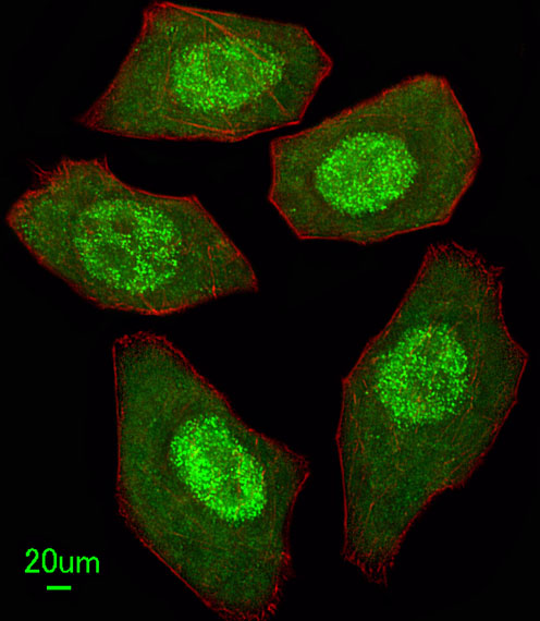
| IF/ICC Q: What location should I expect? | A: HPA supports nucleoplasmic localization in ICC IF images from A-431, U-251MG, and U2OS (HPA subcellular ICC IF). This is a localization cross check; the tissue IHC profile remains the reference for interpreting paraffin section chromogen (HPA tissue IHC). |
| Situation | Likely cause | Next action |
|---|---|---|
| A known positive testis section has no visible spermatogonial signal. | The result conflicts with the listed High cell specific staining (HPA tissue IHC); weak detection, unsuitable retrieval, or a staining run failure are possible (general IHC practice). | Check section integrity, reagent controls, and the IHC validated antibody's documented retrieval and dilution settings; repeat alongside a working positive control (general IHC practice). |
| Signal covers nuclei, cytoplasm, and empty spaces as a uniform haze. | Nonspecific background can obscure the expected nuclear dominant distribution (UniProt Q00597; general IHC practice). | Compare a no primary control, assess blocking and wash steps, and adjust detection exposure within the validated IHC workflow (general IHC practice). |
| Brown color appears in expected negative cells or persists without primary antibody. | Endogenous chromogen generating activity or nonspecific detection may contribute (general IHC practice). HPA lists adipocytes and germinal center cells as Not detected, cell specifically (HPA tissue IHC). | Use a no primary control and appropriate endogenous activity blocking for the detection chemistry; assess those cells separately from other cells in the section (general IHC practice). |
| A medium signal tissue looks negative while testis stains well. | The expected level differs by cell type: testis spermatogonia are High, while adrenal glandular and bone marrow hematopoietic cells are Medium (HPA tissue IHC). | Inspect the specified cells at comparable development and counterstain settings before changing the run; treat a faint result as uncertain if compartment and controls cannot be resolved (general IHC practice). |
| Color forms strong cell borders with little nuclear staining. | This is discordant with the nuclear major form and no transmembrane segment (UniProt Q00597); nonspecific staining or misread cell boundaries are possible (general IHC practice). | Check nuclear counterstain alignment and control sections, then review antibody specificity before scoring the border signal as FANCC (general IHC practice). |
| IHC and ICC IF seem to give different amounts of cytoplasmic staining. | HPA tissue IHC reports nuclear and cytoplasmic expression in most tissues, whereas its ICC IF summary supports nucleoplasm (HPA tissue IHC; HPA subcellular ICC IF). | Compare localization within each application and its controls; do not use ICC IF intensity to set a paraffin section positivity threshold (general IHC practice). |


Comprehensive Human Protein Atlas IHC scoring per tissue (reliability: Approved — Medium consistency between antibody staining and RNA expression data.). Rows are taken directly from the HPA tissue chart — click any row's HPA link to view the source.
| Tissue | Cell type | Level | Evidence | Source |
|---|---|---|---|---|
| Testis | Spermatogonia cells | High | Protein (IHC) | HPA → |
| Adrenal gland | Glandular cells | Medium | Protein (IHC) | HPA → |
| Appendix | Glandular cells | Medium | Protein (IHC) | HPA → |
| Bone marrow | Hematopoietic cells | Medium | Protein (IHC) | HPA → |
| Breast | Glandular cells | Medium | Protein (IHC) | HPA → |
Troubleshoot FANCC staining in paraffin sections by checking retrieval, compartment, controls, and cell specific scoring (UniProt Q00597; HPA tissue IHC).
The human-reactive anti-FANCC antibody has IHC data from formalin-fixed, paraffin-embedded testis and IF data from U251 cells (catalog: human reactivity; IHC and IF image captions).
A02387-1 will render with an IHC-P figure showing DAB staining of formalin-fixed, paraffin-embedded testis (catalog: A02387-1 IHC image caption). The same SKU is listed for human IF and has an IF image of U251 cells (catalog: A02387-1 applications, reactivity, and IF image caption).
Which to pick: Choose A02387-1 for human tissue IHC-P: its own figure documents formalin-fixed, paraffin-embedded testis, and its listed IHC-P dilution is 1:50–1:100 (catalog: A02387-1 IHC image caption and dilution). For IF/ICC, the same rabbit polyclonal antibody has an IF image of U251 cells at 1:25 (catalog: A02387-1 host, dilution_raw, and IF image caption). No cross-species choice is supported by this catalog; A02387-1 lists human reactivity only (catalog: A02387-1 reactivity).