FBL / rRNA 2'-O-methyltransferase fibrillarin · IHC design guide

Design Immunohistochemistry for FBL

Plan chromogenic IHC for FBL in paraffin sections using nucleolar staining as the expected tissue pattern (HPA tissue IHC). Cerebellar Bergmann glia show high nuclear staining, while adipocytes have no detected signal (HPA tissue IHC).

Evidence assembled Oct 2026 · For research use; verify linked source records and product datasheet before use
Immunohistochemistry protocol sheet for FBL (IHC for FBL): expected localisation Nucleolar tissue staining; nucleoplasm also annotated (HPA tissue IHC; UniProt), antibody M03178-6, validated IHC image, and IHC protocol steps
Printable FBL IHC protocol sheet — expected localisation Nucleolar tissue staining; nucleoplasm also annotated (HPA tissue IHC; UniProt), antibody M03178-6, controls and protocol steps. Open the full FBL IHC guide →

FBL Immunohistochemistry Experimental Design Guide

Expected localisation, validated protocols, controls and antibodies — the at-a-glance facts below, then the full design guide.

Must know before staining
Expected localisation Nucleolar tissue staining; nucleoplasm also annotated (HPA tissue IHC; UniProt)
Staining pattern Nucleolar signal across many tissue cell types (HPA tissue IHC)
Antigen retrieval Tris-EDTA pH 9.0 HIER, 95–98 °C, 20 min (rule: nuclear antigen)
Positive control ⓘ Cerebellum+4 more · see all
Negative control ⓘ Adipose tissue+4 more · see all
Important caveats
Reasons your staining may differ from the expected pattern.
Fixation Keep paraffin-section fixation consistent. (standard IHC practice; not target-specific)
Caveat Staining may reflect protein from more than one gene (HPA tissue IHC)
Regulation Low tissue specificity; induction unreported (HPA tissue IHC; UniProt)
Isoform / epitope No annotated isoforms; one 1–321 chain (UniProt)
Section 1

Recommended FBL IHC & IF Protocols

Compare the catalog antibody’s IHC-P protocol with 3 published FBL IHC methods (PMC13038753; PMC12730408; PMC9405174).

Recommended immunohistochemistry (IHC-P) protocol parameters
SampleTissue sections; selected-image fixative not specified (standard IHC workflow)
FixationImage fixative and duration unreported (datasheet M03178-6); verify before use.
Sectioning4–5 µm sections on charged slides (standard)
DeparaffinisationXylene, graded ethanol series to water (standard)
Antigen retrievalHeat-induced epitope retrieval in Tris-EDTA buffer, pH 9.0, 20 min at 95–98 °C (standard rule: nuclear antigen)
Peroxidase block3% H2O2, 10 min, room temperature (standard)
Blocking10% normal serum of the secondary host, 30 min, room temperature (standard)
Primary antibodyRabbit anti-FBL, 1:200-1:1000 (datasheet M03178-6)
Primary incubationOvernight at 4 °C (standard)
DetectionHRP-polymer secondary, DAB chromogen 5–10 min (standard)
CounterstainHematoxylin, blue, dehydrate and mount (standard)
Expected resultFBL-positive staining in bergmann glia - nucleus of cerebellum (HPA tissue IHC: High). HPA tissue profile: General nucleolar expression. No signal in the no-primary control.
💡Decision noteStart with Tris-EDTA pH 9.0 HIER at 95–98 °C for 20 min (page antigen-retrieval setting).
Section 2

What Is the Expected FBL Staining Pattern?

FBL stains nucleoli widely (HPA IHC: Supported); it is nuclear, with no membrane span (UniProt P22087).

What am I looking at on my slide?
Strong nucleolar stain in Bergmann glia (HPA IHC: High).Fits FBL's fibrillar nucleolar location (UniProt P22087).
Predominantly cytoplasmic stain (UniProt P22087: nuclear).Suspect nonspecific staining or precipitate (general IHC practice).
Strong adipocyte stain (HPA IHC: Not detected).Check for cross-reactivity or endogenous detection activity (general IHC practice).
Diffuse stain across the section (general IHC practice).Background obscures nucleolar scoring (general IHC practice).
No stain in Bergmann glia (HPA IHC: High).Check assay performance before calling the sample negative (general IHC practice).
💡Expected FBL appearancePositive: strong Bergmann glial nucleoli (HPA IHC: High); diffuse cytoplasmic stain is suspect (UniProt P22087).
How each factor affects the staining
Tissue and cell type (HPA tissue IHC).Bergmann glia: High; adipocytes: Not detected (HPA tissue IHC).
IHC antibody validation (HPA antibodies).HPA077724 and CAB001514 are IHC Supported (HPA antibodies).
Interpretation limit (HPA tissue IHC).Medium RNA concordance; HPA cautions the target may reflect more than one gene (HPA tissue IHC).
IF/ICC: where should FBL appear? (HPA ICC-IF)Fibrillar-center nucleoli, with some nucleoplasm (HPA ICC-IF).
Why is my staining missing, weak or wrong?
SituationLikely causeNext action
No stain in Bergmann glia (HPA IHC: High).Assay failure is possible (general IHC practice).Check retrieval, dilution and a same-run positive control (general IHC practice).
Cytoplasm dominates (UniProt P22087: nuclear).Nonspecific staining is possible (general IHC practice).Review antibody dilution and blocking (general IHC practice).
Adipocytes stain strongly (HPA IHC: Not detected).Cross-reactivity or endogenous activity is possible (general IHC practice).Run primary-omission and detection controls (general IHC practice).
Diffuse section background (general IHC practice).Insufficient blocking or washing is possible (general IHC practice).Adjust blocking and washing; review detection time (general IHC practice).
Nucleoli are hard to distinguish (UniProt P22087: nucleolar).Heavy counterstain or chromogen can obscure detail (general IHC practice).Lighten counterstain or shorten chromogen development (general IHC practice).

Sample controls for FBL IHC & IF

🧪Run cerebellum first and score nucleolar staining in Bergmann glia nuclei (High; HPA: cerebellum). Use adipose tissue adipocytes as the negative tissue (HPA: adipocytes, Not detected); the supplied HPA rows do not establish a negative cell population within cerebellum, so cells without nucleolar signal on that slide are background references, not validated biological negatives.
Positive control tissue: Cerebellum (Bergmann glia - nucleus, HPA High)
Negative control tissue: Adipose tissue (HPA Not detected)
ICC-IF cell lines (HPA subcellular resource): HPA ICC-IF images show FBL in A-431, U-251MG, U2OS, with annotated localisation: Nucleoli fibrillar center (supported) (HPA subcellular).
Technical controls: Include a no-primary, secondary-only control and a concentration-matched isotype control appropriate to the primary antibody’s host species and clonality; confirm specificity with FBL knockout material or a validated peptide-block experiment (standard IHC control practice). For chromogenic IHC, quench endogenous peroxidase and check endogenous biotin if using avidin–biotin detection; for IF in cerebellum, assess tissue autofluorescence with an unstained control (standard IHC/IF practice).
⚠️Feasibility: A target-specific fixation window and fixation effect are unreported in the supplied evidence; the selected M03178-6 mouse-testis IHC caption also leaves the fixative unreported (selected-SKU caption). Antigen-retrieval dependence and the relative ease of frozen sections versus paraffin sections are unreported, so optimize retrieval on the positive control (supplied target/application evidence; standard IHC practice). IF/ICC can help resolve the expected nucleolar signal, with additional nucleoplasmic signal possible (HPA: supported nucleoli fibrillar-center and approved nucleoplasm locations); check cerebellar autofluorescence when using IF (standard IF practice).

HPA tissue IHC evidence for FBL

Comprehensive Human Protein Atlas IHC scoring per tissue (reliability: Supported — Medium consistency between antibody staining and RNA expression data. Caution, targets protein from more than one gene.). Rows are taken directly from the HPA tissue chart — click any row's HPA link to view the source.

Positive expression · recommended positive controls

TissueCell typeLevelEvidenceSource
Cerebellum Bergmann glia - nucleus High Protein (IHC) HPA →
Adrenal gland Glandular cells Medium Protein (IHC) HPA →
Appendix Glandular cells Medium Protein (IHC) HPA →
Breast Glandular cells Medium Protein (IHC) HPA →
Bronchus Respiratory epithelial cells Medium Protein (IHC) HPA →

Undetected expression · recommended negative controls

TissueCell typeLevelEvidenceSource
Adipose tissue Adipocytes Not detected Protein (IHC) HPA →
Bone marrow Hematopoietic cells Not detected Protein (IHC) HPA →
Liver Cholangiocytes Not detected Protein (IHC) HPA →
Parathyroid gland Glandular cells Not detected Protein (IHC) HPA →
Smooth muscle Smooth muscle cells Not detected Protein (IHC) HPA →
Section 3

Advanced FBL IHC Tips

Troubleshoot FBL staining by checking nucleolar localisation, tissue controls, and the limits of the antibody evidence before interpreting chromogenic signal.

What retrieval should I use if nucleolar FBL staining is weak in paraffin sections?
Start with Tris-EDTA at pH 9.0 for heat-induced retrieval at 95–98 °C for 20 min (page retrieval rule: nuclear antigen). If nucleoli remain faint, compare a controlled extension of retrieval time while keeping section thickness, antibody concentration, and detection conditions constant (standard IHC practice). Inspect nuclear morphology after each condition: damaged or detached sections make a stronger chromogenic signal difficult to interpret (standard IHC practice). Score improvement in discrete nucleolar staining rather than total nuclear darkness, because FBL is mainly nucleolar with additional nucleoplasmic localisation (UniProt P22087 subcellular; HPA subcellular).
Could fixation explain absent FBL staining despite intact tissue morphology?
Target-specific sensitivity of FBL staining to fixation is unknown from the supplied evidence; the mouse testis caption for SKU M03178-6 does not state a fixative (selected IHC caption). Record the actual fixative and fixation duration for each specimen before comparing staining across blocks (standard IHC practice). Check that section adhesion and nuclear morphology survive the specified 95–98 °C, 20 min retrieval step, then compare matched sections under the same detection conditions (page retrieval rule; standard IHC practice). Use an appropriately processed positive tissue control to distinguish a run failure from a specimen-specific loss of detectable antigen (standard IHC practice).
How should I judge diffuse nuclear staining against the expected FBL pattern?
Look first for discrete nucleolar staining: FBL is assigned to the fibrillar region of the nucleolus, with additional nucleoplasmic localisation (UniProt P22087 subcellular; HPA subcellular). Diffuse nuclear staining alone is less persuasive when nucleoli are not distinguishable in otherwise well-preserved cells (UniProt P22087 subcellular; standard IHC practice). Compare staining with a nuclear counterstain and score nucleolar and nucleoplasmic compartments separately across matched fields (standard IHC practice). For a tissue control, HPA reports high nuclear staining in cerebellar Bergmann glia, but its tissue IHC reliability is only “Supported” and carries a cross-gene caution (HPA tissue IHC).
Could an FBL isoform or modified epitope account for inconsistent staining?
The supplied FBL record lists 0 isoforms and a single chain spanning residues 1–321, so an annotated isoform switch cannot explain the discrepancy (UniProt P22087 processing and isoforms). It does list modified residues, including asymmetrically dimethylated arginines at positions 8, 15, 21, 24, and 27 (UniProt P22087 modified residues). Whether any modification affects this antibody’s binding is unknown because its epitope is not specified in the supplied evidence (catalog antibody evidence). Compare retrieval conditions and a separately validated antibody recognising a documented different epitope before assigning an epitope-based cause (standard IHC practice).
How can IF help resolve ambiguous chromogenic FBL localisation?
Use IF/ICC as a localisation cross-check: FBL is mainly nucleolar and can also appear in the nucleoplasm (HPA subcellular; UniProt P22087 subcellular). Multiplex with a validated marker of the expected cell type, such as Bergmann glia in cerebellum, and include a nuclear counterstain to identify the stained cells and compartments (HPA tissue IHC; standard IF practice). Choose fluorophores after checking tissue autofluorescence in an unstained section, using a spectrally separated channel when background overlaps the FBL signal (standard IF practice). Because the target is intracellular and nucleolar, IF permeabilisation must provide access across the plasma and nuclear membranes; optimise it against preserved nuclear morphology (UniProt P22087 subcellular; standard IF practice).
What should I check when chromogenic FBL staining obscures nucleoli?
First compare a no-primary control with the stained section to identify signal from the detection system (standard IHC practice). Check the peroxidase block before DAB development, and inspect endogenous pigment or precipitate in the same tissue region (standard chromogenic IHC practice). Titrate the catalog antibody and shorten chromogen development if the whole nucleus becomes uniformly dark, while retaining a matched positive control (standard IHC practice). The interpretable pattern should preserve discrete nucleoli, with possible additional nucleoplasmic staining, rather than widespread cytoplasmic deposits (UniProt P22087 subcellular; HPA subcellular). Record exposure or development time consistently when comparing runs (standard IHC practice).
How should I quantify FBL staining across paraffin sections? ⚠ ANSWER MARKED FOR VERIFICATION
Define the cell population and region before scoring, then report the percentage of cells with discrete nucleolar staining and an intensity-based H-score if intensity is reproducible (standard IHC practice; UniProt P22087 subcellular). Normalise positive-cell counts to all evaluable nuclei in that population, or report positive-cell density per mm² when cell abundance itself matters (standard IHC practice). Keep nucleolar and diffuse nucleoplasmic scores separate because both locations are reported for FBL (HPA subcellular; UniProt P22087 subcellular). Exclude folded, necrotic, and detached areas using the same rules for every section, and document counterstain and detection settings (standard IHC practice).
When is an apparent FBL-positive cell more likely to be an artefact?
Treat discrete nucleolar signal in an intact nucleus as the strongest localisation match for FBL, allowing additional nucleoplasmic signal (UniProt P22087 subcellular; HPA subcellular). Recheck staining dominated by cytoplasm, tissue edges, or necrotic regions against morphology and a no-primary control (UniProt P22087 subcellular; standard IHC practice). For DAB detection, a signal that persists without primary antibody warrants investigation of endogenous peroxidase or detection background (standard chromogenic IHC practice). Do not equate a stained cell with specific FBL detection solely from tissue identity: HPA describes general nucleolar expression, rates its tissue IHC evidence “Supported,” and cautions that its antibody targets protein from more than one gene (HPA tissue IHC).
Boster reagents

Best FBL / rRNA 2'-O-methyltransferase fibrillarin IHC Antibodies

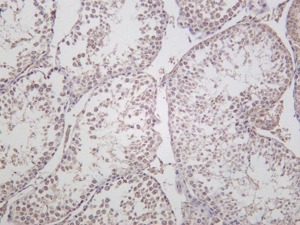
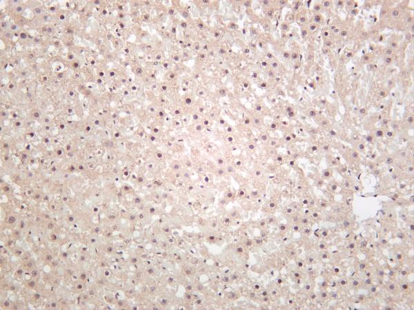
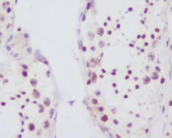
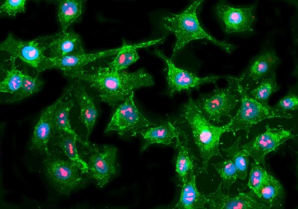
IHC images show mouse testis and paraffin-embedded human testis; an IF image shows A549 cells (catalog: image captions). Listed reactivity spans human, mouse, rat, and monkey (catalog: reactivity).

Real IHC data Mouse testis was stained with Anti-Fibrillarin rabbit antibody
Anti-Fibrillarin Rabbit Monoclonal Antibody
Cat # M03178-6
Real IHC data Immunohistochemical analysis of paraffin-embedded human testis, using Fibrillarin Antibody.
Anti-Fibrillarin FBL Rabbit Monoclonal Antibody
Cat # M03178
Real IF data IF analysis of Fibrillarin/FBL using anti-Fibrillarin/FBL antibody (A03178-1). Fibrillarin/FBL was detected in an immunocytochemical section of A549 cells. Enzyme antigen retrieval was performed using IHC enzyme antigen retrieval reagent (AR0022) for 15 mins. The cells were blocked with 10% goat serum. And then incubated with 5 μg/mL rabbit anti-Fibrillarin/FBL Antibody (A03178-1) overnight at 4°C. DyLight®594 Conjugated Goat Anti-Rabbit IgG (BA1142) was used as secondary antibody at 1:100 dilution and incubated for 30 minutes at 37°C. The tissue section was developed using Phalloidin-iFluor 488 Conjugated. Visualize using a fluorescence microscope and filter sets appropriate for the label used.
Anti-Fibrillarin/FBL Antibody ®
Cat # A03178-1

M03178-6 has IHC images of mouse testis and human, rat, and mouse liver; M03178 has an IHC image of paraffin-embedded human testis (catalog: IHC image captions). A03178-1 has an IF/ICC image of A549 cells and lists IF/ICC among its applications (catalog: A03178-1 IF image caption and applications).

Which to pick: For paraffin-section tissue IHC, choose M03178, illustrated on paraffin-embedded human testis; the fixative is unreported (catalog: M03178 IHC image caption). For IF/ICC in A549 cells, choose A03178-1, which has an IF cell image and lists IF/ICC applications (catalog: A03178-1 IF image caption and applications). For cross-species tissue IHC, choose M03178-6: this recombinant rabbit monoclonal lists human, mouse, and rat reactivity and has IHC images of mouse testis and human, rat, and mouse liver; the fixatives are unreported (catalog: M03178-6 short description, reactivity, and IHC image captions).

Each figure is that product's own IHC / IF validation image from its datasheet.

References

  1. UniProt Consortium. UniProt entry P22087 (FBRL_HUMAN, rRNA 2'-O-methyltransferase fibrillarin).
  2. Human Protein Atlas. FBL tissue IHC expression (reliability: Supported).
  3. Human Protein Atlas. FBL subcellular location (ICC-IF): Mainly localized to the nucleoli fibrillar center. In addition localized to the nucleoplasm..
  4. Human Protein Atlas. FBL antibody validation summary (3 antibodies).
  5. Low level of Fibrillarin, a ribosome biogenesis factor, is a new independent marker of poor outcome in breast cancer. BMC cancer 2022 — PMC9092774.
  6. TRIM21-Mediated ubiquitination of FBL suppresses PI3K/AKT signaling and tumor progression in clear cell renal cell carcinoma. Cellular and molecular life sciences : CMLS 2026 — PMC13038753.
  7. Fibrillarin Contributes to the Oncogenic Characteristics of Colorectal Cancer Cells and Reduces Sensitivity to 5-Fluorouracil. Cancers 2025 — PMC12730408.
  8. Bioinformatic Analysis and In Vitro and In Vivo Experiments Reveal That Fibrillarin Participates in the Promotion of Lung Metastasis in Hepatocellular Carcinoma. Bioengineering (Basel, Switzerland) 2022 — PMC9405174.
  9. PubMed PMID:1846968 — UniProt-cited evidence.
  10. PubMed PMID:2026646 — UniProt-cited evidence.
  11. PubMed PMID:19054851 — UniProt-cited evidence.