FDX1 / Adrenodoxin, mitochondrial · IHC design guide

Design Immunohistochemistry for FDX1

Plan FDX1 paraffin-section IHC around cytoplasmic staining in adrenal zona fasciculata and Leydig cells (HPA tissue IHC). This guide covers fixation consistency, antibody dilution, chromogenic detection and interpretation of the expected mitochondrial location (UniProt).

Evidence assembled Oct 2026 · For research use; verify linked source records and product datasheet before use
Immunohistochemistry protocol sheet for FDX1 (IHC for FDX1): expected localisation Cytoplasmic staining (HPA tissue IHC); mitochondrial matrix expected (UniProt), antibody A05441-1, validated IHC image, and IHC protocol steps
Printable FDX1 IHC protocol sheet — expected localisation Cytoplasmic staining (HPA tissue IHC); mitochondrial matrix expected (UniProt), antibody A05441-1, controls and protocol steps. Open the full FDX1 IHC guide →

FDX1 Immunohistochemistry Experimental Design Guide

Expected localisation, validated protocols, controls and antibodies — the at-a-glance facts below, then the full design guide.

Must know before staining
Expected localisation Cytoplasmic staining (HPA tissue IHC); mitochondrial matrix expected (UniProt)
Staining pattern Cytoplasmic in adrenal zona fasciculata and Leydig cells (HPA tissue IHC)
Antigen retrieval EDTA pH 8.0 HIER, heat-mediated (datasheet A05441-1)
Positive control ⓘ Adrenal gland+3 more · see all
Negative control ⓘ Adipose tissue+4 more · see all
Important caveats
Reasons your staining may differ from the expected pattern.
Fixation Keep fixation consistent across sections. (standard IHC practice; not target-specific)
Caveat Chromogenic cytoplasmic staining cannot resolve the matrix (HPA tissue IHC; UniProt)
Regulation Adrenal highest at protein level (UniProt)
Isoform / epitope No listed isoforms; mature chain 61–184; epitope unspecified (UniProt)
Section 1

Recommended FDX1 IHC & IF Protocols

The catalog antibody’s IHC-P protocol (datasheet: A05441-1) sits alongside four published FDX1 IHC protocols (PMC11156449; PMC13414201; PMC10344858; PMC11552402).

Recommended immunohistochemistry (IHC-P) protocol parameters
SampleParaffin-embedded human liver cancer tissue; fixative not specified (datasheet A05441-1)
FixationImage fixative and duration unreported (datasheet A05441-1); verify before use.
Sectioning4–5 µm sections on charged slides (standard)
DeparaffinisationXylene, graded ethanol series to water (standard)
Antigen retrievalHeat retrieval: EDTA pH 8.0 (datasheet A05441-1); 20 min, 95–100 °C (standard)
Peroxidase block3% H2O2, 10 min, room temperature (standard)
Blocking10% goat serum (datasheet A05441-1)
Primary antibodyRabbit anti-FDX1, 1:50 recommended; image 1:100 (datasheet A05441-1)
Primary incubationOvernight at 4 °C (datasheet A05441-1)
DetectionHRP-conjugated secondary, DAB chromogen (datasheet A05441-1)
CounterstainHematoxylin, blue, dehydrate and mount (standard)
Expected resultFDX1-positive staining in cells in zona fasciculata of adrenal gland (HPA tissue IHC: High). HPA tissue profile: Cytoplasmic expression in adrenal gland and Leydig cells. No signal in the no-primary control.
💡Decision noteStart with heat-mediated EDTA pH 8.0 for the catalog antibody (datasheet: A05441-1); citrate pH 6.0 is documented for HCC sections (PMC11552402).
Section 2

What Is the Expected FDX1 Staining Pattern?

FDX1 is a soluble mitochondrial matrix protein without a transmembrane segment (UniProt P10109: location and topology). In paraffin IHC, expect cytoplasmic staining in adrenal zona fasciculata cells and testicular Leydig cells (HPA: High in both; cytoplasmic profile). Placental syncytiotrophoblast cell bodies also stain strongly, while kidney proximal tubule cell bodies show medium staining (HPA: High; Medium). The tissue IHC profile has Enhanced reliability (HPA: tissue IHC).

What am I looking at on my slide?
Strong cytoplasmic stain in adrenal zona fasciculata cells or Leydig cells.This matches the expected cell types and intensity (HPA: High in both). Score the named cells rather than treating every cell in the section as equally positive (general IHC practice).
Cytoplasmic stain appears concentrated around nuclei or granular at higher magnification.A cytoplasmic distribution is consistent with tissue IHC (HPA: cytoplasmic profile). Mitochondrial location supports a granular appearance, but chromogenic IHC alone cannot prove organelle identity (UniProt P10109: matrix; HPA: mitochondria by ICC-IF).
Predominantly nuclear or crisp cell-surface staining, with little cytoplasmic signal.That distribution conflicts with the supported location (UniProt P10109: mitochondrial matrix; HPA: mitochondria by ICC-IF). Treat it as suspect and review controls and antibody specificity before scoring it as FDX1 (general IHC practice).
Strong stain in cells listed as undetected, such as adipocytes or bronchial respiratory epithelium.Those cells are reported as Not detected (HPA: adipocytes; bronchial respiratory epithelial cells). Consider nonspecific binding or endogenous chromogenic activity; check whether the signal persists without primary antibody (general IHC practice).
No stain in an otherwise interpretable adrenal zona fasciculata positive control.That conflicts with the reported High signal (HPA: adrenal zona fasciculata). First assess control performance, detection and tissue handling; a blank slide alone does not establish biological absence (general IHC practice).
💡Expected FDX1 appearanceCall a result positive when cytoplasmic stain is strong in adrenal zona fasciculata cells or Leydig cells (HPA: High in both); dominant nuclear or surface stain is discordant with mitochondrial matrix localization (UniProt P10109: location).
How each factor affects the staining
Cell and tissue choiceUse adrenal zona fasciculata or Leydig cells as strong reference areas (HPA: High in both). Placental syncytiotrophoblast cell bodies are also High, whereas kidney proximal tubule cell bodies are Medium (HPA: tissue IHC); compare like cell types when judging intensity (general IHC practice).
Compartment and image resolutionFDX1 resides in the mitochondrial matrix and has no transmembrane segment (UniProt P10109: location and topology). Tissue IHC reports cytoplasmic expression (HPA: tissue IHC); use that compartment for chromogenic scoring without requiring individually resolved mitochondria (general IHC practice).
Antibody evidenceThe tissue profile has Enhanced reliability, described as high consistency between antibody staining and RNA expression (HPA: tissue IHC). HPA041630 and HPA062087 each have Enhanced IHC status (HPA: antibody validation); this supports the reported pattern but does not validate every assay condition.
Processing and epitope uncertaintyUniProt lists the mature adrenodoxin chain as residues 61–184 (UniProt P10109: processing). The supplied evidence does not locate either antibody's epitope, so do not infer whether processing affects its staining. Target-specific fixation sensitivity is unreported in the supplied sources.
IF/ICC Q&A: should fluorescence be mitochondrial?Yes: ICC-IF reports mitochondria as the enhanced main location (HPA: subcellular). This supports compartment interpretation; IF/ICC methods belong in the separate guide. The present tissue scoring and controls concern paraffin IHC (HPA: tissue IHC).
Why is my staining missing, weak or wrong?
SituationLikely causeNext action
The adrenal positive control is blank while the counterstain and tissue are visible.An assay failure is possible because zona fasciculata cells are reported High (HPA: adrenal gland). A blank result does not identify which step failed (general IHC practice).Check antibody application, detection reagents, chromogen development and the run control; repeat with a known-positive section (general IHC practice). Do not score the unknown sample as negative until the positive control works.
Signal is weak in kidney but clear in the adrenal control.Kidney proximal tubule cell bodies are reported Medium, while adrenal zona fasciculata cells are High (HPA: tissue IHC). The difference can reflect the reported tissue pattern.Score proximal tubule cell bodies against the working positive control and local background (general IHC practice). Avoid increasing detection solely to make kidney match adrenal intensity.
Brown stain spreads across stroma, lumina or many unrelated cell types.Diffuse deposition may reflect background from staining or detection conditions (general IHC practice). It is harder to reconcile with the reported cell-specific cytoplasmic profile (HPA: tissue IHC).Inspect a no-primary control, wash performance and detection background; retain only cell-associated staining that can be interpreted above background (general IHC practice).
Adipocytes or bronchial epithelium stain as strongly as the positive control.These cells are listed as Not detected (HPA: adipocytes; bronchial respiratory epithelial cells). Nonspecific antibody binding or endogenous enzyme activity is possible (general IHC practice).Compare no-primary and positive controls; review blocking and endogenous enzyme suppression appropriate to the chromogen system (general IHC practice). Confirm a cell-specific pattern before calling FDX1 positive.
The dominant signal is nuclear or sharply outlines cell membranes.This conflicts with mitochondrial matrix localization (UniProt P10109: location and topology) and the cytoplasmic tissue profile (HPA: tissue IHC). The image alone cannot identify the artefact.Review control sections and staining distribution at higher magnification; repeat or corroborate with an independently validated antibody if the discordant pattern persists (general IHC practice; HPA: two Enhanced IHC antibodies).
A placenta section stains strongly, but the adrenal section does not.Syncytiotrophoblast cell bodies and adrenal zona fasciculata cells are both reported High (HPA: tissue IHC). The difference calls for a section-level and run-level check, not an assumption that adrenal FDX1 is absent.Verify that the adrenal section includes intact zona fasciculata cells, then review section quality and the shared run controls (general IHC practice). Reassess cell-specific cytoplasmic signal before interpreting the discrepancy.

Sample controls for FDX1 IHC & IF

🧪Run adrenal gland first: cells in the zona fasciculata should stain strongly (HPA: High). Use adipose tissue adipocytes as the negative tissue (HPA: Not detected); other cells on the adrenal slide can provide a background comparison, but the supplied HPA rows do not establish an internal negative cell type.
Positive control tissue: Adrenal gland (Cells in zona fasciculata, HPA High)
Negative control tissue: Adipose tissue (HPA Not detected)
ICC-IF cell lines (HPA subcellular resource): HPA ICC-IF images show FDX1 in A-431, U-251MG, U2OS, CACO-2, with annotated localisation: Mitochondria (enhanced) (HPA subcellular).
Technical controls: Include no-primary (secondary-only) and host-matched rabbit IgG isotype controls (caption: rabbit anti-FDX1 antibody), plus FDX1 knockout material or validated peptide competition as a biological specificity control. For HRP/DAB detection, block endogenous peroxidase and compare any adrenal pigment with the no-primary slide (caption: HRP/DAB detection; standard IHC practice).
⚠️Feasibility: A target-specific fixation window or fixation effect is unreported, and the selected paraffin-section caption does not state its fixative (caption: fixative not stated). The reported IHC procedure uses heat-mediated retrieval in EDTA at pH 8.0 before a 1:100 primary antibody incubation (caption: retrieval and dilution); no matched evidence establishes frozen sections or IF as easier. Interpret adrenal chromogenic staining with the pigment and no-primary comparison in mind (standard IHC practice).

HPA tissue IHC evidence for FDX1

Comprehensive Human Protein Atlas IHC scoring per tissue (reliability: Enhanced — High consistency between antibody staining and RNA expression data.). Rows are taken directly from the HPA tissue chart — click any row's HPA link to view the source.

Positive expression · recommended positive controls

TissueCell typeLevelEvidenceSource
Adrenal gland Cells in zona fasciculata High Protein (IHC) HPA →
Placenta Syncytiotrophoblasts - cell body High Protein (IHC) HPA →
Testis Leydig cells High Protein (IHC) HPA →
Kidney Proximal tubules (cell body) Medium Protein (IHC) HPA →

Undetected expression · recommended negative controls

TissueCell typeLevelEvidenceSource
Adipose tissue Adipocytes Not detected Protein (IHC) HPA →
Appendix Glandular cells Not detected Protein (IHC) HPA →
Bone marrow Hematopoietic cells Not detected Protein (IHC) HPA →
Breast Adipocytes Not detected Protein (IHC) HPA →
Bronchus Respiratory epithelial cells Not detected Protein (IHC) HPA →
Section 3

Advanced FDX1 IHC Tips

Troubleshoot chromogenic FDX1 IHC in paraffin sections using the catalog antibody’s tissue staining conditions and the expected mitochondrial, cell specific pattern.

Which retrieval condition should I start with for weak FDX1 staining?
Start with heat mediated antigen retrieval in EDTA at pH 8.0 for paraffin sections (datasheet A05441-1). The catalog antibody stained a paraffin embedded human liver cancer section after this retrieval, followed by 1:100 primary antibody overnight at 4°C (datasheet A05441-1). If staining is weak, verify that the retrieval bath reached its intended temperature and that sections remained covered throughout heating (standard IHC practice). Compare any adjusted heating duration on adjacent sections while holding detection and exposure to DAB constant (standard IHC practice). Interpret improvement against the expected mitochondrial, cytoplasmic distribution rather than signal intensity alone (UniProt P10109; HPA tissue IHC).
How should I investigate fixation when FDX1 staining varies between sections?
Target specific sensitivity of FDX1 to fixation is unknown from the supplied evidence; the catalog tissue caption identifies paraffin embedding but does not report a fixative (datasheet A05441-1). Record the fixative, fixation duration, section age and processing history for each specimen before comparing staining (standard IHC practice). Process matched sections together using EDTA at pH 8.0 and the same 1:100 primary antibody incubation overnight at 4°C (datasheet A05441-1). If signal differs, compare preserved morphology and internal tissue staining before assigning the difference to fixation (standard IHC practice). Keep fixation effects as an experimental hypothesis until matched material supports them (standard IHC practice).
What staining pattern is plausible for FDX1 in paraffin sections?
Expect cytoplasmic staining compatible with mitochondria, because FDX1 resides in the mitochondrial matrix (UniProt P10109) and HPA reports cytoplasmic tissue staining (HPA tissue IHC). Its lack of a transmembrane segment gives no basis for scoring a sharp plasma membrane rim as the expected pattern (UniProt P10109 topology). Use adrenal zona fasciculata cells and testicular Leydig cells as positive pattern references when those tissues are available (HPA tissue IHC). Kidney proximal tubule cell bodies offer a medium staining reference (HPA tissue IHC). Check cellular morphology and distribution at high magnification before interpreting diffuse nuclear, luminal or edge confined DAB as FDX1 (standard IHC practice; UniProt P10109).
Could processing or modifications explain a changed FDX1 IHC pattern?
The record lists 0 isoforms and a mature adrenodoxin chain spanning residues 61–184, so an epitope’s position relative to processing matters (UniProt P10109). The supplied antibody caption does not map its epitope, so do not assign weak paraffin staining to loss of a specific segment (datasheet A05441-1). FDX1 also has recorded modifications, including phosphoserine at residue 63 and alternate lysine modifications at residue 66 (UniProt P10109). These annotations alone do not establish whether the catalog antibody recognizes modified protein in sections (UniProt P10109; datasheet A05441-1). If staining varies, compare matched sections under the documented retrieval and primary incubation before proposing an epitope mechanism (standard IHC practice; datasheet A05441-1).
How can IF help assess a questionable chromogenic FDX1 pattern?
Use IF as a separate localization check: HPA reports mitochondrial FDX1 localization from subcellular imaging (HPA subcellular). For multiplexing, pair FDX1 with a validated marker of the expected cell population, such as CYP11A1 for steroidogenic cells, and include a mitochondrial marker when channel capacity permits (UniProt P10109 function and interaction; standard IF practice). Choose spectrally separated fluorophores after checking tissue autofluorescence and single stained controls (standard IF practice). Because FDX1 is in the mitochondrial matrix, permeabilisation must allow antibody access across cellular and mitochondrial membranes; optimize it against morphology and marker retention (UniProt P10109 topology; standard IF practice). Compare compartment agreement with the chromogenic result without treating IF conditions as an IHC protocol (standard IF and IHC practice).
What should I check when DAB obscures the FDX1 signal?
The catalog example used 10% goat serum blocking and peroxidase based DAB detection in a paraffin section (datasheet A05441-1). First inspect a no primary control to separate secondary reagent or endogenous peroxidase signal from primary dependent staining (standard IHC practice). Apply a peroxidase block as part of the general chromogenic workflow, and check whether extended DAB development is producing widespread deposit (standard IHC practice). Compare matched sections with the documented 1:100 primary incubation overnight at 4°C before changing several variables together (datasheet A05441-1; standard IHC practice). Retain signal that follows expected cell bodies and mitochondrial compatible cytoplasm rather than uniform tissue haze (UniProt P10109; HPA tissue IHC).
How should I score FDX1 IHC across samples? ⚠ ANSWER MARKED FOR VERIFICATION
Define the anatomical compartment and eligible cell population before scoring, because HPA reports high staining in adrenal zona fasciculata cells and Leydig cells but medium staining in kidney proximal tubules (HPA tissue IHC). For chromogenic sections, record the percentage of positive eligible cells and intensity, then calculate an H-score with a prespecified 0–3 intensity scale (standard IHC practice). Alternatively, report positive cell density per mm² of viable analyzed tissue when cell counts suit the question (standard IHC practice). Normalize comparisons to eligible cells or viable tissue area, and exclude necrosis, folds and section edges consistently (standard IHC practice). Keep staining, imaging and thresholds consistent across the comparison set (standard IHC practice).
How can I distinguish true FDX1 positivity from staining artefact?
A credible positive signal lies in cytoplasm compatible with mitochondrial localization and follows the expected cell population, such as adrenal zona fasciculata or Leydig cells (UniProt P10109; HPA tissue IHC). Adipocytes and bronchial respiratory epithelial cells are reported as not detected and can help contextualize unexpected widespread staining when present in a specimen (HPA tissue IHC). Treat nuclear only signal, section edge accentuation and staining concentrated in necrotic areas as reasons to inspect morphology and controls (UniProt P10109; standard IHC practice). Use a no primary control and peroxidase blocking check to investigate endogenous enzyme driven DAB deposition (standard IHC practice). Reassess questionable signal on adjacent sections using the documented EDTA pH 8.0 retrieval (datasheet A05441-1; standard IHC practice).
Boster reagents

Best FDX1 / Adrenodoxin, mitochondrial IHC Antibodies

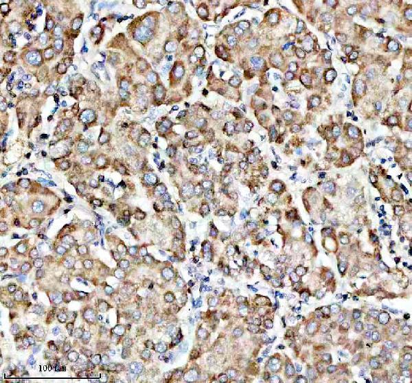
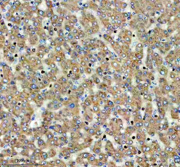
A05441-1 has human paraffin-section IHC images (catalog IHC captions); M05441 has an IF image with no specimen identified (catalog IF caption). Both list human and mouse reactivity (catalog reactivity).

Real IHC data IHC analysis of FDX1 using anti-FDX1 antibody (A05441-1). FDX1 was detected in a paraffin-embedded section of human liver cancer tissue. Heat mediated antigen retrieval was performed in EDTA buffer (pH 8.0, epitope retrieval solution). The tissue section was blocked with 10% goat serum. The tissue section was then incubated with 1:100 rabbit anti-FDX1 Antibody (A05441-1) overnight at 4°C. Peroxidase Conjugated Goat Anti-rabbit IgG was used as secondary antibody and incubated for 30 minutes at 37°C. The tissue section was developed using HRP Conjugated Rabbit IgG Super Vision Assay Kit (Catalog # SV0002) with DAB as the chromogen.
Anti-FDX1 Antibody ®
Cat # A05441-1

A05441-1 shows IHC staining in paraffin-embedded human liver and liver cancer sections and lists IF/ICC use (catalog IHC captions; catalog applications). M05441 lists IHC and IF/ICC use in human, mouse and rat, but its IF caption identifies no specimen (catalog applications/reactivity; catalog IF caption).

Which to pick: For tissue IHC, choose A05441-1: its own captions show paraffin-embedded human sections after EDTA pH 8.0 retrieval, with the fixative unreported (catalog IHC captions). For IF/ICC, M05441 is a rabbit monoclonal with an IF image, though that caption gives no specimen; A05441-1 is a rabbit polyclonal that lists IF/ICC without an IF image (catalog clone/applications/IF caption; catalog dilution_raw/applications/IF image alts). For rat samples, M05441 lists reactivity; both SKUs list human and mouse reactivity (catalog reactivity).

Each figure is that product's own IHC / IF validation image from its datasheet.

References

  1. UniProt Consortium. UniProt entry P10109 (ADX_HUMAN, Adrenodoxin, mitochondrial).
  2. Human Protein Atlas. FDX1 tissue IHC expression (reliability: Enhanced).
  3. Human Protein Atlas. FDX1 subcellular location (ICC-IF): Localized to the mitochondria..
  4. Human Protein Atlas. FDX1 antibody validation summary (2 antibodies).
  5. Cuproptosis mediates copper-induced testicular spermatogenic cell death. Asian journal of andrology 2024 — PMC11156449.
  6. Adipose-derived mesenchymal stem cell exosomes ameliorate copper metabolism dysregulation and reduce cuproptosis caused by liver IRI. Frontiers in veterinary science 2026 — PMC13414201.
  7. A novel cuproptosis-related diagnostic gene signature and differential expression validation in atherosclerosis. Molecular biomedicine 2023 — PMC10344858.
  8. Prognostic value of FDX1, the cuprotosis key gene, and its prediction models across imaging modalities and histology. BMC cancer 2024 — PMC11552402.
  9. PubMed PMID:3229285 — UniProt-cited evidence.
  10. PubMed PMID:3343244 — UniProt-cited evidence.
  11. PubMed PMID:2969697 — UniProt-cited evidence.