This website uses cookies to ensure you get the best experience on our website.
- Table of Contents

Facts about Fer-1-like protein 6.

| Human | |
|---|---|
| Gene Name: | FER1L6 |
| Uniprot: | Q2WGJ9 |
| Entrez: | 654463 |

| Belongs to: |
|---|
| ferlin family |

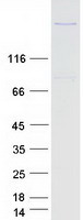
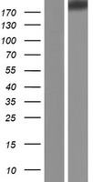
Fer-1-like protein 6
Mass (kDA):
209.308 kDA

| Human | |
|---|---|
| Location: | 8q24.13 |
| Sequence: | 8; NC_000008.11 (123851987..124120061) |
Membrane; Single-pass membrane protein.

PMID: 16421571 by Nusbaum C., et al. DNA sequence and analysis of human chromosome 8.