FGG / Fibrinogen gamma chain · IHC design guide

Design Immunohistochemistry for FGG

Plan paraffin-section FGG IHC using the catalog antibody at 0.5–1 μg/mL (datasheet A00790-1). Compare staining with selective plasma positivity and high proximal-tubule microvillus staining, while accounting for FGG secretion (HPA tissue IHC; UniProt).

Evidence assembled Oct 2026 · For research use; verify linked source records and product datasheet before use
Immunohistochemistry protocol sheet for FGG (IHC for FGG): expected localisation Secreted (UniProt); plasma and proximal-tubule microvilli (HPA tissue IHC), antibody A00790-1, validated IHC image, and IHC protocol steps
Printable FGG IHC protocol sheet — expected localisation Secreted (UniProt); plasma and proximal-tubule microvilli (HPA tissue IHC), antibody A00790-1, controls and protocol steps. Open the full FGG IHC guide →

FGG Immunohistochemistry Experimental Design Guide

Expected localisation, validated protocols, controls and antibodies — the at-a-glance facts below, then the full design guide.

Must know before staining
Expected localisation Secreted (UniProt); plasma and proximal-tubule microvilli (HPA tissue IHC)
Staining pattern Selective plasma positivity; high proximal-tubule microvilli (HPA tissue IHC)
Antigen retrieval Citrate pH 6 HIER, heat-mediated (datasheet A00790-1)
Positive control ⓘ Kidney
Negative control ⓘ Adipose tissue+4 more · see all
Important caveats
Reasons your staining may differ from the expected pattern.
Fixation Keep fixation consistent across sections (standard IHC practice; not target-specific)
Caveat Plasma staining may not indicate local synthesis (HPA tissue IHC)
Regulation Fibrin deposition occurs during infection (UniProt)
Isoform / epitope 2 isoforms, Gamma-A/B; mature chain starts at aa 27 (UniProt)
Section 1

Recommended FGG IHC & IF Protocols

The catalog antibody protocol is accompanied by published FGG IHC methods for human liver, lung, and inflammatory lesion sections (PMC6389006; PMC8756227; PMC5011235).

Recommended immunohistochemistry (IHC-P) protocol parameters
SampleParaffin-embedded human liver cancer tissues; fixative not specified (datasheet A00790-1)
FixationImage fixative and duration unreported (datasheet A00790-1); verify before use.
Sectioning4–5 µm sections on charged slides (standard)
DeparaffinisationXylene, graded ethanol series to water (standard)
Antigen retrievalHeat retrieval: Citrate pH 6, 20 min (datasheet A00790-1)
Peroxidase block3% H2O2, 10 min, room temperature (standard)
Blocking10% goat serum (datasheet A00790-1)
Primary antibodyRabbit anti-FGG, 0.5-1μg/ml (datasheet A00790-1)
Primary incubationOvernight at 4 °C (datasheet A00790-1)
DetectionStreptavidin-biotin complex (SABC), DAB chromogen (datasheet A00790-1)
CounterstainHematoxylin, blue, dehydrate and mount (standard)
Expected resultFGG-positive staining in proximal tubules (microvilli) of kidney (HPA tissue IHC: High). HPA tissue profile: Selective positivity in plasma. No signal in the no-primary control.
💡Decision noteStart with citrate pH 6 heat retrieval for the catalog antibody (datasheet A00790-1); published lung and inflammatory lesion protocols also use pH 6 retrieval (PMC8756227; PMC5011235).
Section 2

What Is the Expected FGG Staining Pattern?

FGG is secreted and has no transmembrane segment (UniProt P02679). In paraffin-section IHC, expect selective positivity in plasma and high staining at kidney proximal-tubule microvilli (HPA tissue IHC: Supported). The listed negative cell populations are reported as not detected (HPA tissue IHC). These observations identify where staining appears; they do not establish which cells synthesized FGG (UniProt P02679; HPA tissue IHC).

What am I looking at on my slide?
Selective plasma staining, with strong staining at kidney proximal-tubule microvilli.This matches the reported tissue pattern (HPA tissue IHC: Supported; High in proximal-tubule microvilli). FGG is a secreted plasma protein (UniProt P02679). Microvillar staining alone does not establish synthesis by tubular cells.
Predominantly nuclear staining, or a uniform membrane outline around many cell types.That distribution is inconsistent with the reported selective plasma and microvillar pattern (HPA tissue IHC) and with secreted FGG lacking a transmembrane segment (UniProt P02679). Treat it as suspect until controls and slide morphology support specificity (general IHC practice).
Strong staining in cells listed as negative, such as adipocytes or adrenal glandular cells.HPA reports FGG as not detected in those sampled populations (HPA tissue IHC). Check for cross-reactivity or endogenous detection activity using appropriate control sections before assigning the signal to FGG (general IHC practice).
Diffuse color across tissue, empty spaces, and the slide background.An unstructured haze cannot be scored as the selective plasma or proximal-tubule microvillar pattern (HPA tissue IHC). It suggests background from the staining or detection workflow; a no-primary control helps locate its source (general IHC practice).
No staining in plasma or in kidney proximal-tubule microvilli.The expected reference pattern is absent (HPA tissue IHC: selective plasma positivity; High in proximal-tubule microvilli). First assess tissue preservation, the positive control, and the antibody and detection steps (general IHC practice); absence alone does not establish a biological negative.
💡Expected FGG appearanceCall the IHC result positive when staining is selective in plasma and high at kidney proximal-tubule microvilli (HPA tissue IHC: Supported); widespread nuclear staining or uniform staining of listed negative cells is suspect (HPA tissue IHC; UniProt P02679 topology).
How each factor affects the staining
Secretion and tissue locationFGG is secreted and detected in blood plasma (UniProt P02679). HPA describes selective plasma positivity and high proximal-tubule microvillar staining (HPA tissue IHC). An extracellular or surface-associated signal cannot, by itself, identify the producing cell.
Precursor processing and epitope positionThe signal peptide spans residues 1–26, while the annotated FGG chain spans 27–453 (UniProt P02679). Without the antibody's epitope, staining cannot be predicted from either region; do not infer recognition of a processed segment.
Isoforms and modified sitesUniProt lists 2 isoforms, Gamma-A and Gamma-B, plus glycosylation and other modified residues (UniProt P02679). The supplied record gives no antibody epitope or isoform specificity, so it cannot establish which forms contribute to an IHC signal.
Evidence across assay typesThe tissue IHC pattern has Supported reliability, and the listed IHC antibodies are Supported (HPA tissue IHC; HPA antibodies). HPA separately reports endoplasmic reticulum and vesicles in ICC-IF (HPA subcellular); that intracellular observation does not redefine the paraffin tissue pattern.
Why is my staining missing, weak or wrong?
SituationLikely causeNext action
Kidney microvilli are weak or absent.The observed result falls short of the reported high proximal-tubule microvillar level (HPA tissue IHC); the cause is undetermined.Verify that the section contains intact proximal tubules, then check the IHC-validated antibody, positive control, retrieval, dilution, and detection steps against the assay instructions (general IHC practice).
Plasma appears negative across the section.The reported selective plasma signal is missing (HPA tissue IHC). This may reflect a section with little visible plasma or a staining-run problem; the slide alone cannot distinguish them.Review morphology for plasma-containing areas and compare with a suitable positive-control section processed in the same run (general IHC practice).
Nuclei or many unrelated cell membranes stain.This differs from the supported tissue pattern (HPA tissue IHC) and is difficult to reconcile with a secreted protein lacking a transmembrane segment (UniProt P02679).Inspect a no-primary control and the distribution of staining before interpreting those compartments as FGG (general IHC practice).
Listed negative cells stain strongly.Adipocytes and adrenal glandular cells are reported as not detected in the supplied HPA tissue observations (HPA tissue IHC). Cross-reactivity or endogenous detection activity is possible.Compare negative tissue controls with a no-primary control and review blocking and detection conditions (general IHC practice).
Diffuse chromogen obscures the selective pattern.Background across unrelated structures prevents comparison with plasma and microvilli (HPA tissue IHC). The responsible staining step is unknown.Check no-primary and detection-only controls, then review blocking, washes, and chromogen development for the run (general IHC practice).
Q: Should an ICC-IF image show the same pattern as paraffin IHC?HPA reports endoplasmic reticulum as the main ICC-IF location and vesicles as an additional location (HPA subcellular), while tissue IHC reports plasma and microvilli (HPA tissue IHC).A: Interpret each assay against its own HPA observation. Use the separate IF/ICC guide for that assay; do not use its intracellular pattern as an IHC-P scoring rule (HPA subcellular; HPA tissue IHC).

Sample controls for FGG IHC & IF

🧪Run kidney first: proximal tubule microvilli should stain (High; HPA: Kidney, proximal tubules). Use adipose tissue as the negative tissue, focusing on adipocytes (HPA: Adipose tissue, adipocytes, Not detected); on the kidney slide, use nuclei and areas without proximal tubule microvillar staining to judge background, while interpreting luminal or vascular signal cautiously (UniProt P02679: detected in blood plasma).
Positive control tissue: Kidney (Proximal tubules (microvilli), HPA High)
Negative control tissue: Adipose tissue (HPA Not detected)
ICC-IF cell lines (HPA subcellular resource): HPA ICC-IF images show FGG in A-549, Hep-G2, U2OS, with annotated localisation: Endoplasmic reticulum (approved) (HPA subcellular).
Technical controls: Include a no-primary, secondary-only control; concentration-matched rabbit IgG isotype control for the rabbit catalog antibody; and FGG knockout tissue as a biological negative if available (caption: rabbit anti-FGG antibody; standard IHC practice). Check endogenous peroxidase and biotin in kidney before interpreting DAB signal from the biotin-based detection system (caption: SABC with DAB; standard IHC practice).
⚠️Feasibility: A target-specific fixation window or fixation effect is unreported, and the selected A00790-1 paraffin-section caption does not report a fixative (caption: fixative not stated). The caption uses citrate retrieval at pH 6 for 20 minutes, but does not establish whether retrieval is required (caption: heat-mediated antigen retrieval). The supplied evidence does not establish that frozen sections or IF are easier; blood-derived FGG in kidney vessels or lumens could complicate tissue staining interpretation (UniProt P02679: secreted and detected in blood plasma).

HPA tissue IHC evidence for FGG

Comprehensive Human Protein Atlas IHC scoring per tissue (reliability: Supported — At least one protein variant secreted, tissue location of RNA and protein might differ and correlation is complex.). Rows are taken directly from the HPA tissue chart — click any row's HPA link to view the source.

Positive expression · recommended positive controls

TissueCell typeLevelEvidenceSource
Kidney Proximal tubules (microvilli) High Protein (IHC) HPA →

Undetected expression · recommended negative controls

TissueCell typeLevelEvidenceSource
Adipose tissue Adipocytes Not detected Protein (IHC) HPA →
Adrenal gland Glandular cells Not detected Protein (IHC) HPA →
Appendix Glandular cells Not detected Protein (IHC) HPA →
Bone marrow Hematopoietic cells Not detected Protein (IHC) HPA →
Breast Adipocytes Not detected Protein (IHC) HPA →
Section 3

Advanced FGG IHC Tips

Troubleshoot FGG chromogenic IHC in paraffin sections by checking retrieval, extracellular signal, antibody specificity, and scoring against tissue context.

What retrieval should I try when FGG staining is weak?
Start with heat-mediated citrate buffer at pH 6 for 20 minutes on paraffin sections (datasheet A00790-1). The selected antibody stained a human liver cancer section after this treatment, followed by 1 μg/ml primary antibody overnight at 4°C (datasheet A00790-1). If staining remains weak, check deparaffinisation, reagent activity, and incubation consistency before changing retrieval conditions (standard IHC practice). Compare any adjusted retrieval condition with the documented citrate condition on adjacent sections, since stronger signal can also increase background or alter tissue morphology (standard IHC practice).
Could fixation explain weak or uneven FGG staining?
The selected tissue IHC caption identifies a paraffin section but does not state its fixative, so target-specific fixation sensitivity is unknown (datasheet A00790-1). Record the fixative, fixation duration, section thickness, and processing history for each specimen before comparing staining intensity (standard IHC practice). Run sections from differently processed samples together with the same citrate pH 6, 20-minute retrieval and antibody incubation to locate processing-related variation (datasheet A00790-1; standard IHC practice). Do not infer fixation tolerance from tissue staining patterns or FGG’s secreted topology; neither provides a controlled fixation comparison (HPA: tissue IHC; UniProt P02679: secreted).
Where should convincing FGG signal appear in a tissue section?
Interpret extracellular or plasma-associated staining in light of FGG being secreted and detected in blood plasma (UniProt P02679: localisation and tissue specificity; HPA: selective positivity in plasma). Intracellular staining can also merit review because cell imaging places FGG mainly in the endoplasmic reticulum, with additional vesicular localisation (HPA: subcellular). HPA reports high staining at kidney proximal-tubule microvilli, which provides a defined tissue pattern for comparison (HPA: kidney proximal tubules, high). Assess cellular, luminal, vascular, and deposited signal separately; a stained tissue area alone does not establish which cells produced the protein (HPA: secreted-variant reliability note; standard IHC interpretation).
How can FGG isoforms and processing affect antibody interpretation?
FGG has Gamma-A and Gamma-B isoforms, so check whether the antibody’s documented epitope is shared before claiming that staining represents both (UniProt P02679: isoforms; standard antibody validation practice). The protein has a signal peptide at residues 1–26 and a mature chain at 27–453; an epitope within the removed signal peptide needs particular scrutiny (UniProt P02679: processing). Glycosylation at residues 78 and 334, plus reported phosphorylation and sulfation, may matter if an epitope overlaps those positions (UniProt P02679: modifications). Without an epitope map, describe the result as antibody-detected FGG rather than assigning an isoform or modification state (standard IHC interpretation).
How should I examine FGG localisation by multiplex IF?
Use IF as a separate validation experiment, since the selected product evidence describes chromogenic staining of a paraffin section (datasheet A00790-1). Pair FGG with a marker identifying the cell population under study, and inspect channels separately before interpreting overlap with intracellular signal (standard IF practice). Choose a fluorophore whose emission is distinguishable from autofluorescence in the specimen, using an unstained control to judge that background (standard IF practice). For extracellular FGG, test staining without permeabilisation; for a putative endoplasmic-reticulum or vesicular epitope, compare gentle permeabilisation after fixation, because the expected accessible compartment differs (UniProt P02679: secreted, no transmembrane segment; HPA: subcellular; standard IF practice).
How do I reduce diffuse or vascular FGG background?
First distinguish nonspecific reagent staining from plausible plasma-associated FGG, since HPA describes selective positivity in plasma and UniProt identifies the protein as secreted (HPA: tissue IHC; UniProt P02679: localisation). The selected workflow used 10% goat serum, a biotinylated secondary antibody, streptavidin–biotin detection, and DAB (datasheet A00790-1). Include a no-primary control, assess endogenous peroxidase blocking, and compare vascular regions with surrounding tissue before changing antibody concentration (standard chromogenic IHC practice). If background persists, titrate the primary around the documented 1 μg/ml condition while keeping retrieval and development time constant (datasheet A00790-1; standard IHC practice).
What should I measure when comparing FGG staining between sections? ⚠ ANSWER MARKED FOR VERIFICATION
Define the tissue compartment and analysis region before scoring, because secreted FGG can appear in plasma-associated spaces as well as tissue structures (UniProt P02679: secreted; HPA: selective positivity in plasma). For cellular staining, report the percentage of positive cells and an H-score; for extracellular deposits, measure positive area or deposit density per mm² of evaluable tissue (standard IHC quantification practice). Normalise cellular counts to the relevant cell population and deposit measurements to viable tissue area, excluding blank space and necrosis consistently (standard IHC quantification practice). Keep retrieval, DAB development, imaging settings, and scoring thresholds consistent across the comparison (standard IHC quantification practice).
How can I distinguish true FGG staining from an artefact?
Treat plasma-associated or deposited signal as plausible because FGG is secreted and participates in fibrin-matrix formation (UniProt P02679: localisation and function; HPA: selective positivity in plasma). Check whether cellular staining fits the question being tested; intracellular endoplasmic-reticulum or vesicular signal is reported in cell imaging, but tissue production cannot be assigned from extracellular staining alone (HPA: subcellular and secreted-variant reliability note). Compare suspicious staining at section edges or necrotic areas with intact interior tissue, and inspect a no-primary control for detection-system signal (standard IHC practice). Review endogenous peroxidase blocking when DAB appears broadly, since the selected chromogenic workflow uses DAB (datasheet A00790-1; standard IHC practice).
Boster reagents

Best FGG / Fibrinogen gamma chain IHC Antibodies

Anti-FGG options include paraffin-section IHC in human liver cancer tissue (A00790-1 image caption) and IF/ICC in HEP3B cells (M00790-3 image caption); listed reactivity spans human, mouse and rat (catalog reactivity).

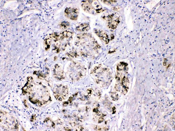
Real IHC data IHC analysis of FGG using anti-FGG antibody (A00790-1). FGG was detected in paraffin-embedded section of human liver cancer tissues. Heat mediated antigen retrieval was performed in citrate buffer (pH6, epitope retrieval solution) for 20 mins. The tissue section was blocked with 10% goat serum. The tissue section was then incubated with 1μg/ml rabbit anti-FGG Antibody (A00790-1) overnight at 4°C. Biotinylated goat anti-rabbit IgG was used as secondary antibody and incubated for 30 minutes at 37°C. The tissue section was developed using Strepavidin-Biotin-Complex (SABC)(Catalog # SA1022) with DAB as the chromogen.
Anti-Fibrinogen gamma chain/FGG Antibody ®
Cat # A00790-1
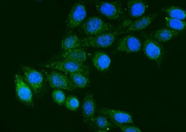
Real IF data IF analysis of FGG using anti-FGG antibody (M00790-3). FGG was detected in an immunocytochemical section of HEP3B cells. Enzyme antigen retrieval was performed using IHC enzyme antigen retrieval reagent (AR0022) for 15 mins. The cells were blocked with 10% goat serum. And then incubated with 5 μg/mL mouse anti-FGG Antibody (M00790-3) overnight at 4°C. DyLight®488 Conjugated Goat Anti-Mouse IgG (BA1126) was used as secondary antibody at 1:100 dilution and incubated for 30 minutes at 37°C. The section was counterstained with DAPI. Visualize using a fluorescence microscope and filter sets appropriate for the label used.
Anti-FGG Antibody ® (monoclonal, 5H9)
Cat # M00790-3

A00790-1 has IHC imagery from paraffin-embedded human liver cancer tissue and lists human, mouse and rat reactivity (A00790-1 image caption; catalog reactivity). M00790-3 has IF imagery from HEP3B cells and lists human reactivity with IF/ICC applications (M00790-3 image caption; catalog applications/reactivity).

Which to pick: Choose A00790-1 for tissue IHC: its caption documents paraffin-embedded human liver cancer tissue, citrate retrieval at pH 6 for 20 minutes, and 1 μg/mL primary antibody; the fixative is unreported (A00790-1 image caption). Choose M00790-3 for human-cell IF/ICC: it is mouse monoclonal clone 5H9 with HEP3B IF imagery (M00790-3 catalog; image caption). For cross-species planning, A00790-1 lists human, mouse and rat reactivity, while its IHC dilution and image evidence are for human samples (A00790-1 catalog dilution; image caption).

Each figure is that product's own IHC / IF validation image from its datasheet.

References

  1. UniProt Consortium. UniProt entry P02679 (FIBG_HUMAN, Fibrinogen gamma chain).
  2. Human Protein Atlas. FGG tissue IHC expression (reliability: Supported).
  3. Human Protein Atlas. FGG subcellular location (ICC-IF): Mainly localized to the endoplasmic reticulum. In addition localized to vesicles..
  4. Human Protein Atlas. FGG antibody validation summary (4 antibodies).
  5. FGG promotes migration and invasion in hepatocellular carcinoma cells through activating epithelial to mesenchymal transition. Cancer management and research 2019 — PMC6389006.
  6. Identification of an Osteochondral Erosion Process in Gout Driven by Fibrinogen-Integrin Activation and Local MSU Crystallization. Advanced science (Weinheim, Baden-Wurttemberg, Germany) 2026 — PMC13360169.
  7. Integrated analysis reveals lung fibrinogen gamma chain as a biomarker for chronic obstructive pulmonary disease. Annals of translational medicine 2021 — PMC8756227.
  8. Visualization of Neutrophil Extracellular Traps and Fibrin Meshwork in Human Fibrinopurulent Inflammatory Lesions: I. Light Microscopic Study. Acta histochemica et cytochemica 2016 — PMC5011235.
  9. PubMed PMID:6688357 — UniProt-cited evidence.
  10. PubMed PMID:2990550 — UniProt-cited evidence.
  11. PubMed PMID:14702039 — UniProt-cited evidence.