This website uses cookies to ensure you get the best experience on our website.


- Table of Contents
Plan FHL1 staining in paraffin sections using skeletal muscle myocytes or thyroid glandular cells as positive controls (HPA tissue IHC). The catalog antibody’s IHC starting concentration is 0.5–1 μg/mL (datasheet A01258-1); assess staining by cell type because tissue compartments remain unconfirmed (HPA tissue IHC; UniProt).

Expected localisation, validated protocols, controls and antibodies — the at-a-glance facts below, then the full design guide.
| Expected localisation | Cytoplasm or nucleus; tissue compartment unconfirmed (UniProt) | |
| Staining pattern | Strong myocyte and thyroid glandular-cell staining; compartment unreported (HPA tissue IHC) | |
| Antigen retrieval | EDTA pH 8.0 HIER, heat-mediated (datasheet A01258-1) | |
| Positive control | Endometrium+4 more · see all | |
| Negative control | Adrenal gland+4 more · see all |
| Fixation | Keep fixation consistent across paraffin sections. (standard IHC practice; not target-specific) | |
| Caveat | Cell-type differences can complicate tissue-level scoring (HPA tissue IHC) | |
| Regulation | Isoform 1 is enriched in skeletal muscle (UniProt) | |
| Isoform / epitope | Five isoforms; epitope coverage is undetermined (UniProt) |
The catalog antibody has an IHC-P protocol (datasheet A01258-1); the published options below describe FHL1 staining in glioma, thyroid carcinoma, and human myocardial tissue (PMC7202308; PMC11551809; PMC6687666).
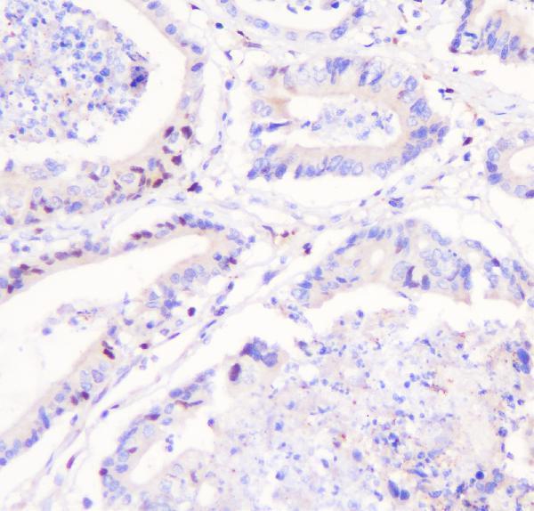
| Sample | Paraffin-embedded human rectal cancer tissue; fixative not specified (datasheet A01258-1) |
| Fixation | Image fixative and duration unreported (datasheet A01258-1); verify before use. |
| Sectioning | 4–5 µm sections on charged slides (standard) |
| Deparaffinisation | Xylene, graded ethanol series to water (standard) |
| Antigen retrieval | Heat retrieval: EDTA pH 8.0 (datasheet A01258-1); 20 min, 95–100 °C (standard) |
| Peroxidase block | 3% H2O2, 10 min, room temperature (standard) |
| Blocking | 10% goat serum (datasheet A01258-1) |
| Primary antibody | Rabbit anti-FHL1, 0.5-1μg/ml (datasheet A01258-1) |
| Primary incubation | Overnight at 4 °C (datasheet A01258-1) |
| Detection | Streptavidin-biotin complex (SABC), DAB chromogen (datasheet A01258-1) |
| Counterstain | Hematoxylin, blue, dehydrate and mount (standard) |
| Expected result | FHL1-positive staining in smooth muscle cells of endometrium (HPA tissue IHC: High). HPA tissue profile: Distinct expression in muscle tissue and thyroid glands. No signal in the no-primary control. |
FHL1 staining is expected mainly in the cytoplasm, with nuclear localisation possible in some cell states (UniProt Q13642: subcellular location). In paraffin sections, skeletal myocytes are a strong positive reference; smooth muscle cells and thyroid glandular cells are also reported as high staining (HPA: High in these cells). FHL1 has no transmembrane segment (UniProt Q13642: topology). HPA rates the tissue IHC profile Enhanced, with medium consistency between staining and RNA expression (HPA: reliability).
| Strong staining in skeletal myocytes, with signal mainly in the cell body. | This fits the high skeletal-myocyte staining reported by HPA and FHL1's cytoplasmic localisation (HPA: High in skeletal myocytes; UniProt Q13642: cytoplasm). Assess the muscle cells themselves: a dark field without identifiable positive cells is insufficient to establish the expected pattern (general IHC interpretation). |
| Signal appears exclusively in nuclei, or as a sharp rim around many unrelated cells. | Review the cell type and controls before calling this positive. Nuclear FHL1 is possible, especially in myoblasts (UniProt Q13642: subcellular location), and plasma-membrane signal is reported in ICC-IF (HPA: supported additional location). Neither source establishes a universal nuclear-only or membrane-rim pattern for paraffin tissue. |
| Strong colour appears in cell types expected to be unstained, including bronchial respiratory epithelium. | The reported lack of staining in that cell type makes cross-reactivity or endogenous detection activity plausible explanations, subject to controls (HPA: Not detected in bronchial respiratory epithelial cells; general IHC practice). Identify the stained cells before treating nearby positive structures as evidence for epithelial FHL1. |
| Colour spreads across cell-free areas or obscures boundaries between cells. | This prevents a reliable compartment or cell-type call (general IHC interpretation). A diffuse deposit is compatible with excess reagent, incomplete blocking or detection background (general IHC practice); it does not reproduce the cell-resolved high staining reported for skeletal myocytes (HPA: High in skeletal myocytes). |
| No convincing signal appears in a skeletal-muscle section. | This conflicts with the reported high staining in skeletal myocytes (HPA: High in skeletal myocytes), but one negative section cannot establish absent FHL1. Check tissue preservation and the staining run with appropriate controls (general IHC practice). The HPA tissue profile has only medium consistency with RNA data (HPA: reliability description). |
| Cell state and compartment | FHL1 is predominantly nuclear in myoblasts but cytosolic in differentiated myotubes (UniProt Q13642: subcellular location). Interpret nuclear signal with cell identity and controls; the myoblast observation does not define a paraffin-section scoring rule. |
| Tissue and cell identity | HPA reports high staining in skeletal myocytes, endometrial smooth muscle cells and thyroid glandular cells, but no detection in bronchial respiratory epithelial cells (HPA: tissue IHC). These are cell-specific references; do not score an entire organ as uniformly positive or negative. |
| Isoform distribution | Isoform 1 is highly expressed in skeletal muscle; isoforms 2 and 3 have different reported tissue distributions (UniProt Q13642: tissue specificity). The supplied record gives no antibody epitope or isoform coverage, so staining cannot identify a particular isoform. |
| Evidence strength | The tissue IHC profile is Enhanced, while its consistency with RNA expression is medium (HPA: reliability). Three listed antibodies have Enhanced IHC status (HPA: HPA001040, HPA001391, CAB020817); this supports the observed profile without proving every new reagent or section will match it. |
| Topology and processing | FHL1 has no transmembrane segment or signal peptide, and the listed protein chain begins at residue 2 (UniProt Q13642: topology and processing). These annotations offer no basis to expect a secreted extracellular stain or to infer a fixation-sensitive epitope. |
| Situation | Likely cause | Next action |
|---|---|---|
| Skeletal myocytes remain unstained. | A weak run, unsuitable working dilution or ineffective antigen retrieval may reduce IHC signal (general IHC practice); high staining is reported in these cells (HPA: skeletal myocytes). | Check a known-positive section and run controls, then optimise retrieval and antibody dilution within the reagent's validated IHC workflow (general IHC practice). The supplied sources give no FHL1-specific retrieval condition or fixation sensitivity. |
| The whole section looks brown or cell borders cannot be resolved. | Residual detection activity, incomplete blocking or overly concentrated reagents can create broad background (general IHC practice). | Inspect the no-primary control, review blocking and washes, and titrate primary or detection reagents (general IHC practice). Reassess whether staining resolves to skeletal myocytes, where HPA reports high signal (HPA: skeletal myocytes). |
| Bronchial respiratory epithelium stains strongly. | This differs from the HPA observation of no detection in those epithelial cells; cross-reactivity or endogenous activity is possible (HPA: bronchus; general IHC practice). | Confirm cell identity and compare no-primary and detection controls (general IHC practice). If staining persists only with primary antibody, assess reagent specificity before reporting epithelial FHL1; HPA's negative observation is a reference, not proof of universal absence. |
| Only nuclei stain in the putative positive cells. | Nuclear FHL1 can occur in myoblasts, while differentiated myotubes are described as cytosolic (UniProt Q13642: subcellular location). Tissue cell state may be uncertain. | Verify the stained cell population and inspect cytoplasmic signal in a positive reference (HPA: High in skeletal myocytes; general IHC practice). Do not reject every nuclear signal solely on compartment, or accept nuclear-only staining without controls. |
| High staining is patchy across a tissue section. | The HPA entries refer to named cell types, including endometrial smooth muscle cells and thyroid glandular cells (HPA: tissue IHC); their distribution need not fill the section. | Score the specified cells separately from neighbours and document their location and intensity (general IHC interpretation). Compare with an appropriate positive reference; avoid treating a whole-tissue average as the reported HPA cell-level result. |
| IF/ICC Q&A: should cytosolic or membrane signal be expected? | HPA calls cytosol the main location with uncertain support and plasma membrane an additional location with supported evidence (HPA: subcellular ICC-IF). UniProt also records nuclear localisation in myoblasts (UniProt Q13642: subcellular location). | Interpret IF/ICC with its own cell-state and compartment controls (general IF practice). The ICC-IF observations can inform localisation, but do not establish a paraffin IHC pattern or supply an IF/ICC protocol. |


Comprehensive Human Protein Atlas IHC scoring per tissue (reliability: Enhanced — Medium consistency between antibody staining and RNA expression data.). Rows are taken directly from the HPA tissue chart — click any row's HPA link to view the source.
| Tissue | Cell type | Level | Evidence | Source |
|---|---|---|---|---|
| Endometrium | Smooth muscle cells | High | Protein (IHC) | HPA → |
| Lung | Alveolar cells type I | High | Protein (IHC) | HPA → |
| Placenta | Syncytiotrophoblasts - cell body | High | Protein (IHC) | HPA → |
| Skeletal muscle | Myocytes | High | Protein (IHC) | HPA → |
| Testis | Elongated or late spermatids | High | Protein (IHC) | HPA → |
| Tissue | Cell type | Level | Evidence | Source |
|---|---|---|---|---|
| Adrenal gland | Glandular cells | Not detected | Protein (IHC) | HPA → |
| Appendix | Endocrine cells | Not detected | Protein (IHC) | HPA → |
| Bone marrow | Hematopoietic cells | Not detected | Protein (IHC) | HPA → |
| Bronchus | Respiratory epithelial cells | Not detected | Protein (IHC) | HPA → |
| Caudate | Glial cells | Not detected | Protein (IHC) | HPA → |
Use cell type, compartment, and matched controls to troubleshoot FHL1 staining in paraffin section IHC (HPA tissue IHC; UniProt Q13642).
The catalog lists two anti-FHL1 antibodies for IHC; A01258-1 also has human paraffin-section IHC and cell IF images (catalog applications/reactivity; A01258-1 image captions).
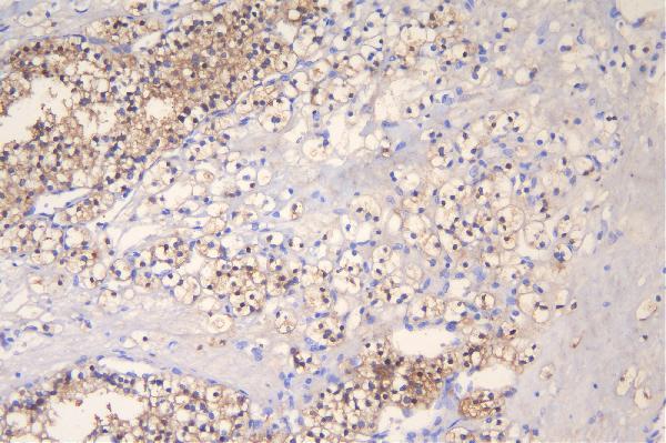
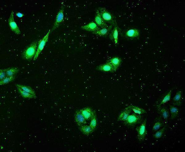
A01258-1 has IHC images from human rectal and renal cancer paraffin sections and an IF image from U20S cells (A01258-1 image captions). M01258 lists IHC as an application and human, mouse and rat reactivity, but has no supplied IHC image (catalog applications/reactivity; M01258 image captions).
Which to pick: For tissue IHC, choose A01258-1 when a documented human paraffin-section example is useful; its caption reports EDTA retrieval at pH 8.0, but does not report the fixative (A01258-1 IHC image captions). For IF/ICC, choose A01258-1 because both applications are listed and its IF caption documents U20S cells (catalog applications; A01258-1 IF image caption). For mouse or rat IHC, M01258 is the catalog-listed option based on its stated reactivity and IHC application; it is monoclonal, and no sample image is supplied (catalog host/clone/applications/reactivity; M01258 image captions).