This website uses cookies to ensure you get the best experience on our website.
- Table of Contents


2 Citations
Facts about Friend leukemia integration 1 transcription factor.

.
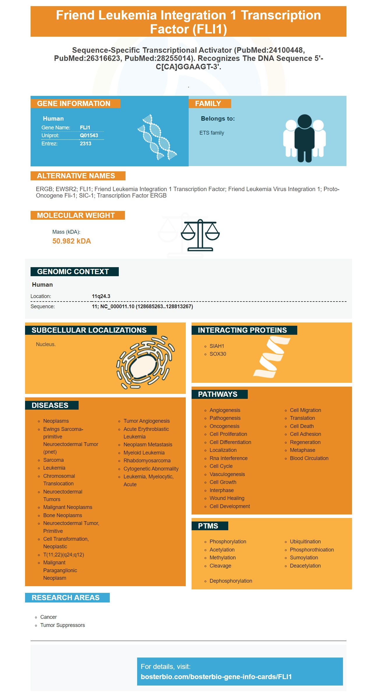
| Human | |
|---|---|
| Gene Name: | FLI1 |
| Uniprot: | Q01543 |
| Entrez: | 2313 |

| Belongs to: |
|---|
| ETS family |

ERGB; EWSR2; FLI1; Friend leukemia integration 1 transcription factor; Friend leukemia virus integration 1; Proto-oncogene Fli-1; SIC-1; Transcription factor ERGB
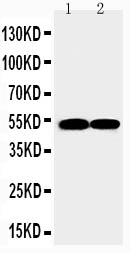
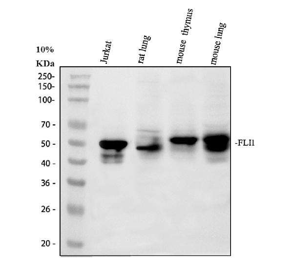
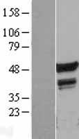
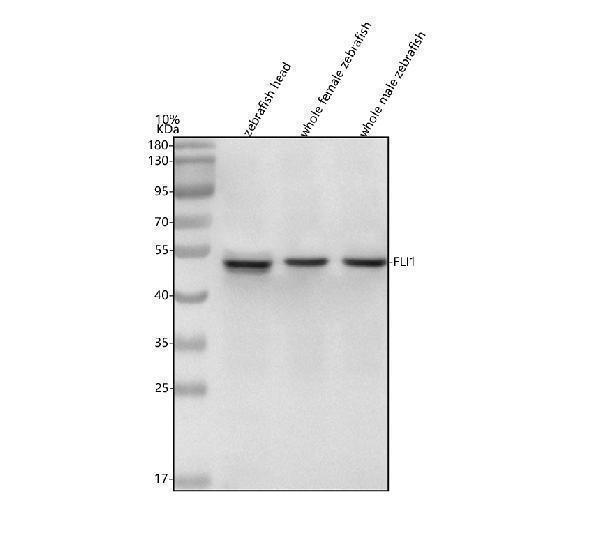
Mass (kDA):
50.982 kDA

| Human | |
|---|---|
| Location: | 11q24.3 |
| Sequence: | 11; NC_000011.10 (128685263..128813267) |
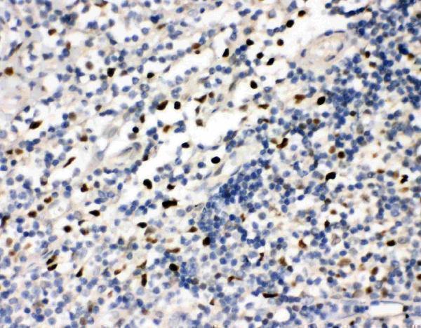
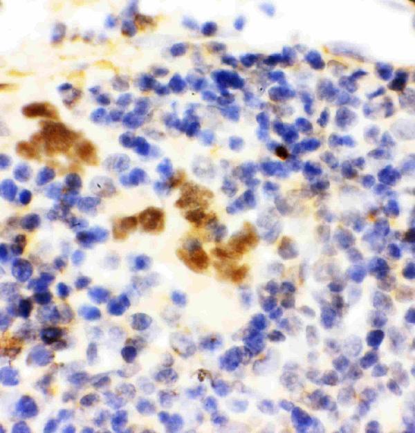
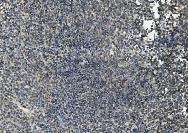
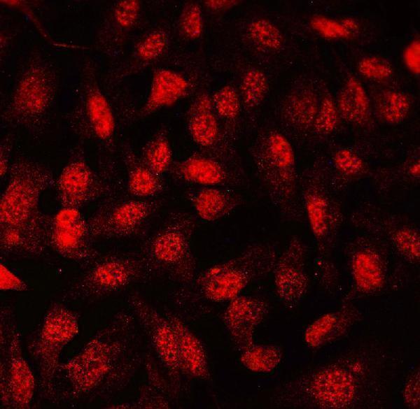
Nucleus.


PMID: 1522903 by Delattre O., et al. Gene fusion with an ETS DNA-binding domain caused by chromosome translocation in human tumours.
PMID: 1445800 by Watson D.K., et al. The ERGB/Fli-1 gene: isolation and characterization of a new member of the family of human ETS transcription factors.