This website uses cookies to ensure you get the best experience on our website.


- Table of Contents
Plan FLNB staining in paraffin sections using its cytoplasmic and membranous tissue pattern and abundant endothelial signal (HPA tissue IHC). This guide covers fixation, antibody selection, chromogenic detection and interpretation, with isoform expression as a consideration (UniProt).

Expected localisation, validated protocols, controls and antibodies — the at-a-glance facts below, then the full design guide.
| Expected localisation | Cytoplasmic and membranous in most cells (HPA tissue IHC) | |
| Staining pattern | Cytoplasmic and membranous; abundant in endothelium (HPA tissue IHC) | |
| Antigen retrieval | EDTA pH 8.0 HIER, heat-mediated (datasheet A02562-1) | |
| Positive control | Adrenal gland+4 more · see all | |
| Negative control | Caudate+4 more · see all |
| Fixation | Keep fixation consistent across sections (standard IHC practice; not target-specific); Matched tissue-IHC evidence does not establish the fixation claim. Validate the specimen-specific method before use. (selected-SKU IHC image A02562-1) | |
| Caveat | Abundant endothelial staining may obscure weaker cells (HPA tissue IHC) | |
| Regulation | Isoform expression varies by tissue (UniProt) | |
| Isoform / epitope | 9 isoforms; check splice-region epitope coverage (UniProt) |
The catalog antibody protocol is paired with a published IHC protocol for human PDAC sections (PMC7611572).
| Sample | Paraffin-embedded human lung cancer tissue; fixative not specified (datasheet A02562-1) |
| Fixation | Image fixative and duration unreported (datasheet A02562-1); verify before use. |
| Sectioning | 4–5 µm sections on charged slides (standard) |
| Deparaffinisation | Xylene, graded ethanol series to water (standard) |
| Antigen retrieval | Heat retrieval: EDTA pH 8.0 (datasheet A02562-1); 20 min, 95–100 °C (standard) |
| Peroxidase block | 3% H2O2, 10 min, room temperature (standard) |
| Blocking | 10% goat serum (datasheet A02562-1) |
| Primary antibody | Rabbit anti-FLNB, 1-2 μg/ml (datasheet A02562-1) |
| Primary incubation | Overnight at 4 °C (datasheet A02562-1) |
| Detection | Streptavidin-biotin complex (SABC), DAB chromogen (datasheet A02562-1) |
| Counterstain | Hematoxylin, blue, dehydrate and mount (standard) |
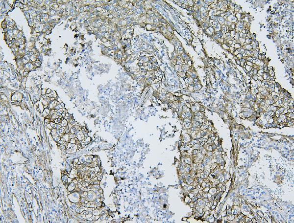
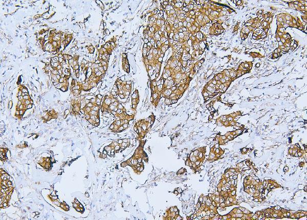
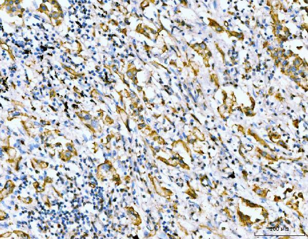
| Expected result | FLNB-positive staining in glandular cells of adrenal gland (HPA tissue IHC: High). HPA tissue profile: Cytoplasmic and membranous expression in most cells. Highly abundant in endothelial cells. No signal in the no-primary control. |
FLNB is a cytoplasmic, cortical and cytoskeletal protein with no transmembrane segment (UniProt O75369 localization and topology). In paraffin sections, expect cytoplasmic and membranous staining in many cell types, especially abundant signal in endothelial cells (HPA tissue IHC). HPA rates the tissue profile Enhanced, with medium agreement between staining and RNA data and support from paired antibodies (HPA tissue IHC reliability).
| Cytoplasmic and cell-edge signal appears in endothelial cells and glandular epithelium. | This fits the reported distribution: endothelial cells are highly abundant, while glandular cells score High in adrenal gland, appendix, breast, colon and other listed tissues (HPA tissue IHC). Judge the stained cell population as well as intensity; a positive vessel does not make every neighboring cell positive (HPA tissue IHC). |
| A sharp nuclear-only pattern appears, without convincing cytoplasmic or cell-edge staining. | Treat this as a localization mismatch requiring investigation, rather than a confirmed FLNB pattern: UniProt places FLNB in the cytoplasm, cortex and cytoskeleton, while HPA reports cytoplasmic and membranous tissue staining (UniProt O75369 localization; HPA tissue IHC). Check antibody specificity and detection background (general IHC practice). |
| Strong signal appears in a cell population reported as undetected, such as cardiomyocytes or germinal center cells. | Question specificity or endogenous detection activity before calling these cells FLNB-positive (HPA: Not detected in cardiomyocytes and germinal center cells; general IHC practice). Interpret the named cell type, not the whole tissue: other cells in the section may stain (HPA tissue IHC). |
| Color spreads across extracellular space or appears uniformly across unrelated cells. | Diffuse deposition obscures the expected cell-associated distribution (HPA tissue IHC; UniProt O75369 localization). Review blocking, washes, antibody concentration and the detection-only control for nonspecific or endogenous signal (general IHC practice). Do not score this haze as positive FLNB staining (general IHC practice). |
| No staining appears in a section containing an expected positive population. | First verify that the positive population is actually present: HPA reports abundant endothelial staining and High glandular staining in several listed tissues (HPA tissue IHC). If those cells are present but blank, check the IHC workflow and antibody performance with a validated positive control (general IHC practice). |
| Cell population and tissue | HPA reports cytoplasmic and membranous expression in most cells, with especially abundant endothelial staining, but its listed intensities vary by cell type (HPA tissue IHC). For example, skeletal myocytes are Low and cardiomyocytes are Not detected; do not turn a tissue-level positive into an expectation for every cell (HPA tissue IHC). |
| Antibody validation | Two listed rabbit polyclonal antibodies, HPA004747 and HPA004886, have Enhanced IHC validation; CAB019322 is Supported for IHC (HPA antibodies). Enhanced supports the reported profile through agreement between independent antibodies or orthogonal evidence, but does not guarantee the same pattern from an unvalidated reagent (HPA antibody-validation summary). |
| Isoform interpretation | UniProt lists 9 FLNB isoforms and tissue-dependent isoform expression (UniProt O75369 isoforms and tissue specificity). The supplied antibody data give no epitope mapping, so staining cannot identify an isoform or establish that every isoform is detected (UniProt O75369; HPA antibodies). |
| Heart muscle interpretation | UniProt reports FLNB isoform expression in heart, whereas HPA reports cardiomyocytes as Not detected by tissue IHC (UniProt O75369 tissue specificity; HPA: Not detected in cardiomyocytes). These observations refer to different measurement contexts; do not use the UniProt statement alone to demand a positive cardiomyocyte stain. |
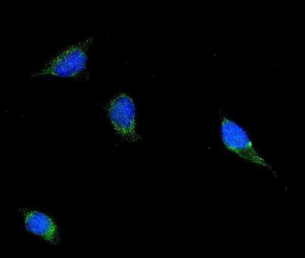
| IF/ICC question: where should FLNB appear? | In ICC-IF, HPA places FLNB mainly at the plasma membrane, with additional Golgi, actin-filament and cytosolic localization (HPA subcellular ICC-IF). This is a localization reference for the separate IF/ICC guide, not an IHC-P protocol or a requirement that every compartment be visible in tissue sections. |
| Situation | Likely cause | Next action |
|---|---|---|
| Expected endothelial or glandular cells are present, but the stain is blank. | The positive population expected from the tissue profile is absent from the result (HPA tissue IHC). A failed staining or detection step is possible (general IHC practice). | Confirm section identity and cell preservation, then check antibody incubation, retrieval conditions and detection with a known positive section (general IHC practice). No FLNB-specific retrieval condition is supplied. |
| A section appears negative because its cardiomyocytes do not stain. | Cardiomyocytes are listed as Not detected by HPA tissue IHC, despite UniProt reporting heart isoform expression (HPA: Not detected in cardiomyocytes; UniProt O75369 tissue specificity). | Assess endothelial and other identifiable cells separately; use a listed High-staining glandular population as a positive reference when available (HPA tissue IHC). |
| Strong color is confined to nuclei. | The compartment conflicts with cytoplasmic, cortical and cytoskeletal localization and with the HPA tissue profile (UniProt O75369 localization; HPA tissue IHC). | Compare a detection-only control and a second IHC-validated antibody, then reassess whether cytoplasmic or membranous signal is present (general IHC practice; HPA antibodies). |
| Chromogen appears in cells HPA lists as Not detected. | Cross-reactivity or endogenous detection activity may explain a discordant cell-type pattern; neither is established by the image alone (HPA tissue IHC; general IHC practice). | Use a detection-only control, inspect tissue pigment and compare the pattern with an independently validated antibody (general IHC practice; HPA antibodies). |
| The whole section has diffuse color that prevents cell-by-cell scoring. | Excess reagent signal, incomplete washing or endogenous detection activity can produce background (general IHC practice). | Review blocking and washes, titrate the antibody and detection reagents, and score FLNB only after cell-associated signal is distinguishable (general IHC practice; HPA tissue IHC). |
| Cell-edge staining is seen, but fine actin-associated detail is unclear. | HPA tissue IHC describes broad cytoplasmic and membranous staining, while actin filaments are an additional ICC-IF location (HPA tissue IHC; HPA subcellular ICC-IF). | Score the IHC section against its tissue and cell-type pattern; consult the separate IF/ICC guide if filament-level localization is the experimental question (HPA tissue IHC; HPA subcellular ICC-IF). |
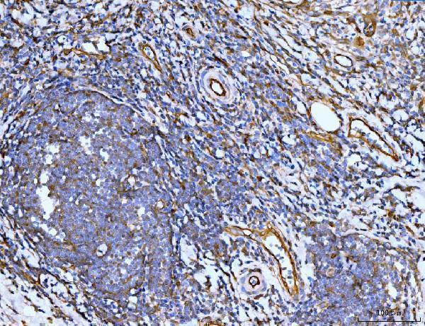
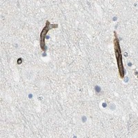


Comprehensive Human Protein Atlas IHC scoring per tissue (reliability: Enhanced — Medium consistency between antibody staining and RNA expression data. Paired antibodies with high similarity supports the protein expression profile.). Rows are taken directly from the HPA tissue chart — click any row's HPA link to view the source.
| Tissue | Cell type | Level | Evidence | Source |
|---|---|---|---|---|
| Adrenal gland | Glandular cells | High | Protein (IHC) | HPA → |
| Appendix | Glandular cells | High | Protein (IHC) | HPA → |
| Breast | Glandular cells | High | Protein (IHC) | HPA → |
| Bronchus | Respiratory epithelial cells | High | Protein (IHC) | HPA → |
| Colon | Glandular cells | High | Protein (IHC) | HPA → |
| Tissue | Cell type | Level | Evidence | Source |
|---|---|---|---|---|
| Caudate | Glial cells | Not detected | Protein (IHC) | HPA → |
| Cerebellum | Cells in granular layer | Not detected | Protein (IHC) | HPA → |
| Heart muscle | Cardiomyocytes | Not detected | Protein (IHC) | HPA → |
| Hippocampus | Glial cells | Not detected | Protein (IHC) | HPA → |
| Lymph node | Germinal center cells | Not detected | Protein (IHC) | HPA → |
Troubleshoot FLNB staining in paraffin sections by checking retrieval, tissue handling, cell compartment and controls before interpreting chromogenic signal.
Both anti-FLNB antibodies have human paraffin-section IHC images and IF/ICC cell images; A02562-1 also lists mouse and rat reactivity (catalog applications, reactivity and image captions).
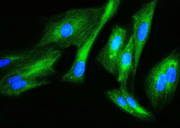
A02562-1 has IHC images from human lung cancer, mammary cancer, placenta and rectal cancer sections, plus an IF/ICC image from U2-OS cells (A02562-1 image captions). M02562-1 has IHC images from human lung cancer, rectal-cancer lymph node, renal-pelvis squamous metaplasia and spleen sections, plus an IF/ICC image from HeLa cells (M02562-1 image captions).
Which to pick: For human tissue IHC, either SKU has its own paraffin-section lung cancer image using EDTA retrieval at pH 8.0 and DAB detection; neither caption reports the fixative (A02562-1 and M02562-1 IHC captions). For IF/ICC, choose by host or cell-image precedent: rabbit A02562-1 is shown in U2-OS cells, while mouse monoclonal M02562-1, clone 11E2D2, is shown in HeLa cells (catalog host/clone and IF captions). For mouse or rat tissue IHC, choose A02562-1 based on its listed species reactivity and IHC application; its supplied IHC images show human tissue (A02562-1 catalog reactivity/applications and IHC captions).