This website uses cookies to ensure you get the best experience on our website.


- Table of Contents
Plan FOSB IHC in paraffin sections using nuclear staining as the expected pattern (HPA tissue IHC). Compare positive tissue controls with your specimens and score staining in the relevant cell populations.

Expected localisation, validated protocols, controls and antibodies — the at-a-glance facts below, then the full design guide.
| Expected localisation | Nuclear staining in several tissues (HPA tissue IHC) | |
| Staining pattern | Nuclear staining in trophoblastic and urothelial cells (HPA tissue IHC) | |
| Antigen retrieval | EDTA pH 8.0 HIER, heat-mediated (datasheet RP1086) | |
| Positive control | Placenta+4 more · see all | |
| Negative control | Adrenal gland+4 more · see all |
| Fixation | Matched tissue-IHC evidence does not establish the fixation claim. Validate the specimen-specific method before use. (selected-SKU IHC image RP1086) | |
| Caveat | Staining has low consistency with RNA expression (HPA tissue IHC) | |
| Regulation | Staining regulation is unreported (UniProt) | |
| Isoform / epitope | 11 isoforms; epitope coverage needs validation (UniProt) |
The catalog antibody’s IHC-P protocol is paired with 4 published FOSB IHC protocols (PMC3503985; PMC5678090; PMC7085481; PMC12196134).
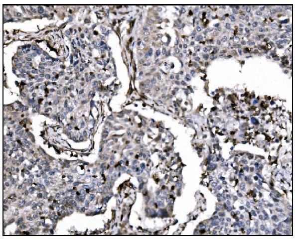
| Sample | Paraffin-embedded human renal cancer tissue; fixative not specified (datasheet RP1086) |
| Fixation | Image fixative and duration unreported (datasheet RP1086); verify before use. |
| Sectioning | 4–5 µm sections on charged slides (standard) |
| Deparaffinisation | Xylene, graded ethanol series to water (standard) |
| Antigen retrieval | Heat retrieval: EDTA pH 8.0 (datasheet RP1086); 20 min, 95–100 °C (standard) |
| Peroxidase block | 3% H2O2, 10 min, room temperature (standard) |
| Blocking | 10% goat serum (datasheet RP1086) |
| Primary antibody | Rabbit anti-FOSB, 0.5-1μg/ml (datasheet RP1086) |
| Primary incubation | Overnight at 4 °C (datasheet RP1086) |
| Detection | Streptavidin-biotin complex (SABC), DAB chromogen (datasheet RP1086) |
| Counterstain | Hematoxylin, blue, dehydrate and mount (standard) |
| Expected result | FOSB-positive staining in trophoblastic cells of placenta (HPA tissue IHC: High). HPA tissue profile: Nuclear expression in several tissues. No signal in the no-primary control. |
FOSB is a nuclear transcription factor with no transmembrane segment (UniProt P53539). In paraffin-section IHC, expect nuclear staining in trophoblastic cells of placenta and urothelial cells of urinary bladder, both scored High by HPA (HPA tissue IHC). HPA reports nuclear expression across several tissues, but rates its IHC data Approved with low consistency between antibody staining and RNA expression (HPA tissue IHC).
| Distinct nuclear chromogen in placental trophoblastic cells or bladder urothelial cells (HPA: High in both). | This fits the expected compartment and cell populations (UniProt P53539: nucleus; HPA tissue IHC). Score the relevant cells separately from surrounding tissue; HPA's High designation describes its observed staining, not a required intensity for every specimen (HPA tissue IHC). |
| Predominantly cytoplasmic or extracellular staining, with little nuclear signal. | This conflicts with the established nuclear location (UniProt P53539; HPA: nuclear tissue expression). Consider nonspecific binding or detection artefact, then check morphology and controls (general IHC practice). HPA reports uncertain vesicle localisation in ICC-IF; that does not validate a cytoplasmic IHC pattern (HPA subcellular). |
| Strong staining in cells listed as Not detected, such as adrenal glandular or marrow hematopoietic cells (HPA tissue IHC). | Treat an unexpected cell-type pattern as possible cross-reactivity or endogenous detection activity, not proof of FOSB expression (general IHC practice). HPA's Not detected calls describe the cited samples and antibody assessment; they are not universal biological negatives (HPA tissue IHC). |
| Diffuse chromogen over nuclei, cytoplasm and stroma, obscuring cell boundaries. | The distribution cannot establish the expected nuclear pattern (UniProt P53539; HPA tissue IHC). Excess antibody, incomplete blocking or endogenous detection activity can produce background (general IHC practice). Compare an appropriately processed negative control before scoring nuclei (general IHC practice). |
| No nuclear staining in placental trophoblasts or bladder urothelium (HPA: High in both). | First assess assay failure using tissue integrity and detection controls (general IHC practice). HPA's High observations make these reasonable positive comparators, but HPA reports low staining–RNA consistency; a blank section alone does not establish biological absence (HPA tissue IHC). |
| Cell and tissue context (HPA tissue IHC). | Placental trophoblasts and bladder urothelium are High; several other cell populations are Medium or Low (HPA tissue IHC). Judge signal in the specified cells, since HPA reports low tissue RNA specificity and low staining–RNA consistency (HPA tissue IHC). |
| Antibody validation (HPA antibodies; HPA tissue IHC). | CAB010267 has Approved IHC status; HPA054663 has Supported ICC status with no IHC status listed (HPA antibodies). These labels are application-specific, and Approved IHC does not remove HPA's stated staining–RNA consistency caveat (HPA tissue IHC). |
| Isoforms and epitope coverage (UniProt P53539). | UniProt lists 11 FOSB isoforms (UniProt P53539). The supplied record does not map the catalog antibody's epitope, so which isoforms it recognizes cannot be inferred; interpret a negative stain within that limitation (UniProt P53539; general assay interpretation). |
| Antigen retrieval (general IHC practice). | Use the IHC-validated antibody's documented paraffin-section conditions as a starting point, and compare retrieval conditions only with suitable controls (general IHC practice). The supplied UniProt and HPA records establish no FOSB-specific retrieval response or fixation sensitivity. |
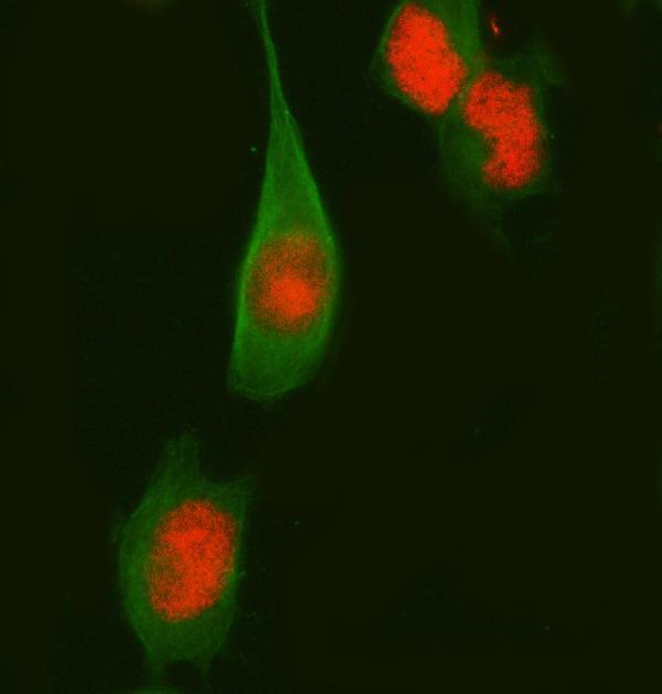
| IF/ICC Q: What compartment should be checked? (HPA subcellular). | A: Nucleoplasm is the supported main location; vesicles are an uncertain additional location (HPA subcellular). This informs localisation checks only: ICC-IF evidence does not supply an IF protocol option for this IHC-P guide (HPA subcellular; HPA antibodies). |
| Situation | Likely cause | Next action |
|---|---|---|
| Positive comparator is blank: no nuclear color in placental trophoblasts or bladder urothelium (HPA: High). | A staining-run problem, weak detection or unsuitable antibody conditions are possibilities (general IHC practice); HPA does not identify the cause for an individual section. | Check tissue morphology, reagent and detection controls, then review the IHC-validated antibody's documented IHC-P conditions (general IHC practice). Repeat with a matched positive section before calling the specimen negative (general IHC practice). |
| Predominantly cytoplasmic signal obscures nuclei (UniProt P53539: nucleus). | Nonspecific signal or excess background is possible (general IHC practice). HPA's uncertain vesicle observation comes from ICC-IF and does not establish this as an IHC-positive pattern (HPA subcellular). | Inspect nuclear counterstain and cell boundaries; compare a negative control and adjust antibody concentration within documented IHC conditions if needed (general IHC practice). Score only interpretable nuclear staining (UniProt P53539; HPA tissue IHC). |
| Color appears broadly in stromal or cell populations outside the expected positive cells (HPA tissue IHC). | Cross-reactivity or endogenous enzyme activity may contribute in chromogenic IHC (general IHC practice). HPA's tissue calls are cell-specific and its staining–RNA consistency is low (HPA tissue IHC). | Compare a no-primary control, verify blocking and inspect the exact stained cell type (general IHC practice). Avoid assigning FOSB positivity from color alone when the compartment conflicts with nuclear localisation (UniProt P53539). |
| Diffuse background prevents reliable nuclear scoring. | Incomplete blocking, excessive antibody or inadequate washing can elevate nonspecific signal (general IHC practice); the supplied sources give no FOSB-specific cause. | Review blocking, washing and antibody concentration against the documented IHC workflow; process positive and negative controls in the same run (general IHC practice). Reassess only after nuclei are distinguishable (general IHC practice). |
| A specimen disagrees with an HPA Not detected example, such as adrenal glandular cells (HPA tissue IHC). | The sample may differ from HPA's observed material, or the signal may be nonspecific (HPA tissue IHC; general IHC practice). A single discrepant stain cannot distinguish these explanations. | Confirm that staining is nuclear, compare controls and report the cell type and intensity explicitly (UniProt P53539; general IHC practice). Treat the HPA category as a reference observation, not an absolute exclusion (HPA tissue IHC). |
| A clear nuclear signal is present, but its meaning is uncertain because RNA evidence differs (HPA tissue IHC). | HPA rates tissue IHC Approved while explicitly reporting low consistency with RNA expression (HPA tissue IHC). Nuclear localisation supports pattern assessment but does not resolve antibody specificity by itself (UniProt P53539; general IHC practice). | Record compartment, stained cell population and controls; describe the finding as IHC staining with the HPA consistency caveat (HPA tissue IHC). Use independent validation before making a strong biological expression claim (general IHC practice). |




![HPA ICC-IF BJ [Human fibroblast] — FOSB (HPA054663)](https://images.proteinatlas.org/54663/1534_B6_1_blue_red_green.jpg)
![HPA ICC-IF BJ [Human fibroblast] — FOSB (HPA054663)](https://images.proteinatlas.org/54663/1534_B6_2_blue_red_green.jpg)
Comprehensive Human Protein Atlas IHC scoring per tissue (reliability: Approved — Low consistency between antibody staining and RNA expression data.). Rows are taken directly from the HPA tissue chart — click any row's HPA link to view the source.
| Tissue | Cell type | Level | Evidence | Source |
|---|---|---|---|---|
| Placenta | Trophoblastic cells | High | Protein (IHC) | HPA → |
| Urinary bladder | Urothelial cells | High | Protein (IHC) | HPA → |
| Adipose tissue | Adipocytes | Medium | Protein (IHC) | HPA → |
| Lymph node | Non-germinal center cells | Medium | Protein (IHC) | HPA → |
| Ovary | Ovarian stroma cells | Medium | Protein (IHC) | HPA → |
| Tissue | Cell type | Level | Evidence | Source |
|---|---|---|---|---|
| Adrenal gland | Glandular cells | Not detected | Protein (IHC) | HPA → |
| Bone marrow | Hematopoietic cells | Not detected | Protein (IHC) | HPA → |
| Breast | Adipocytes | Not detected | Protein (IHC) | HPA → |
| Caudate | Glial cells | Not detected | Protein (IHC) | HPA → |
| Cerebellum | Cells in granular layer | Not detected | Protein (IHC) | HPA → |
Troubleshoot FOSB staining in paraffin sections by checking retrieval, nuclear localisation, cell identity and controls before interpreting signal intensity.
Anti-FOSB antibodies have paraffin-section IHC images from human and mouse tissues and IF/ICC images from SiHa and HeLa cells (catalog image captions).
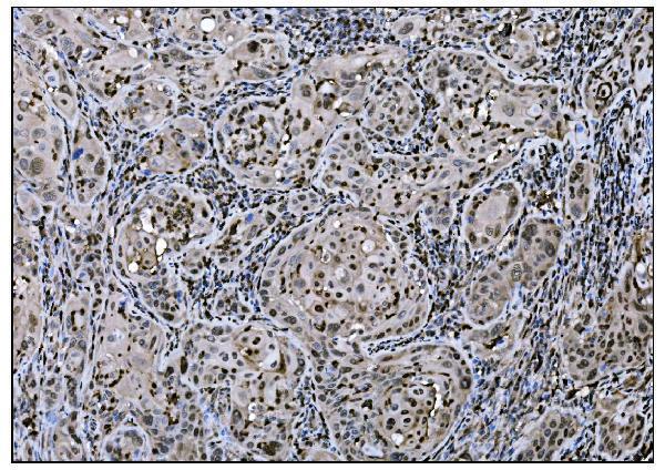
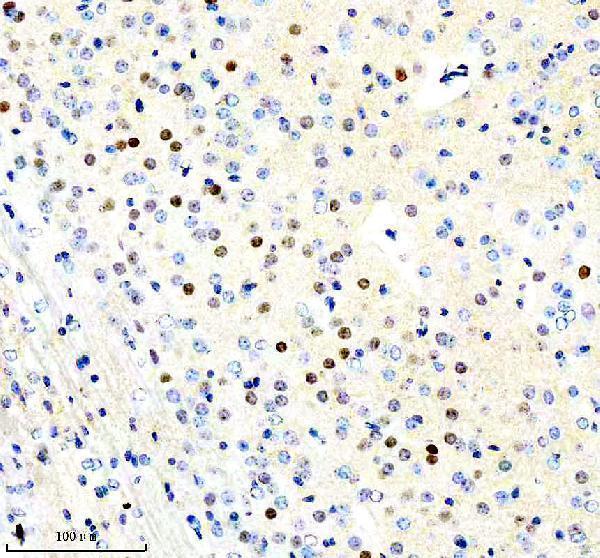
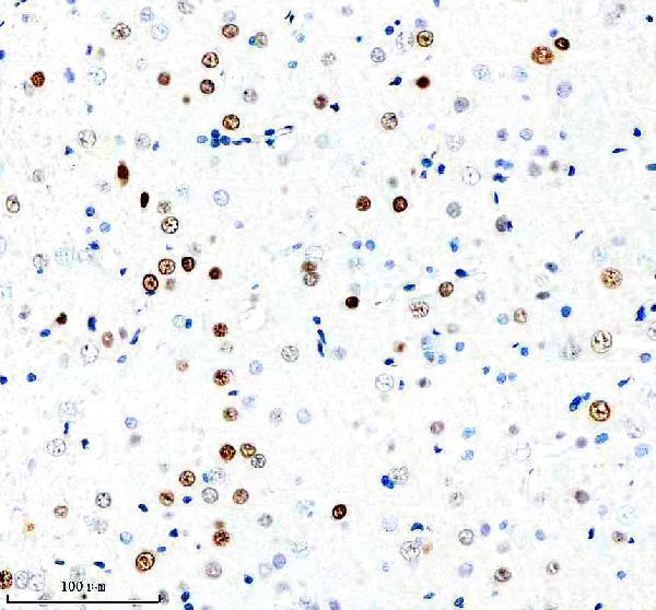
RP1086 has IHC images from human renal cancer and mouse intestine; A01569-1 has IHC images from human lung cancer, gallbladder adenocarcinoma, and placenta (respective IHC captions). M01569 has IHC images from mouse and rat brain, while A01569-1 and M01569 have IF/ICC images from SiHa and HeLa cells, respectively (respective image captions).
Which to pick: For tissue IHC, choose RP1086 for human renal cancer or mouse intestine, or A01569-1 for the pictured human tissues; each SKU’s IHC caption documents paraffin sections and EDTA pH 8 retrieval (respective IHC captions). For IF/ICC, choose A01569-1 for human SiHa cells or monoclonal M01569 for HeLa cells; both list IF/ICC, while RP1086 does not (catalog applications; IF captions; M01569 clone DFO-6). For cross-species IHC, M01569 has mouse and rat brain images, and RP1086 has human and mouse tissue images; both list human, mouse, and rat reactivity, but their IHC captions do not report the fixative (catalog reactivity; respective IHC captions).