This website uses cookies to ensure you get the best experience on our website.
- Table of Contents
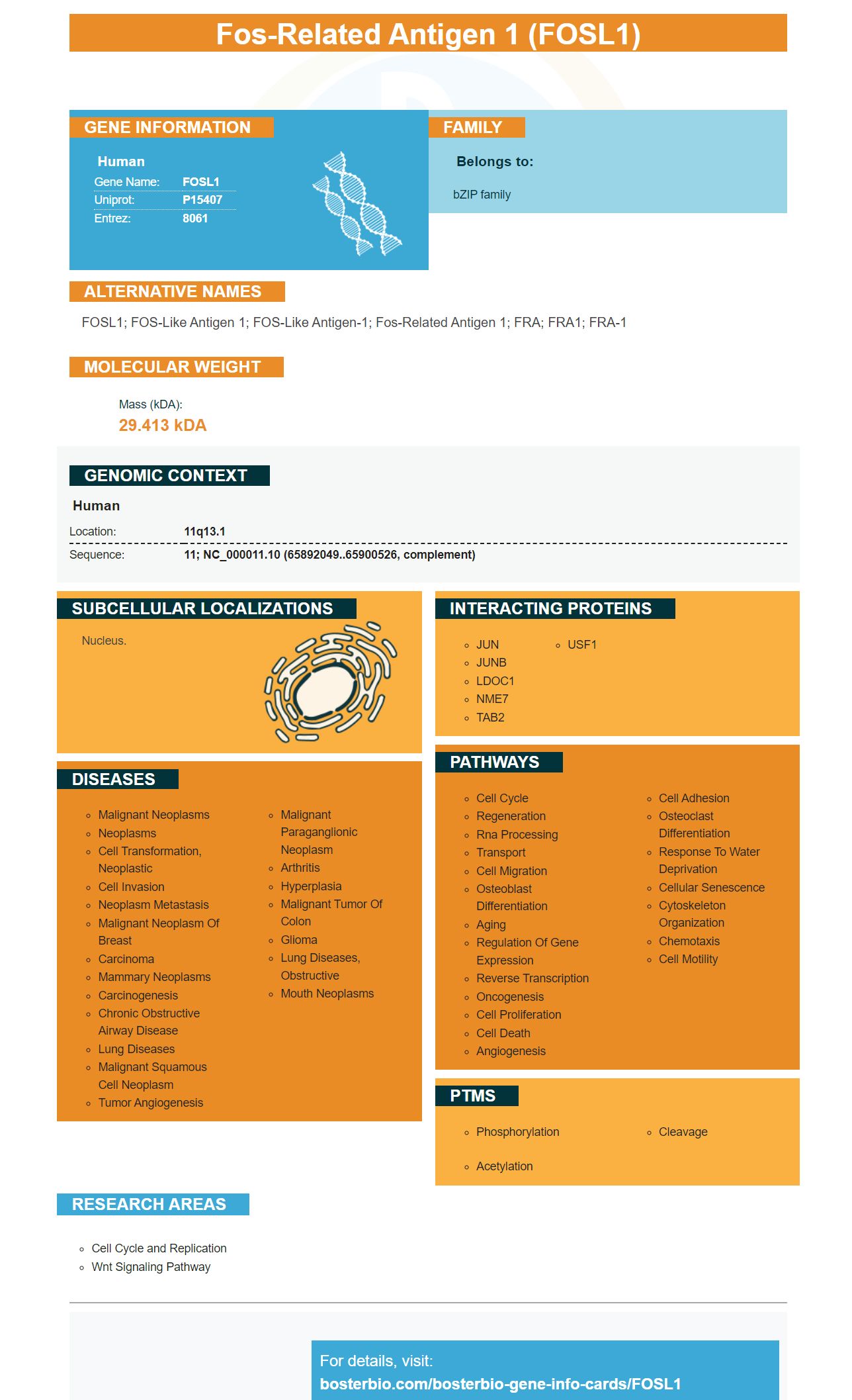
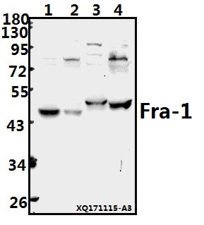
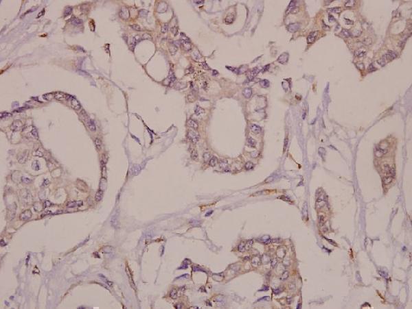
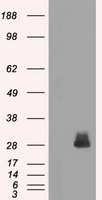
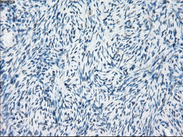
Facts about Fos-related antigen 1.
Human | |
---|---|
Gene Name: | FOSL1 |
Uniprot: | P15407 |
Entrez: | 8061 |
Belongs to: |
---|
bZIP family |
FOSL1; FOS-like antigen 1; FOS-like antigen-1; fos-related antigen 1; FRA; FRA1; FRA-1
Mass (kDA):
29.413 kDA
Human | |
---|---|
Location: | 11q13.1 |
Sequence: | 11; NC_000011.10 (65892049..65900526, complement) |
Nucleus.
This article will provide examples of FOSL1 markers and explain how they can be used in research. This article will cover the following topics: incubation and detection of target protein using western blotting. It also covers PCR amplification for mouse Psg gene sequences. Refer to the references below for more information. This is an excerpt from "Best Uses For The FOSL1Marker".
This article describes incubation of FOSL1 markers using a boster bioengineering technique. CCK-8 solution, which is manufactured by Wuhan Boster Biological Technology, Ltd., Wuhan, China, was used for this procedure. After incubation at 37°C, the cells are measured for optical density using an Epoch plate reader at 405 nm.
Detection of target protein using western-blotting involves a multistep procedure involving SDS-PAGE to separate the proteins and electrophoresis to transfer them to a membrane. Before using the target protein, it must be thoroughly researched. The sample is then transferred to the membrane by electrophoresis, and then immobilized. A high-quality antibody is required to detect target proteins.
The proteins used in the western-blotting process are purified, denatured, or a mixture of both. The target proteins are then transferred onto a membrane and detected with antibodies specific to their target. Over the years, the process has been improved and improved upon. Numerous reports have been written on various aspects of this procedure. These are some important guidelines:
- Western-blotting, which detects small-scale signals from small samples, is an effective way to detect target proteins. Although western-blotting may not be as sensitive as immunoblotting it can quickly detect a single protein. Detection of a single protein using western-blotting is a good solution for small-scale experiments and for research that requires multiple samples.
- Detection of target protein using western-blotting requires primary antibodies. These antibodies can be diluted with wash buffer to minimize background and unbound antibody. A reduced signal will result if the wash buffer is left on for too long. - The detection method you use to detect target protein in western-blotting depends on what it is. Stripping is a good choice if you need to analyze multiple protein samples or have high-value samples.
- It is important to block nonspecific binding sites. It prevents non-specific binding between proteins and membranes, and reduces background noise. It also eliminates false-positive results. This process is simple and cost-effective. This method can destroy some proteins, so it is important to choose the right primary antibody. Use western blotting with caution to detect target protein.
- The western-blotting technique is useful for identifying and separating proteins from various samples. It can also be used to detect posttranslational modifications in proteins. It is also an excellent tool to determine the molecular mass of proteins. It helps determine the expression status of a protein. These results can be crucial in diagnosing a condition. If you suspect a condition or illness, Western-blotting can be a great option.
- When performing western-blotting, the primary antibodies should be validated. The choice of primary antibody will depend on the antigen to be detected. Monoclonal antibodies have a higher specificity and a lower background level than polyclonal antibody. Polyclonal antibody, on the contrary, recognizes many epitopes and is often more effective at detecting protein. They can be used with diluted secondary antibodies.
The mouse Psg genes are located on proximal Chromome 7 between cytobands B1 and B2 and interspersed by other genes, such as Ceacams. Contig NT_039395 contains information about the mouse Psgs' genomic sequence. The amplifications of mouse Psg gene sequences were performed to identify the Psg gene subclusters. Two-colour FISH prophase maps on mouse m5S and C57BL/6CrSlc mice lymphocytes were used to determine the relative orientations. Fluorosceine and rhodamine were used to label BAC 310D2 in subcluster 1.
The amplifications of mouse Psg sequences showed that the precursor of Psg31 had undergone duplication in the N1 domain. These analyses support the hypothesis that human and mouse Psg genes may have evolved independently. Although they have a common ancestor, PSG genes from mice differ significantly from those found in humans. Scientists have suggested that these two gene family may have evolved independently from each other through gene duplication, exon shuffling events, and possibly gene duplication.
PCR amplifications of mouse Pg gene sequences showed that these genes contain alternative exons. This was confirmed via BLAST searches in the mouse genome database and the mouse High Throughput Genomic Sequences databases (HTGS). Sequences with a score of greater than 90% on BLAST were considered hits. Sequences greater than 150 bp are considered hits. Mismatches to other sequences were called mismatches.
Molecular analysis of the mouse Psg gene sequences revealed that the exon structure of this gene has undergone extensive evolution. Moreover, the gene is expressed in trophoblasts during pregnancy. Once the mouse Psg locus genome map has been established, detailed studies on the functional roles and functions of the Psg gene family will be possible.
While the function of PSGs is not yet clear, functional experiments are starting to give some clues. Studies in mice show that low levels of the hormone can cause threatened abortion, intrauterine retardation, foetal hypoxia, and intrauterine enlargement. Anti-PSG antibodies and anti–PSG vaccination in monkeys induce abortion and decrease fertility in non-pregnant animals. Most studies on PSGs' functional roles in mammals have focused only on modulating the immune response.
The complexity of mouse genome is 2x10⁹. A PCR amplification with mouse Psg gene sequences should yield 18,626, 116.7, and 0.45 respectively. The easiest resolution would be achieved by random 10-mers. But N =11 would result in no resolvable Bands.
SSCP can be used to detect recombination errors in a mixture or sequences. When using SSCP, a single PCR product can split into four bands if it has a single base substitution. This protocol is highly effective in determining Psg proteins. This amplification method is widely used for the analysis of various diseases in humans.
PMID: 2107490 by Matsui M., et al. Isolation of human fos-related genes and their expression during monocyte-macrophage differentiation.
PMID: 8230424 by Tsuchiya H., et al. Human T-cell leukemia virus type 1 Tax activates transcription of the human fra-1 gene through multiple cis elements responsive to transmembrane signals.
*More publications can be found for each product on its corresponding product page