FOXI1 / Forkhead box protein I1 · IHC design guide

Design Immunohistochemistry for FOXI1

Plan chromogenic FOXI1 IHC around nuclear staining in subsets of renal tubules and salivary gland (HPA tissue IHC). Use kidney collecting ducts, where staining is medium, as a positive control and disregard cytoplasmic positivity when interpreting the tissue pattern (HPA tissue IHC).

Evidence assembled Oct 2026 · For research use; verify linked source records and product datasheet before use
Immunohistochemistry protocol sheet for FOXI1 (IHC for FOXI1): expected localisation Nuclear in subsets of renal tubules and salivary gland (HPA tissue IHC), antibody A07512-1, validated IHC image, and IHC protocol steps
Printable FOXI1 IHC protocol sheet — expected localisation Nuclear in subsets of renal tubules and salivary gland (HPA tissue IHC), antibody A07512-1, controls and protocol steps. Open the full FOXI1 IHC guide →

FOXI1 Immunohistochemistry Experimental Design Guide

Expected localisation, validated protocols, controls and antibodies — the at-a-glance facts below, then the full design guide.

Must know before staining
Expected localisation Nuclear in subsets of renal tubules and salivary gland (HPA tissue IHC)
Staining pattern Subset nuclei stain in renal tubules and salivary gland (HPA tissue IHC)
Antigen retrieval Tris-EDTA pH 9.0 HIER, 95–98 °C, 20 min (rule: nuclear antigen)
Positive control ⓘ Kidney
Negative control ⓘ Adipose tissue+4 more · see all
Important caveats
Reasons your staining may differ from the expected pattern.
Fixation Keep fixation conditions consistent across sections. (standard IHC practice; not target-specific)
Caveat Cytoplasmic positivity was observed and disregarded (HPA tissue IHC)
Regulation No expression regulation annotated (UniProt)
Isoform / epitope 2 isoforms; no membrane topology; epitope maps unknown (UniProt)
Section 1

Recommended FOXI1 IHC & IF Protocols

Compare the catalog antibody’s IHC-P protocol with four published FOXI1 IHC protocols (PMC8424250; PMC10869675; PMC11110679; PMC10200853).

Recommended immunohistochemistry (IHC-P) protocol parameters
SampleParaffin-embedded human breast carcinoma tissue; fixative not specified (datasheet A07512-1)
FixationImage fixative and duration unreported (datasheet A07512-1); verify before use.
Sectioning4–5 µm sections on charged slides (standard)
DeparaffinisationXylene, graded ethanol series to water (standard)
Antigen retrievalHeat-induced epitope retrieval in Tris-EDTA buffer, pH 9.0, 20 min at 95–98 °C (standard rule: nuclear antigen)
Peroxidase block3% H2O2, 10 min, room temperature (standard)
Blocking10% normal serum of the secondary host, 30 min, room temperature (standard)
Primary antibodyRabbit anti-FOXI1, 1:100–1:500 starting range (standard)
Primary incubationOvernight at 4 °C (standard)
DetectionHRP-polymer secondary, DAB chromogen 5–10 min (standard)
CounterstainHematoxylin, blue, dehydrate and mount (standard)
Expected resultFOXI1-positive staining in collecting ducts of kidney (HPA tissue IHC: Medium). HPA tissue profile: Distinct nuclear expression in subsets of cells in renal tubuli and salivary gland. No signal in the no-primary control.
💡Decision noteStart with Tris-EDTA pH 9.0 HIER at 95–98 °C for 20 min (page retrieval); use each paper’s stated retrieval when reproducing its protocol.
Section 2

What Is the Expected FOXI1 Staining Pattern?

FOXI1 is a nuclear transcription factor with no transmembrane segment (UniProt Q12951). In paraffin-section IHC, expect distinct nuclear staining in subsets of renal tubular and salivary gland cells; kidney collecting ducts show medium staining (HPA tissue IHC). HPA rates the tissue pattern Enhanced, while noting medium consistency with RNA data and disregarding observed cytoplasmic positivity (HPA tissue IHC).

What am I looking at on my slide?
Discrete nuclear staining in some collecting-duct cells, with neighbouring nuclei unstained.This fits the subset pattern and medium collecting-duct level (HPA tissue IHC). Assess nuclear signal cell by cell; uniform staining throughout a tubule is not the reported pattern (HPA tissue IHC).
Predominantly cytoplasmic staining, with little convincing nuclear signal.Do not score this as FOXI1 positivity: HPA observed and disregarded cytoplasmic positivity, while UniProt places FOXI1 in the nucleus (HPA tissue IHC; UniProt Q12951). Review nuclear counterstain alignment and detection background (general IHC practice).
Strong staining in cells outside the reported subset pattern.Treat this as unconfirmed; cross-reactivity or endogenous detection activity is possible (general IHC practice). Compare cell identity and nuclear localisation against the HPA tissue pattern before calling the slide positive (HPA tissue IHC).
Haze or precipitate obscures both positive and negative areas.The compartment cannot be judged reliably under diffuse background (general IHC practice). Check a no-primary control and inspect reagent deposition before scoring nuclei (general IHC practice).
No stained nuclei in a kidney collecting-duct area.This conflicts with the reported medium collecting-duct signal, but one negative section does not establish absent FOXI1 (HPA tissue IHC). Confirm the relevant cells are present and review assay controls and detection performance (general IHC practice).
💡Expected FOXI1 appearanceCall a result positive when distinct nuclear staining appears in a subset of renal tubular cells, including collecting ducts at about medium intensity; isolated cytoplasmic colour is a false-positive pattern for scoring purposes (HPA tissue IHC; UniProt Q12951).
How each factor affects the staining
Which tissue and cells provide the clearest reference?Kidney collecting ducts have reported medium staining; salivary gland has distinct nuclear staining in a subset despite a low glandular-cell summary (HPA tissue IHC). Use cell-level localisation when interpreting either tissue (HPA tissue IHC).
How strong is the IHC evidence?The tissue profile is Enhanced, and antibody HPA071469 has Enhanced IHC validation (HPA tissue IHC; HPA antibodies). HPA still reports only medium staining–RNA consistency and explicitly disregards cytoplasmic positivity (HPA tissue IHC).
What do protein features imply for scoring?UniProt assigns FOXI1 to the nucleus and reports no transmembrane segment, signal peptide, propeptide or glycosylation sites (UniProt Q12951). Those annotations support a nuclear scoring rule; they do not predict antigen-retrieval or fixation sensitivity (UniProt Q12951).
Do isoforms change the expected pattern?Two isoforms are listed, but the supplied record gives no isoform-specific tissue distribution or antibody epitope (UniProt Q12951; HPA antibodies). Do not assign differing staining patterns to isoforms from these data (UniProt Q12951; HPA tissue IHC).
What about IF/ICC?HPA reports mainly nucleolar ICC-IF staining, with additional vesicle localisation marked uncertain; its image lines are HaCaT, MCF-7 and U2OS (HPA subcellular). That cell-line observation does not replace the nuclear tissue-IHC scoring pattern (HPA subcellular; HPA tissue IHC).
Why is my staining missing, weak or wrong?
SituationLikely causeNext action
Kidney section shows no convincing nuclear signal.The expected collecting-duct cells may be absent from the field, or the IHC run may have failed (HPA tissue IHC; general IHC practice).Locate collecting ducts, check a known-positive control in the same run, then review retrieval and detection settings using the antibody's validated IHC instructions (general IHC practice).
Nuclear colour is weak or hard to distinguish.Collecting-duct staining is reported as medium and occurs in a subset, so sparse signal may be missed; counterstain can also obscure it (HPA tissue IHC; general IHC practice).Inspect several appropriate fields and compare nuclear colour with an unstained-cell reference; adjust counterstain or detection only with appropriate controls (general IHC practice).
Cytoplasm stains more clearly than nuclei.HPA observed cytoplasmic positivity and disregarded it; its tissue call is nuclear (HPA tissue IHC).Exclude cytoplasmic colour from the FOXI1 score. Check no-primary background and reassess compartment boundaries against the nuclear counterstain (general IHC practice).
Nearly every cell in a gland or tubule stains.This exceeds HPA's reported subset pattern and may reflect nonspecific binding or excess detection signal (HPA tissue IHC; general IHC practice).Check cell identity, compare with a no-primary control and review antibody concentration and detection exposure under standard IHC optimisation (general IHC practice).
A reported negative tissue stains strongly.HPA lists adipocytes and several other sampled cell types as not detected; positive colour there needs independent assessment (HPA tissue IHC).Confirm the stained cell type, nuclear compartment and control behaviour before attributing the result to FOXI1; investigate cross-reactivity or endogenous activity (general IHC practice).
Diffuse colour or precipitate prevents scoring.Background from blocking, washing or detection can obscure discrete nuclear signal (general IHC practice).Inspect no-primary and reagent controls, improve washing or blocking as indicated, and repeat scoring only when individual nuclei can be resolved (general IHC practice).

Sample controls for FOXI1 IHC & IF

🧪Run kidney first and look for staining in collecting ducts, where FOXI1 is reported at medium level (HPA: Kidney, collecting ducts, Medium). Use adipose tissue as a negative tissue, with adipocytes reported as not detected; on the kidney slide, cells outside the stained collecting duct population should lack specific signal (HPA: Adipose tissue, adipocytes, Not detected; standard IHC practice).
Positive control tissue: Kidney (Collecting ducts, HPA Medium)
Negative control tissue: Adipose tissue (HPA Not detected)
ICC-IF cell lines (HPA subcellular resource): HPA ICC-IF images show FOXI1 in HaCaT, MCF-7, U2OS, with annotated localisation: Nucleoli (supported) (HPA subcellular).
Technical controls: Include a no-primary (secondary-only) control, an isotype control matched to the primary antibody’s host species and clonality, and a peptide-blocked antibody control (standard IHC practice; selected-SKU tissue-IHC caption: peptide block). For chromogenic kidney IHC, quench endogenous peroxidase and assess residual background (standard IHC practice).
⚠️Feasibility: A FOXI1-specific fixation window and retrieval dependency are unreported in the supplied evidence; optimize antigen retrieval empirically for paraffin sections (supplied evidence; standard IHC practice). The selected-SKU paraffin-section caption does not state a fixative, so its fixative is unreported (selected-SKU tissue-IHC caption). The evidence does not establish that frozen sections or IF are easier; if testing IF, assess kidney autofluorescence with an unstained control (supplied evidence; standard IF practice).

HPA tissue IHC evidence for FOXI1

Comprehensive Human Protein Atlas IHC scoring per tissue (reliability: Enhanced — Medium consistency between antibody staining and RNA expression data. Cytoplasmic positivity observed and disregarded.). Rows are taken directly from the HPA tissue chart — click any row's HPA link to view the source.

Positive expression · recommended positive controls

TissueCell typeLevelEvidenceSource
Kidney Collecting ducts Medium Protein (IHC) HPA →

Undetected expression · recommended negative controls

TissueCell typeLevelEvidenceSource
Adipose tissue Adipocytes Not detected Protein (IHC) HPA →
Adrenal gland Glandular cells Not detected Protein (IHC) HPA →
Appendix Glandular cells Not detected Protein (IHC) HPA →
Bone marrow Hematopoietic cells Not detected Protein (IHC) HPA →
Caudate Glial cells Not detected Protein (IHC) HPA →
Section 3

Advanced FOXI1 IHC Tips

Troubleshoot FOXI1 chromogenic IHC in paraffin sections by prioritising nuclear staining, matched controls and cell type specific scoring.

What retrieval should I try first if FOXI1 nuclear staining is weak?
For paraffin sections, begin with Tris-EDTA pH 9.0 heat-induced epitope retrieval at 95–98 °C for 20 min (page protocol: nuclear-antigen rule). Let slides cool in retrieval buffer before washing, and keep section thickness, antibody incubation and detection conditions constant while comparing runs (standard IHC practice). Assess nuclear signal in renal collecting ducts, where FOXI1 staining is reported at a medium level (HPA: kidney tissue IHC). If staining remains weak, compare a milder retrieval condition on adjacent sections as a fallback, while checking whether it reduces background (standard IHC practice). Score nuclear signal separately from cytoplasmic colour, which HPA disregarded (HPA: tissue IHC reliability description).
Could fixation explain weak or uneven FOXI1 staining?
Target-specific FOXI1 sensitivity to fixative type or fixation duration is unknown from the supplied evidence; the catalog caption reports paraffin embedding but does not state a fixative (catalog caption: A07512-1). Record the fixative, duration and tissue thickness for each specimen before attributing uneven staining to retrieval or antibody concentration (standard IHC practice). Compare similarly processed sections within a run, using renal collecting ducts as a relevant staining reference when available (HPA: kidney tissue IHC). If fixation histories differ, interpret intensity differences cautiously and assess morphology alongside nuclear staining (standard IHC practice). Do not use tissue expression patterns to infer a FOXI1-specific fixation effect.
Should FOXI1 staining be nuclear, nucleolar or cytoplasmic in tissue sections?
Prioritise nuclear staining in chromogenic tissue IHC: FOXI1 is annotated as nuclear (UniProt Q12951: subcellular location), and HPA reports distinct nuclear staining in subsets of renal tubule and salivary gland cells (HPA: tissue IHC profile). HPA reports nucleoli as its supported main location in cell imaging, which can guide close inspection of nuclear detail without requiring every tissue section to show a distinct nucleolus (HPA: subcellular). FOXI1 has no annotated transmembrane segment, so a membrane-only pattern needs scrutiny (UniProt Q12951: topology). Record nuclear and cytoplasmic staining separately (standard IHC practice). HPA observed cytoplasmic positivity but disregarded it when interpreting tissue staining (HPA: tissue IHC reliability description).
How can I check whether the antibody detects both FOXI1 isoforms?
FOXI1 has 2 listed isoforms, but the supplied evidence does not locate this antibody’s epitope relative to their sequences (UniProt Q12951: isoforms; supplied catalog evidence). Check the antibody’s immunogen or epitope documentation before claiming that staining represents both isoforms (standard IHC practice). The catalog image for SKU A07512-1 includes a synthesized-peptide blocking comparison in paraffin-embedded human breast carcinoma (catalog caption: A07512-1). A matched peptide competition control can help assess binding under those image conditions, but it cannot establish which isoform contributes to tissue staining (standard IHC interpretation). Report results as FOXI1 immunoreactivity unless isoform coverage has been established independently.
How should I adapt the FOXI1 localisation check for multiplex IF?
For the separate IF/ICC workflow, pair FOXI1 with a marker identifying the expected renal collecting-duct cell population and inspect whether FOXI1 signal occupies its nuclei (HPA: kidney tissue IHC; UniProt Q12951: nuclear location). Choose fluorophores and filter sets after measuring tissue autofluorescence in an unstained control, favouring a channel with clear signal separation (standard IF practice). FOXI1 has no transmembrane segment and is nuclear, so permeabilisation must permit antibody access across the plasma and nuclear membranes (UniProt Q12951: topology and subcellular location; standard IF practice). Start with 0.1% Triton X-100 for 5 min as a general IF trial, then check morphology and nuclear signal (standard IF practice).
What should I investigate when FOXI1 IHC shows diffuse brown staining?
First distinguish diffuse colour from discrete nuclear FOXI1 staining; HPA disregarded cytoplasmic positivity in its tissue assessment (HPA: tissue IHC reliability description). Run a no-primary control through the same chromogenic detection steps to assess secondary-reagent background and endogenous enzyme signal (standard IHC practice). Include a peroxidase block before DAB detection, and compare blocked and unblocked controls if background persists (standard chromogenic IHC practice). Inspect section edges, folds and poorly preserved areas before changing antibody conditions, because local staining artefacts can mimic intensity differences (standard IHC practice). If nuclear signal is retained in collecting ducts while diffuse colour falls, evaluate that improvement using matched processing conditions (HPA: kidney tissue IHC; standard IHC practice).
How should I quantify FOXI1 staining across renal sections? ⚠ ANSWER MARKED FOR VERIFICATION
Define the anatomical compartment and count evaluable nuclei in it before scoring FOXI1, using collecting ducts as a documented positive cell population (HPA: kidney tissue IHC; standard IHC practice). Report the percentage of positive nuclei and an H-score from intensity categories 0–3, with the scoring thresholds held constant across slides (standard IHC scoring practice). For spatial comparisons, report FOXI1-positive nuclei per mm² of the prespecified compartment and state how that area was delineated (standard image-analysis practice). Normalise counts to all evaluable nuclei in the same compartment, and report excluded folds or damaged regions (standard IHC practice). Keep cytoplasmic colour out of the nuclear score because HPA disregarded it (HPA: tissue IHC reliability description).
When is a FOXI1-positive IHC result convincing rather than artefactual?
A convincing result places staining in nuclei of an anatomically plausible cell subset, particularly renal collecting ducts, where HPA reports medium staining (UniProt Q12951: nuclear location; HPA: kidney tissue IHC). Compare the pattern with adjacent preserved tissue and a no-primary control before interpreting isolated edge staining, necrotic areas or diffuse DAB colour (standard IHC practice). Cytoplasmic-only signal is weak evidence for FOXI1 because HPA observed cytoplasmic positivity and disregarded it (HPA: tissue IHC reliability description). The SKU A07512-1 paraffin breast carcinoma image includes a synthesized-peptide blocking comparison, which offers a product-specific control example (catalog caption: A07512-1). Record compartment, cell population and control outcomes together when reporting the result (standard IHC practice).
Boster reagents

Best FOXI1 / Forkhead box protein I1 IHC Antibodies

The catalog shows FOXI1 staining in paraffin-embedded human breast carcinoma tissue and IF staining in U20S cells (catalog image captions). Listed reactivity covers human/mouse and human/rat, respectively (catalog).

Real IHC data Immunohistochemistry analysis of paraffin-embedded human breast carcinoma tissue, using FOXI1 Antibody. The picture on the right is blocked with the synthesized peptide.
Anti-Forkhead box protein I1 FOXI1 Antibody
Cat # A07512-1
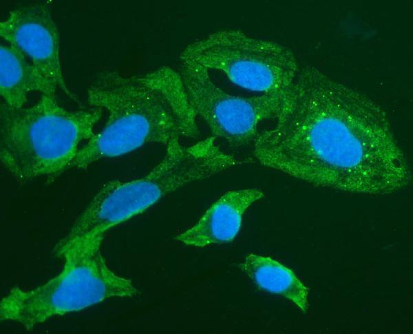
Real IF data IF analysis of FOXI1 using anti-FOXI1 antibody (A07512-3). FOXI1 was detected in immunocytochemical section of U20S cells. Enzyme antigen retrieval was performed using IHC enzyme antigen retrieval reagent (AR0022) for 15 mins. The cells were blocked with 10% goat serum. And then incubated with 2μg/mL rabbit anti-FOXI1 Antibody (A07512-3) overnight at 4°C. DyLight®488 conjugated Goat Anti-Rabbit IgG (BA1127) was used as secondary antibody at 1:100 dilution and incubated for 30 minutes at 37°C. The section was counterstained with DAPI. Visualize using a fluorescence microscope and filter sets appropriate for the label used.
Anti-Forkhead box protein I1 FOXI1 Antibody ®
Cat # A07512-3

A07512-1 lists IHC and IF and has an IHC image of paraffin-embedded human breast carcinoma tissue, with a peptide-blocked comparison (catalog applications; A07512-1 image caption). A07512-3 lists IF/ICC and has an IF image of U20S cells; it does not list IHC (catalog applications; A07512-3 image caption).

Which to pick: Choose A07512-1 for paraffin-section tissue IHC: it is a rabbit polyclonal antibody with an IHC image in human breast carcinoma tissue; the caption does not report the fixative (catalog; A07512-1 image caption). Choose A07512-3 for IF/ICC: its U20S-cell IF image reports 2 μg/mL primary antibody (A07512-3 image caption). For cross-species planning, A07512-1 lists mouse reactivity and A07512-3 lists rat reactivity, but their respective images document human tissue IHC and cell IF only (catalog reactivity; image captions).

Each figure is that product's own IHC / IF validation image from its datasheet.

References

  1. UniProt Consortium. UniProt entry Q12951 (FOXI1_HUMAN, Forkhead box protein I1).
  2. Human Protein Atlas. FOXI1 tissue IHC expression (reliability: Enhanced).
  3. Human Protein Atlas. FOXI1 subcellular location (ICC-IF): Mainly localized to the nucleoli. In addition localized to vesicles..
  4. Human Protein Atlas. FOXI1 antibody validation summary (1 antibodies).
  5. Forkhead Box I1 in Breast Carcinoma as a Potent Prognostic Factor. Acta histochemica et cytochemica 2021 — PMC8424250.
  6. Expression of FOXI1 and POU2F3 varies among different salivary gland neoplasms and is higher in Warthin tumor. Discover oncology 2024 — PMC10869675.
  7. Transcriptomics reveals age-related changes in ion transport-related factors in yak lungs. Frontiers in veterinary science 2024 — PMC11110679.
  8. Comparative analyses define differences between BHD-associated renal tumour and sporadic chromophobe renal cell carcinoma. EBioMedicine 2023 — PMC10200853.
  9. PubMed PMID:15489334 — UniProt-cited evidence.
  10. PubMed PMID:9153225 — UniProt-cited evidence.
  11. PubMed PMID:7957066 — UniProt-cited evidence.