This website uses cookies to ensure you get the best experience on our website.


- Table of Contents
Plan FOXP1 paraffin IHC around nuclear staining in most cells (HPA tissue IHC) and the catalog antibody at 2 μg/ml (datasheet A00723). This guide covers positive tissue choice, fixation consistency, and the reported risk of staining proteins from other genes (HPA tissue IHC).

Expected localisation, validated protocols, controls and antibodies — the at-a-glance facts below, then the full design guide.
| Expected localisation | Nucleus; excluded from nucleolus (UniProt) | |
| Staining pattern | Nuclear staining in most cells (HPA tissue IHC) | |
| Antigen retrieval | EDTA pH 8.0 HIER, heat-mediated (datasheet A00723) | |
| Positive control | Appendix+4 more · see all | |
| Negative control | None in HPA (detected in all 45 tissues); use no-primary + isotype controls |
| Fixation | Keep fixation consistent across sections. (standard IHC practice; not target-specific) | |
| Caveat | Staining may include proteins from other genes (HPA tissue IHC) | |
| Regulation | Low tissue specificity (HPA tissue RNA) | |
| Isoform / epitope | 7 isoforms; isoform 8 is embryonic stem cell-specific (UniProt) |
Compare the catalog antibody’s IHC-P protocol (datasheet: A00723) with published FOXP1 staining protocols for oesophageal tissue, extrahepatic cholangiocarcinoma and mouse brain sections (PMC11037412; PMC6489656; PMC4920670).
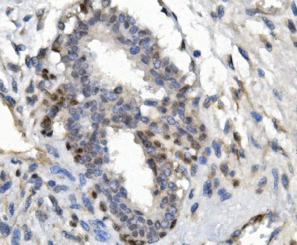
| Sample | Paraffin-embedded human mammary cancer tissue; fixative not specified (datasheet A00723) |
| Fixation | Image fixative and duration unreported (datasheet A00723); verify before use. |
| Sectioning | 4–5 µm sections on charged slides (standard) |
| Deparaffinisation | Xylene, graded ethanol series to water (standard) |
| Antigen retrieval | Heat retrieval: EDTA pH 8.0 (datasheet A00723); 20 min, 95–100 °C (standard) |
| Peroxidase block | 3% H2O2, 10 min, room temperature (standard) |
| Blocking | 10% goat serum (datasheet A00723) |
| Primary antibody | Rabbit anti-FOXP1, 2μg/ml (datasheet A00723) |
| Primary incubation | Overnight at 4 °C (datasheet A00723) |
| Detection | Streptavidin-biotin complex (SABC), DAB chromogen (datasheet A00723) |
| Counterstain | Hematoxylin, blue, dehydrate and mount (standard) |
| Expected result | FOXP1-positive staining in endocrine cells of appendix (HPA tissue IHC: High). HPA tissue profile: Nuclear expression in most cells. No signal in the no-primary control. |
FOXP1 is a nuclear protein, absent from the nucleolus (UniProt Q9H334). In paraffin sections, expect nuclear staining in many cell types, including respiratory epithelium, breast glands, neurons, and hematopoietic cells (HPA tissue IHC: nuclear expression in most cells; High in the listed populations). Interpret intensity cautiously: HPA rates tissue IHC reliability as Supported, with medium agreement between staining and RNA data and a warning that the antibody signal may include protein from another gene (HPA tissue IHC).
| Distinct nuclear staining in expected cells, with little signal outside nuclei. | This matches FOXP1 localisation (UniProt Q9H334: nucleus; HPA tissue IHC: nuclear expression in most cells). Nucleoplasmic staining with a spared nucleolus is also consistent with the independent ICC-IF location (HPA subcellular: nucleoplasm; UniProt Q9H334: not found in nucleolus). Nuclear placement supports the interpretation, but staining alone does not establish that every positive nucleus contains FOXP1 (HPA tissue IHC: antibody signal may include protein from another gene). |
| Predominantly cytoplasmic, membrane-like, or nucleolar staining. | These compartments conflict with the expected nuclear pattern (UniProt Q9H334: nucleus, not nucleolus; HPA tissue IHC: nuclear expression). FOXP1 has no transmembrane segment (UniProt Q9H334 topology). Treat a dominant signal there as suspect; review the counterstain, morphology, detection controls, and antibody validation before scoring it as FOXP1 (general IHC practice). |
| Signal is concentrated in an unexpected cell population while expected high-staining cells are blank. | Compare cell identity on the same section before calling the result positive: HPA reports High staining in bronchial respiratory epithelial cells, breast glandular cells, bone marrow hematopoietic cells, and several listed endocrine or neuronal populations (HPA tissue IHC). An isolated pattern elsewhere could reflect cross-reactivity or endogenous detection activity (general IHC practice), especially given HPA's warning that the antibody signal may include protein from another gene (HPA tissue IHC). |
| Diffuse colour covers nuclei and surrounding tissue without clear cell boundaries. | This obscures the nuclear pattern needed for interpretation (UniProt Q9H334: nucleus; HPA tissue IHC: nuclear expression). Excess primary or detection reagent, incomplete blocking, or inadequate washing can cause nonspecific background (general IHC practice). Reassess controls and staining conditions before assigning positive cells; diffuse colour has no reliable compartment or cell-type information (general IHC practice). |
| No nuclear signal appears in a section chosen for a reported High-staining cell population. | First confirm that the relevant cells are present and preserved on the slide; a tissue label alone does not ensure the sampled field contains them (general IHC practice). HPA reports High staining in specific cells, such as bone marrow hematopoietic cells and bronchial respiratory epithelial cells (HPA tissue IHC). If those cells are present but blank, check the staining run and controls before interpreting the absence biologically (general IHC practice). |
| Cell population and comparison tissue | HPA describes nuclear expression in most cells and low tissue specificity at the RNA level, while naming particular High and Low staining populations (HPA tissue IHC). Judge intensity within identified cell populations; a Low designation, such as hippocampal neurons, is not a universal negative control (HPA tissue IHC). |
| Antibody validation and specificity | Two listed antibodies have Supported IHC status, but HPA describes only medium agreement with RNA data and cautions that antibody signal may include protein from another gene (HPA antibodies; HPA tissue IHC). Nuclear staining is therefore compatible with FOXP1, not proof of molecular specificity (HPA tissue IHC; general IHC practice). |
| Isoform and epitope coverage | UniProt lists seven FOXP1 isoforms and reports isoform 8 specifically in embryonic stem cells (UniProt Q9H334). No epitope or isoform coverage is supplied for the IHC antibody here; a stain cannot distinguish those isoforms without additional evidence (general IHC practice). |
| Processing and compartment | FOXP1 is annotated as a nuclear protein with no signal peptide, propeptide, or transmembrane segment (UniProt Q9H334). Those features support a nuclear interpretation; they do not establish a particular retrieval method or a fixation sensitivity (UniProt Q9H334; general IHC practice). Target-specific fixation effects are unreported in the supplied evidence. |
| Situation | Likely cause | Next action |
|---|---|---|
| High-staining reference cells show no nuclear signal. | The expected cells may be absent from the sampled field, or a reagent, retrieval, or detection step may have failed (HPA tissue IHC: cell-specific High staining; general IHC practice). The supplied sources give no FOXP1-specific fixation or retrieval sensitivity. | Verify the cell population on the counterstained section, then review a run control, reagent preparation, and the established IHC-P procedure for the catalog antibody (general IHC practice). Do not infer biological absence from one blank section. |
| Colour is mainly cytoplasmic or outlines cell membranes. | The distribution disagrees with nuclear FOXP1 and its lack of a transmembrane segment (UniProt Q9H334; HPA tissue IHC). Nonspecific binding or detection artefact is possible (general IHC practice). | Inspect morphology and the counterstain, compare the expected nuclear compartment, and check appropriate staining controls (general IHC practice). Withhold a FOXP1-positive call if the abnormal compartment dominates. |
| Scattered deposits or widespread colour make nuclei hard to score. | Background from blocking, reagent concentration, washing, or the chromogenic detection system can obscure cellular localisation (general IHC practice). | Review a no-primary control and the established blocking, dilution, washing, and detection steps (general IHC practice). Score only interpretable cells with nuclear signal; do not count diffuse deposits as positive. |
| An unexpected population stains strongly while the expected population is blank. | Cell identity may have been misread, or the signal may arise from cross-reactivity or endogenous detection activity (general IHC practice). HPA specifically cautions that antibody signal may include protein from another gene (HPA tissue IHC). | Recheck the cell population against the counterstain and HPA's cell-specific examples, then compare appropriate detection controls and independent antibody evidence where available (HPA tissue IHC; general IHC practice). |
| A purported negative tissue contains some nuclear-positive cells. | FOXP1 has broad reported nuclear expression, and HPA's Low entries describe particular cell populations rather than absence throughout a tissue (HPA tissue IHC). | Record which cells stain and compare intensity with a reported High population processed in the same run (HPA tissue IHC; general IHC practice). Avoid treating a Low population as an absolute negative control. |
| IF/ICC: what pattern should be expected? | The separate ICC-IF evidence places FOXP1 in the nucleoplasm, consistent with the nuclear IHC pattern (HPA subcellular: supported nucleoplasm; HPA tissue IHC: nuclear expression). | Look for nucleoplasmic signal aligned with the nuclear counterstain and assess background with appropriate IF controls (HPA subcellular; general IF practice). Interpret IF/ICC using its separate guide; this IHC section supplies no IF protocol. |



Comprehensive Human Protein Atlas IHC scoring per tissue (reliability: Supported — Medium consistency between antibody staining and RNA expression data. Caution, targets protein from more than one gene.). Rows are taken directly from the HPA tissue chart — click any row's HPA link to view the source.
| Tissue | Cell type | Level | Evidence | Source |
|---|---|---|---|---|
| Appendix | Endocrine cells | High | Protein (IHC) | HPA → |
| Bone marrow | Hematopoietic cells | High | Protein (IHC) | HPA → |
| Breast | Glandular cells | High | Protein (IHC) | HPA → |
| Bronchus | Respiratory epithelial cells | High | Protein (IHC) | HPA → |
| Caudate | Neuronal cells | High | Protein (IHC) | HPA → |
| Tissue | Cell type | Level | Evidence | Source |
|---|---|---|---|---|
| None in HPA: FOXP1 is detected in all 45 scored tissues. Use a no-primary (secondary-only) and an isotype control instead. | ||||
Use nuclear staining, matched controls and the A00723 paraffin-section example to troubleshoot FOXP1 chromogenic IHC; assess IF/ICC separately.
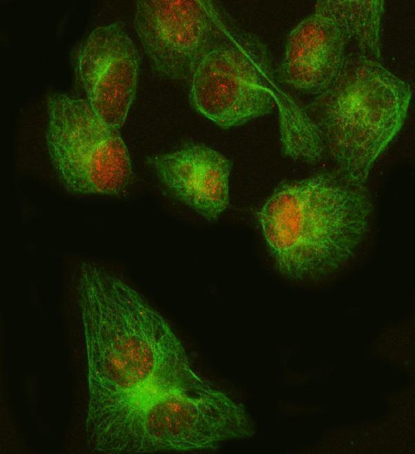
The catalog includes FOXP1 antibodies with IHC images from human paraffin sections and an IF image from HeLa cells; listed reactivity also covers mouse, rat, and, for A00723, monkey (catalog applications/reactivity; image captions).
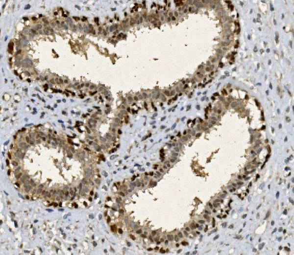
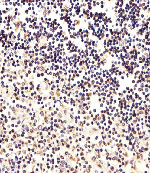
A00723 shows IHC in human mammary and rectal cancer sections, while A00723-1 shows IHC in human thymus and lung sections (A00723 and A00723-1 image captions). PA1593 shows IHC in human rectal cancer tissue, and M00723 shows IF/ICC in HeLa cells (PA1593 and M00723 image captions).
Which to pick: For tissue IHC, A00723 has paraffin-section images with EDTA retrieval, while A00723-1 has a paraffin-section, paraffin-embedded lung image; the fixative is unreported for A00723 and for A00723-1’s thymus image (A00723 and A00723-1 image captions). PA1593 is another IHC option with a human rectal cancer image, but its fixative is unreported; choose M00723 for IF/ICC based on its HeLa IF image and listed IF/ICC applications (PA1593 and M00723 image captions; M00723 catalog applications). For cross-species planning, A00723 lists human, monkey, mouse, and rat reactivity and IHC/IF applications, while the monoclonal M00723 lists human, mouse, and rat reactivity and IF/ICC applications; the pictured IHC and IF results are human only (catalog reactivity/applications; image captions). The selected A00723 tissue-IHC caption documents paraffin sections, but does not specify the fixative (selected-SKU IHC image A00723).