FUNDC1 / FUN14 domain-containing protein 1 · IHC design guide

Design Immunohistochemistry for FUNDC1

Plan chromogenic FUNDC1 IHC in paraffin sections with the catalog antibody at 2–5 μg/ml (datasheet A08688-2). Compare high staining in caudate neuronal cells with undetected staining in lymph node germinal center cells, while accounting for uncertain tissue IHC reliability (HPA tissue IHC).

Evidence assembled Oct 2026 · For research use; verify linked source records and product datasheet before use
Immunohistochemistry protocol sheet for FUNDC1 (IHC for FUNDC1): expected localisation Cytoplasmic tissue staining (HPA tissue IHC); outer mitochondrial membrane (UniProt), antibody A08688-2, validated IHC image, and IHC protocol steps
Printable FUNDC1 IHC protocol sheet — expected localisation Cytoplasmic tissue staining (HPA tissue IHC); outer mitochondrial membrane (UniProt), antibody A08688-2, controls and protocol steps. Open the full FUNDC1 IHC guide →

FUNDC1 Immunohistochemistry Experimental Design Guide

Expected localisation, validated protocols, controls and antibodies — the at-a-glance facts below, then the full design guide.

Must know before staining
Expected localisation Cytoplasmic tissue staining (HPA tissue IHC); outer mitochondrial membrane (UniProt)
Staining pattern Ubiquitous cytoplasmic staining across cell types (HPA tissue IHC)
Antigen retrieval EDTA pH 8.0 HIER, heat-mediated (datasheet A08688-2)
Positive control ⓘ Caudate+4 more · see all
Negative control ⓘ Liver+4 more · see all
Important caveats
Reasons your staining may differ from the expected pattern.
Fixation Keep fixation consistent across paraffin sections. (standard IHC practice; not target-specific)
Caveat Tissue staining has uncertain reliability (HPA tissue IHC)
Regulation Intensity regulation unreported (UniProt)
Isoform / epitope No annotated isoforms or cleavage; chain 1–155 (UniProt)
Section 1

Recommended FUNDC1 IHC & IF Protocols

The catalog antibody’s IHC-P protocol is accompanied by 4 published FUNDC1 IHC protocols (PMC10951295; PMC8989429; PMC6442990; PMC11632979).

Recommended immunohistochemistry (IHC-P) protocol parameters
SampleParaffin-embedded human cervical cancer tissue; fixative not specified (datasheet A08688-2)
FixationImage fixative and duration unreported (datasheet A08688-2); verify before use.
Sectioning4–5 µm sections on charged slides (standard)
DeparaffinisationXylene, graded ethanol series to water (standard)
Antigen retrievalHeat retrieval: EDTA pH 8.0 (datasheet A08688-2); 20 min, 95–100 °C (standard)
Peroxidase block3% H2O2, 10 min, room temperature (standard)
Blocking10% goat serum (datasheet A08688-2)
Primary antibodyRabbit anti-FUNDC1, 2-5 μg/ml (datasheet A08688-2)
Primary incubationOvernight at 4 °C (datasheet A08688-2)
DetectionHRP-conjugated secondary, DAB chromogen (datasheet A08688-2)
CounterstainHematoxylin, blue, dehydrate and mount (standard)
Expected resultFUNDC1-positive staining in neuronal cells of caudate (HPA tissue IHC: High). HPA tissue profile: Ubiquitous cytoplasmic expression. No signal in the no-primary control.
💡Decision noteStart with heat-mediated EDTA retrieval at pH 8.0 for the catalog antibody (datasheet A08688-2); published protocols also report citrate retrieval (PMC8989429; PMC6442990).
Section 2

What Is the Expected FUNDC1 Staining Pattern?

FUNDC1 is an integral mitochondrial outer-membrane protein with three transmembrane segments and cytoplasmic-facing regions (UniProt Q8IVP5 topology). In paraffin-section IHC, expect cytoplasmic staining, particularly in caudate and hippocampal neurons and cerebellar Purkinje cells (HPA: High in these cells; ubiquitous cytoplasmic profile). Interpret this as a provisional pattern: HPA rates tissue IHC reliability Uncertain, and antibody HPA038773 has Uncertain IHC validation (HPA).

What am I looking at on my slide?
Cytoplasmic staining is stronger in caudate or hippocampal neurons and cerebellar Purkinje cells than in nearby weakly stained cells.This agrees with the reported High staining in those cell types (HPA). FUNDC1's mitochondrial outer-membrane location supports a cytoplasmic distribution (UniProt Q8IVP5); chromogenic IHC alone cannot prove mitochondrial colocalisation (general IHC practice).
Predominantly nuclear or extracellular deposits replace the expected cytoplasmic pattern.Treat this as a localisation mismatch: UniProt places FUNDC1 in the mitochondrial outer membrane (UniProt Q8IVP5). Review morphology and controls before scoring; nonspecific binding or detection artefact can mimic a positive stain (general IHC practice).
Strong staining appears in cholangiocytes, germinal-center cells or oral squamous epithelium.These specific cells were Not detected in the respective liver, lymph-node and oral-mucosa HPA sections (HPA). Check for cross-reactivity or endogenous detection activity (general IHC practice), while allowing for HPA's Uncertain reliability rather than declaring every discordant sample false (HPA).
Uniform chromogen covers tissue edges, empty spaces and unrelated cell types.A diffuse, anatomy-independent deposit does not resemble HPA's cell-resolved cytoplasmic profile (HPA). Examine the no-primary control, wash steps and detection background before interpreting intensity (general IHC practice).
No stain is visible in a caudate or cerebellar section processed with the test samples.HPA reports High neuronal or Purkinje-cell staining in these tissues, making them useful reference sections (HPA). An absent signal warrants a run-control check; the Uncertain HPA validation prevents treating one negative run as proof of no FUNDC1 expression (HPA).
💡Expected FUNDC1 appearanceA plausible positive is cell-resolved cytoplasmic chromogen, strongest in caudate or hippocampal neurons or cerebellar Purkinje cells (HPA: High); dominant nuclear or anatomy-independent staining is suspect given FUNDC1's outer-membrane location (UniProt Q8IVP5; general IHC practice).
How each factor affects the staining
Cell-specific reference patternUse named cells when comparing sections: adipocytes and bronchial respiratory epithelial cells were Medium, while caudate neurons and Purkinje cells were High (HPA). A tissue-level positive or negative label cannot substitute for identifying its stained cell population (general IHC practice).
Topology and epitope accessFUNDC1 has three transmembrane segments and cytoplasmic-facing residues 1–47 and 96–133 (UniProt Q8IVP5 topology). The supplied sources do not identify this antibody's epitope, so topology alone cannot prescribe antigen retrieval or predict epitope accessibility.
Evidence strengthHPA calls the tissue profile ubiquitous cytoplasmic but rates its reliability Uncertain; HPA038773 also has Uncertain IHC validation (HPA). Use morphology and controls to assess a result, especially when a specimen differs from the reported cell-specific pattern (general IHC practice).
Processing and isoformsUniProt lists one 1–155 chain, no signal peptide or propeptide, and no annotated isoforms (UniProt Q8IVP5). The supplied record therefore offers no processing-based explanation for a changed tissue pattern; do not infer shedding or isoform-specific staining.
IF/ICC Q: What pattern is established?A: UniProt supports mitochondrial outer-membrane localisation (UniProt Q8IVP5). HPA supplies no ICC-IF images or main subcellular location and lists no ICC validation for HPA038773 (HPA); an observed IF pattern therefore needs its own controls and colocalisation assessment (general IF practice).
Why is my staining missing, weak or wrong?
SituationLikely causeNext action
Known High reference cells show no chromogen.The run may have failed, or the antibody may not yield a detectable signal under the current IHC conditions (general IHC practice); HPA's validation is Uncertain (HPA).Check the positive reference section, no-primary control and detection reagents together; review the antibody's documented IHC-P conditions before repeating (general IHC practice).
Signal is mainly nuclear.This conflicts with FUNDC1's mitochondrial outer-membrane localisation (UniProt Q8IVP5); nonspecific staining or misplaced precipitate is possible (general IHC practice).Compare nuclear counterstain and cytoplasmic boundaries, then inspect the no-primary control before assigning a positive score (general IHC practice).
A purported negative control cell population stains strongly.HPA reports Not detected in cholangiocytes, lymph-node germinal-center cells and oral squamous epithelium (HPA); cross-reactivity or endogenous detection activity may explain discordance (general IHC practice).Confirm cell identity and compare no-primary and detection controls; report persistent discordance with HPA's Uncertain reliability (HPA; general IHC practice).
Brown haze obscures cell boundaries.Diffuse deposit may reflect nonspecific antibody binding, endogenous detection activity or inadequate washing (general IHC practice). It cannot establish the cell-resolved cytoplasmic profile described by HPA (HPA).Inspect no-primary and reagent controls; review blocking, washes and detection exposure using the same tissue (general IHC practice).
Only some cells in an otherwise positive tissue stain.HPA levels are assigned to named cell populations, with Medium adipocytes and High Purkinje cells among its examples (HPA). Mixed tissue does not imply equal staining in every cell (general IHC practice).Score the specified cell population and its cytoplasmic compartment separately; record the sampled tissue and cell type (HPA; general IHC practice).
A changed antigen-retrieval condition changes intensity.Retrieval can alter immunostaining in paraffin sections (general IHC practice); no supplied source establishes FUNDC1-specific fixation sensitivity or an optimal retrieval condition.Compare conditions using the same reference tissue and controls, document the method, and avoid attributing the change to FUNDC1-specific epitope masking (general IHC practice).

Sample controls for FUNDC1 IHC & IF

🧪Run cerebellum first and look for staining in Purkinje cells (HPA: High in Purkinje cells); use oral mucosa squamous epithelial cells as the negative tissue comparison (HPA: Not detected in squamous epithelial cells). On the cerebellum slide, unstained neighboring cells can help assess background, but their FUNDC1-negative status is not established by the supplied HPA rows (HPA: High in Purkinje cells).
Positive control tissue: Caudate (Neuronal cells, HPA High)
Negative control tissue: Liver (HPA Not detected)
ICC-IF cell lines (HPA subcellular resource): HPA carries no ICC-IF cell line for FUNDC1; derive a cell-line control from the positive tissue's cell type (Neuronal cells) and confirm it by RNA or western blot first.
Technical controls: Include a no-primary, secondary-only control; a matched nonimmune rabbit IgG isotype control; and, where available, a FUNDC1 knockout specimen as a biological specificity control (selected-SKU IHC caption: rabbit primary antibody). For chromogenic cerebellum IHC, block endogenous peroxidase and distinguish DAB signal from neuronal pigment (standard IHC practice).
⚠️Feasibility: A target-specific fixation window and fixation effect are unreported, and the selected-SKU tissue-IHC caption does not state the fixative (selected-SKU IHC caption: fixative not stated). The reported paraffin-section staining used heat retrieval in EDTA at pH 8.0, so retrieval should be checked in the chosen tissue (selected-SKU IHC caption: EDTA heat retrieval); the caption does not establish that this condition is required for all specimens. Frozen-section suitability and whether IF is easier are unreported in the supplied application evidence, and no HPA ICC-IF image-bearing cell lines are listed (HPA subcellular payload: cell_lines_with_icc_if_images empty).

HPA tissue IHC evidence for FUNDC1

Comprehensive Human Protein Atlas IHC scoring per tissue (reliability: Uncertain — Medium consistency between antibody staining and RNA expression data. Pending external verification.). Rows are taken directly from the HPA tissue chart — click any row's HPA link to view the source.

Positive expression · recommended positive controls

TissueCell typeLevelEvidenceSource
Caudate Neuronal cells High Protein (IHC) HPA →
Cerebellum Purkinje cells High Protein (IHC) HPA →
Hippocampus Neuronal cells High Protein (IHC) HPA →
Adipose tissue Adipocytes Medium Protein (IHC) HPA →
Adrenal gland Glandular cells Medium Protein (IHC) HPA →

Undetected expression · recommended negative controls

TissueCell typeLevelEvidenceSource
Liver Cholangiocytes Not detected Protein (IHC) HPA →
Lymph node Germinal center cells Not detected Protein (IHC) HPA →
Oral mucosa Squamous epithelial cells Not detected Protein (IHC) HPA →
Ovary Ovarian stroma cells Not detected Protein (IHC) HPA →
Spleen Cells in red pulp Not detected Protein (IHC) HPA →
Section 3

Advanced FUNDC1 IHC Tips

Use the catalog antibody’s paraffin-section IHC conditions as a starting point, then assess cell-level staining against FUNDC1’s mitochondrial location and appropriate controls.

Which antigen retrieval conditions should I start with for FUNDC1 IHC?
Start with heat-mediated antigen retrieval in EDTA buffer at pH 8.0 for paraffin sections (datasheet A08688-2). The selected tissue-IHC image used this condition before staining with 2 μg/ml antibody overnight at 4°C (caption A08688-2). If staining is weak, vary heating duration on adjacent sections while keeping buffer and detection conditions fixed, and check tissue integrity after retrieval (standard IHC practice). Include a no-primary control to identify signal from the detection system, and compare candidate conditions at the same imaging and scoring settings (standard IHC practice).
How should I troubleshoot weak staining when fixation details are missing?
The selected paraffin-section caption does not state a fixative, so FUNDC1-specific sensitivity to fixation is unknown (caption A08688-2). Record the fixative, fixation duration and processing history for each specimen before comparing staining across blocks (standard IHC practice). On adjacent sections, keep the documented EDTA pH 8.0 retrieval and 2 μg/ml antibody concentration constant while assessing whether staining varies with processing history (caption A08688-2; standard IHC practice). Interpret any difference as a specimen-processing association until matched material and appropriate controls distinguish fixation effects from differences in cellular composition or detection (standard IHC practice).
What staining pattern is plausible for FUNDC1 in paraffin sections?
Expect cellular staining compatible with a mitochondrial outer-membrane protein, predominantly within the cytoplasmic compartment at light-microscope resolution (UniProt Q8IVP5 localisation; standard IHC practice). Three transmembrane segments anchor FUNDC1, while residues 1–47 and 96–133 face the cytoplasm (UniProt Q8IVP5 topology). HPA describes ubiquitous cytoplasmic tissue staining, but rates its tissue-IHC reliability uncertain, so that pattern alone cannot establish antibody specificity (HPA tissue IHC). Compare staining with cell morphology and a validated mitochondrial marker on matched material; diffuse nuclear-only signal calls for closer control review (UniProt Q8IVP5 localisation; standard IHC practice).
Could epitope position explain inconsistent FUNDC1 IHC staining?
FUNDC1 has no annotated isoforms in the supplied record, so an isoform-specific explanation is unsupported here (UniProt Q8IVP5 record). Its three membrane-spanning segments separate cytoplasmic residues 1–47 and 96–133 from short intermembrane regions, making documented epitope position useful when assessing accessibility (UniProt Q8IVP5 topology; standard IHC practice). Residues 13, 17 and 18 are annotated phosphorylation sites, but the catalog antibody’s epitope and modification sensitivity are unspecified (UniProt Q8IVP5 modified residues; caption A08688-2). Ask for epitope information and compare controlled retrieval conditions before attributing staining differences to phosphorylation or membrane orientation (standard IHC practice).
How can I assess FUNDC1 by multiplex immunofluorescence?
Treat IF performance as unestablished by the supplied catalog evidence, which shows a paraffin-section chromogenic IHC application (caption A08688-2). For a cell-type check, multiplex FUNDC1 with a validated neuronal marker in caudate sections, where HPA reports high staining in neuronal cells but uncertain overall IHC reliability (HPA tissue IHC). Choose a far-red channel when tissue autofluorescence affects shorter wavelengths, and examine single-stain and no-primary controls for bleed-through and background (standard IF practice). Select permeabilisation only after identifying the antibody epitope: cytoplasmic regions 1–47 and 96–133 differ in accessibility from the intermembrane regions (UniProt Q8IVP5 topology; standard IF practice).
How do I separate FUNDC1 signal from chromogenic background?
The selected IHC procedure used 10% goat serum, a peroxidase-conjugated secondary antibody and DAB development (caption A08688-2). Include a no-primary section to assess secondary or detection-system staining, and use a peroxidase block when endogenous enzyme activity contributes to DAB signal (standard IHC practice). Compare adjacent sections at a fixed 2 μg/ml primary concentration before changing blocking or wash conditions, since that is the reported concentration for this antibody (caption A08688-2; standard IHC practice). Check whether apparent positivity follows tissue edges, damaged areas or precipitate rather than intact cell cytoplasm (UniProt Q8IVP5 localisation; standard IHC practice).
How should I quantify FUNDC1 staining across sections? ⚠ ANSWER MARKED FOR VERIFICATION
Define the cell population and tissue region before scoring, then record the percentage of positive cells and an intensity-based H-score using consistent thresholds (standard IHC practice). Report positive-cell counts per mm² when cellular density differs, and normalise regional counts to viable tissue area or the number of eligible cells (standard IHC practice). Keep section processing, DAB development, imaging and threshold settings matched across comparison groups, with blinded scoring when feasible (standard IHC practice). Because HPA rates its FUNDC1 tissue-IHC reliability uncertain, treat staining scores as assay-dependent until specificity controls support the measured signal (HPA tissue IHC; standard IHC practice).
Which findings support true FUNDC1 staining rather than artefact?
A credible pattern should occur in intact cell cytoplasm and be compatible with FUNDC1’s mitochondrial outer-membrane location (UniProt Q8IVP5 localisation; standard IHC practice). HPA reports high staining in caudate neuronal cells and cerebellar Purkinje cells, but its overall tissue-IHC reliability is uncertain; use these observations as context rather than proof (HPA tissue IHC). Reassess isolated nuclear-only staining, strong section-edge signal, necrotic areas and DAB deposits, and inspect a no-primary control for endogenous or detection-system signal (UniProt Q8IVP5 localisation; standard IHC practice). Compare cell-level morphology and matched mitochondrial-marker staining before interpreting differences in intensity as changes in FUNDC1 abundance (standard IHC practice).
Boster reagents

Best FUNDC1 / FUN14 domain-containing protein 1 IHC Antibodies

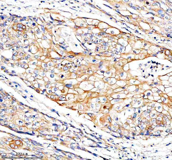
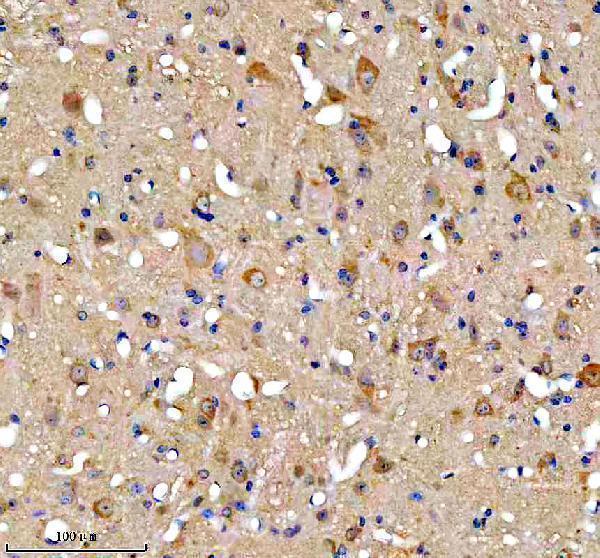
A08688-2 has IHC images from human cancer and mouse and rat midbrain paraffin sections (catalog image captions); no IF/ICC data are supplied (catalog: applications and image captions).

Real IHC data IHC analysis of FUNDC1 using anti-FUNDC1 antibody (A08688-2). FUNDC1 was detected in a paraffin-embedded section of human cervical cancer tissue. Heat mediated antigen retrieval was performed in EDTA buffer (pH 8.0, epitope retrieval solution). The tissue section was blocked with 10% goat serum. The tissue section was then incubated with 2 μg/ml rabbit anti-FUNDC1 Antibody (A08688-2) overnight at 4°C. Peroxidase Conjugated Goat Anti-rabbit IgG was used as secondary antibody and incubated for 30 minutes at 37°C. The tissue section was developed using HRP Conjugated Rabbit IgG Super Vision Assay Kit (Catalog # SV0002) with DAB as the chromogen.
Anti-FUNDC1 Antibody ®
Cat # A08688-2

A08688-2 will render with its IHC image of a human cervical cancer paraffin section (catalog: figure caption). Additional IHC captions show human pancreas cancer and mouse and rat midbrain paraffin sections; the catalog lists Human, Mouse and Rat reactivity (catalog: image captions and reactivity).

Which to pick: Choose A08688-2 for paraffin-section tissue IHC, starting within 2–5 μg/ml; its IHC caption uses 2 μg/ml after EDTA pH 8.0 retrieval (datasheet: IHC dilution; A08688-2 figure caption). It is the listed cross-species option for Human, Mouse and Rat, with paraffin-section images in each species; the captions do not report a fixative, and clonality is unreported (catalog: reactivity, image captions and clone field). No listed SKU is validated for IF/ICC (catalog: applications and IF image captions).

Each figure is that product's own IHC / IF validation image from its datasheet.

References

  1. UniProt Consortium. UniProt entry Q8IVP5 (FUND1_HUMAN, FUN14 domain-containing protein 1).
  2. Human Protein Atlas. FUNDC1 tissue IHC expression (reliability: Uncertain).
  3. Human Protein Atlas. FUNDC1 subcellular location (ICC-IF): Highest expression in GAMG: 65.8 nTPM.
  4. Human Protein Atlas. FUNDC1 antibody validation summary (1 antibodies).
  5. Thread-structural microneedles loaded with engineered exosomes for annulus fibrosus repair by regulating mitophagy recovery and extracellular matrix homeostasis. Bioactive materials 2024 — PMC10951295.
  6. Hypoxia‑induced mitophagy regulates proliferation, migration and odontoblastic differentiation of human dental pulp cells through FUN14 domain‑containing 1. International journal of molecular medicine 2022 — PMC8989429.
  7. FUN14 domain-containing 1 promotes breast cancer proliferation and migration by activating calcium-NFATC1-BMI1 axis. EBioMedicine 2019 — PMC6442990.
  8. FUNDC1 predicts Poor Prognosis and promotes Progression and Chemoresistance in Endometrial Carcinoma. Journal of Cancer 2024 — PMC11632979.
  9. PubMed PMID:15772651 — UniProt-cited evidence.
  10. PubMed PMID:15489334 — UniProt-cited evidence.
  11. PubMed PMID:18669648 — UniProt-cited evidence.