This website uses cookies to ensure you get the best experience on our website.


- Table of Contents
Real validated FUNDC1 Western blot protocols, expected-band and isoform facts, troubleshooting for weak or shifted signal, and recommended anti-FUNDC1 WB antibodies. Everything you need to plan the experiment before you commit precious samples.

Expected bands, validated protocols, controls and antibodies — the at-a-glance facts below, then the full design guide.
| Expected band | ~17.2 kDa | |
| Observed band | ~17 kDa | |
| Gel | 12% (catalog A08688-2) | |
| Positive control | Caudate (IHC candidate; verify WB) +4 more | |
| Negative control | Liver (IHC candidate; verify WB) |
| PTM | Phosphorylated + Ubl conjugation | |
| Caveat | Phosphorylation-state controls | |
| Gene-set association | MSigDB C7 membership | |
| Isoform | 1 isoform(s) |
The A08688-2 protocol combines labelled catalog values with standard starting conditions. Published comparisons retain their own sample, reagent and detection scope.
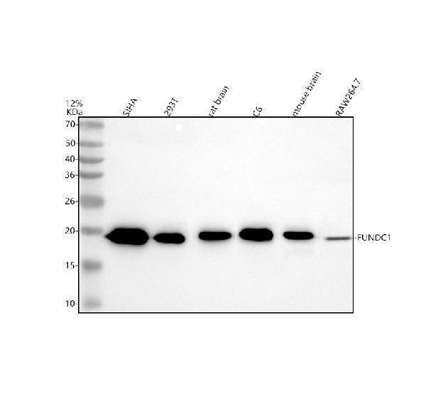
| Sample / lysate | human SiHa, human 293T, rat brain (catalog A08688-2) |
| Gel % | 12% (catalog A08688-2) |
| Load | 30 ug; reducing conditions (catalog A08688-2) |
| Transfer | a nitrocellulose membrane at 150 mA for 50-90 minutes (catalog A08688-2) |
| Membrane | nitrocellulose membrane (catalog A08688-2) |
| Blocking | 5% non-fat milk/TBS for 1.5 hour at RT (catalog A08688-2) |
| Primary antibody | A08688-2 · 0.5 μg/mL (catalog A08688-2) |
| Primary incubation | overnight at 4°C (catalog A08688-2) |
| Secondary antibody | goat anti-rabbit IgG-HRP, 1:5000 (catalog A08688-2) |
| Secondary incubation | 1.5 hour at RT (catalog A08688-2) |
| Wash | TBS-0.1%Tween 3 times with 5 minutes each (catalog A08688-2) |
| Detection | ECL (catalog A08688-2) |
FUNDC1 is predicted at 17.2 kDa and observed at approximately 17 kDa; the supplied evidence does not establish a cause for the small difference.
| Band near 17 kDa | Matches the observed FUNDC1 band and its 17.2 kDa predicted mass |
| Faint band near 17 kDa in whole-cell lysate | Mitochondrial outer membrane FUNDC1 may be poorly recovered from the sample |
| Little band near 17 kDa in a soluble fraction | FUNDC1 is a multi-pass mitochondrial outer membrane protein |
| Nearby doublet around 17 kDa | Could reflect phosphorylation states at Ser13, Ser17, or Tyr18, but distinct migration is unproven |
| UniProt predicted mass | Places full-length FUNDC1 near 17.2 kDa; the observed band is approximately 17 kDa |
| Ser13 phosphorylation by CK2 | May affect migration, but a visible size shift is not established |
| Ser17 phosphorylation by ULK1 | May affect migration, but a visible size shift is not established |
| Tyr18 phosphorylation by SRC | May affect migration, but a visible size shift is not established |
| Situation | Likely cause | Next action |
|---|---|---|
| No band in lysate | Poor recovery of multi-pass mitochondrial outer membrane FUNDC1 | Check membrane solubilization and a mitochondrial membrane marker |
| Band higher than expected | Incomplete solubilization or altered migration of the membrane protein is possible | Optimize solubilization and compare with the approximately 17 kDa control band |
| Band lower than expected | A smaller signal has no assigned FUNDC1 form in the supplied features | Check sample integrity and antibody specificity |
| Broad smear instead of sharp band | Incomplete solubilization of the multi-pass membrane protein is possible | Optimize extraction and sample preparation |
| Multiple bands | Phosphorylation states are possible, but distinct bands are unproven | Use phosphatase treatment and antibody specificity controls |
| Weak or no signal | Low recovery of mitochondrial outer membrane FUNDC1 | Check membrane enrichment and loading with a mitochondrial membrane marker |
Comprehensive Human Protein Atlas IHC scoring per tissue. Rows are taken directly from the HPA tissue chart — click any row's HPA link to view the source.
| Tissue | Cell type | Level | Evidence | Source |
|---|---|---|---|---|
| Caudate | neuronal cells | High | Protein (IHC) | HPA → |
| Cerebellum | Purkinje cells | High | Protein (IHC) | HPA → |
| Hippocampus | neuronal cells | High | Protein (IHC) | HPA → |
| Adipose tissue | adipocytes | Medium | Protein (IHC) | HPA → |
| Adrenal gland | glandular cells | Medium | Protein (IHC) | HPA → |
| Tissue | Cell type | Level | Evidence | Source |
|---|---|---|---|---|
| Liver | cholangiocytes | Not detected | Protein (IHC) | HPA → |
| Lymph node | germinal center cells | Not detected | Protein (IHC) | HPA → |
| Oral mucosa | squamous epithelial cells | Not detected | Protein (IHC) | HPA → |
| Ovary | ovarian stroma cells | Not detected | Protein (IHC) | HPA → |
| Spleen | cells in red pulp | Not detected | Protein (IHC) | HPA → |
Deeper troubleshooting and optimisation questions for FUNDC1, answered from its protein features.
Catalog antibodies with Western blot application and product-specific WB images. Evaluate suitability with the reported sample, controls and experimental conditions.
The catalog reports one anti-FUNDC1 antibody for Western blotting, A08688-2, with stated human, mouse and rat reactivity. Its WB image shows an approximately 17 kDa band in the human cell, rat brain and cell, and mouse brain and cell lysates named in the caption. No independent validation is supplied.
Which to pick: A08688-2 is the only listed option. Its WB image includes human SiHa and 293T, rat brain and C6, and mouse brain and RAW264.7 lysates, tested at 0.5 μg/mL primary antibody under the stated conditions. Match your sample and protocol to that evidence.