This website uses cookies to ensure you get the best experience on our website.


- Table of Contents
Plan FUT6 paraffin IHC around granular cytoplasmic staining in gastrointestinal tissue (HPA tissue IHC). Start the IHC-validated antibody at 1:100–1:200 (datasheet: A06862), and account for a secreted variant when interpreting tissue staining (HPA tissue IHC).

Expected localisation, validated protocols, controls and antibodies — the at-a-glance facts below, then the full design guide.
| Expected localisation | Granular cytoplasm in tissue (HPA tissue IHC) | |
| Staining pattern | Granular cytoplasm in enterocytes and glandular cells (HPA tissue IHC) | |
| Antigen retrieval | Citrate pH 6.0 HIER, 95–98 °C, 20 min (rule: cytoplasmic / membrane antigen) | |
| Positive control | Duodenum+4 more · see all | |
| Negative control | Adipose tissue+4 more · see all |
| Fixation | Keep fixation consistent across samples. (standard IHC practice; not target-specific) | |
| Caveat | A secreted variant may shift tissue staining (HPA tissue IHC) | |
| Regulation | Regulation not specified (UniProt) | |
| Isoform / epitope | 2 isoforms; verify antibody epitope coverage (UniProt) |
The catalog antibody IHC-P protocol is followed by published FUT6 staining protocols for prostate biopsies and mouse tumor sections (PMC3699712; PMC3847326; PMC7811455).
| Sample | Paraffin-embedded human liver tissue; fixative not specified (datasheet A06862) |
| Fixation | Image fixative and duration unreported (datasheet A06862); verify before use. |
| Sectioning | 4–5 µm sections on charged slides (standard) |
| Deparaffinisation | Xylene, graded ethanol series to water (standard) |
| Antigen retrieval | Heat-induced epitope retrieval in citrate buffer, pH 6.0, 20 min at 95–98 °C (standard rule: cytoplasmic / membrane antigen) |
| Peroxidase block | 3% H2O2, 10 min, room temperature (standard) |
| Blocking | 10% normal serum of the secondary host, 30 min, room temperature (standard) |
| Primary antibody | Rabbit anti-FUT6, 1:100-1:200 (datasheet A06862) |
| Primary incubation | Overnight at 4 °C (standard) |
| Detection | HRP-polymer secondary, DAB chromogen 5–10 min (standard) |
| Counterstain | Hematoxylin, blue, dehydrate and mount (standard) |
| Expected result | FUT6-positive staining in enterocytes - Microvilli of duodenum (HPA tissue IHC: High). HPA tissue profile: Granular cytoplasmic expression in gastrointestinal tract, gallbladder, seminal vesicle and cervix. No signal in the no-primary control. |
FUT6 is a Golgi stack membrane protein with a lumenal catalytic region and a reported secreted form (UniProt P51993 topology; UniProt P51993 subcellular location). In paraffin section IHC, expect granular cytoplasmic staining in gastrointestinal cells and strong staining at duodenal enterocyte microvilli (HPA tissue IHC). HPA rates its tissue IHC profile Approved, with medium consistency against RNA and a caution that antibodies may detect proteins from more than one gene (HPA tissue IHC).
| Granular cytoplasmic staining in gastrointestinal cells, with strong duodenal enterocyte microvilli staining (HPA tissue IHC). | This matches HPA’s tissue profile. Supported Golgi localization gives the cytoplasmic granules a plausible compartment, while the observed microvilli pattern should be recorded separately from Golgi staining (HPA tissue IHC; HPA ICC-IF; UniProt P51993 subcellular location). |
| Predominantly nuclear staining, without the expected gastrointestinal cytoplasmic pattern. | A nuclear-dominant pattern is unsupported by the Golgi localization records (UniProt P51993 subcellular location; HPA ICC-IF). Check the no-primary control and staining specificity before scoring it as FUT6; the observation alone does not identify the artefact (general IHC practice). |
| Prominent staining in a cell population HPA reports as not detected, such as adipocytes in adipose tissue. | Treat this as discordant with the reported IHC pattern, not automatic proof of cross-reactivity (HPA tissue IHC). Compare morphology and controls; off-target binding or endogenous detection activity are possible explanations in chromogenic IHC (general IHC practice). |
| Diffuse color covers cells and surrounding tissue, obscuring the granular pattern. | That appearance is difficult to score against HPA’s granular cytoplasmic profile (HPA tissue IHC). Uneven blocking, residual detection reagent or excessive chromogen development can create background; inspect controls and the detection workflow (general IHC practice). |
| No signal in duodenal enterocytes on a run expected to show FUT6. | HPA reports high staining at duodenal enterocyte microvilli, so a blank result warrants a run check (HPA tissue IHC). Confirm tissue preservation and morphology, then review retrieval, antibody dilution and detection controls; none has a supplied FUT6-specific setting (general IHC practice). |
| Cell and compartment choice | Duodenal enterocyte microvilli are High; gallbladder glandular cells and colonic endocrine cells are Medium (HPA tissue IHC). Golgi localization is supported independently (HPA ICC-IF; UniProt P51993). |
| Antibody interpretation | HPA calls the tissue IHC profile Approved, with medium RNA consistency and a multiple-gene detection caution; its listed IHC antibody is Approved, not Enhanced (HPA tissue IHC; HPA antibodies). |
| Membrane topology and epitope | Residues 15–34 span the membrane; residues 35–359 are lumenal (UniProt P51993 topology). Epitope location is not supplied, so topology alone cannot specify retrieval or staining intensity. |
| Secreted form | UniProt reports both membrane-bound Golgi FUT6 and a secreted form (UniProt P51993 subcellular location). Extracellular color needs cautious interpretation because the tissue IHC profile does not establish its source (HPA tissue IHC). |
| Variants and glycosylation | Two isoforms and glycosylation sites at residues 46, 91, 153 and 184 are annotated (UniProt P51993). Their effects on this antibody’s paraffin section staining are unreported. |
| RNA versus protein | Esophagus is RNA-enhanced, yet its squamous epithelial cells are reported Not detected by IHC (HPA tissue IHC). Choose and score controls using the protein staining record, with HPA’s RNA consistency caveat (HPA tissue IHC). |
| IF/ICC: what pattern is supported? | Golgi localization is supported in ICC-IF images from A-431, Hep-G2 and U2OS (HPA ICC-IF). This is a localization reference, not an IHC-P protocol or evidence of identical intensity in sections. |
| Situation | Likely cause | Next action |
|---|---|---|
| Duodenum is blank. | A run or reagent problem is possible because duodenal enterocyte microvilli are reported High (HPA tissue IHC). | Inspect morphology and a run control; review retrieval, antibody dilution and detection in the catalog antibody’s IHC-P protocol (general IHC practice). |
| Granular cytoplasmic signal is weak but tissue morphology is intact. | The sampled cells may differ from the stronger reference: HPA reports Medium staining in several listed gastrointestinal cell populations and High at duodenal microvilli (HPA tissue IHC). | Confirm the cell type and compare with a duodenum positive control before changing the IHC workflow (HPA tissue IHC; general IHC practice). |
| Nuclei dominate the stain. | That distribution conflicts with supported Golgi localization; its precise cause cannot be assigned from appearance alone (HPA ICC-IF; UniProt P51993). | Check no-primary and detection controls, then verify whether cytoplasmic granules are present on a positive tissue (general IHC practice; HPA tissue IHC). |
| A nominal negative tissue stains strongly. | HPA warns of detection from more than one gene; endogenous chromogenic activity or nonspecific binding can also give color (HPA tissue IHC; general IHC practice). | Compare the same cell type with HPA’s IHC record and review no-primary, blocking and detection controls before calling FUT6 positive (HPA tissue IHC; general IHC practice). |
| Color spreads uniformly across the section. | Diffuse deposit can hide the reported granular pattern; reagent background or excessive development is possible (HPA tissue IHC; general IHC practice). | Review blocking, washes and chromogen development against run controls, then score only localized cellular signal (general IHC practice). |
| RNA suggests a positive tissue, but IHC is negative. | RNA and protein patterns need not agree; HPA specifically reports RNA-enhanced esophagus with squamous epithelial IHC Not detected (HPA tissue IHC). | Use HPA’s cell-level protein pattern for the IHC comparison, and document the RNA/IHC discrepancy rather than treating RNA as a positive stain control (HPA tissue IHC). |


Comprehensive Human Protein Atlas IHC scoring per tissue (reliability: Approved — Medium consistency between antibody staining and RNA expression data. Caution, targets protein from more than one gene. At least one protein variant secreted, tissue location of RNA and protein might differ and correlation is complex.). Rows are taken directly from the HPA tissue chart — click any row's HPA link to view the source.
| Tissue | Cell type | Level | Evidence | Source |
|---|---|---|---|---|
| Duodenum | Enterocytes - Microvilli | High | Protein (IHC) | HPA → |
| Appendix | Endocrine cells | Medium | Protein (IHC) | HPA → |
| Colon | Endocrine cells | Medium | Protein (IHC) | HPA → |
| Gallbladder | Glandular cells | Medium | Protein (IHC) | HPA → |
| Rectum | Endocrine cells | Medium | Protein (IHC) | HPA → |
| Tissue | Cell type | Level | Evidence | Source |
|---|---|---|---|---|
| Adipose tissue | Adipocytes | Not detected | Protein (IHC) | HPA → |
| Adrenal gland | Glandular cells | Not detected | Protein (IHC) | HPA → |
| Bone marrow | Hematopoietic cells | Not detected | Protein (IHC) | HPA → |
| Esophagus | Squamous epithelial cells | Not detected | Protein (IHC) | HPA → |
| Hippocampus | Glial cells | Not detected | Protein (IHC) | HPA → |
Troubleshoot FUT6 staining by checking retrieval, compartment, cell type and controls together (UniProt P51993; HPA tissue IHC).
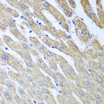
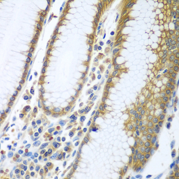
A06862 has IHC images of human paraffin-embedded liver and stomach (catalog image captions); no IF image is supplied (catalog IF images: none).
A06862 will render with its human liver paraffin-section IHC figure at 1:100 (catalog liver image caption). Its application list includes IHC and WB, and a separate caption reports human stomach paraffin-section IHC at 1:100 (catalog applications; catalog stomach image caption).
Which to pick: Choose rabbit antibody A06862 for human paraffin-section IHC; its captions report microwave antigen retrieval in 10 mM PBS, pH 7.2, and the catalog IHC range is 1:100–1:200 (catalog host; catalog liver and stomach image captions; datasheet: IHC dilution). The captions do not report the fixative (catalog liver and stomach image captions). No IF/ICC application or nonhuman reactivity is listed for A06862, so the payload does not support an IF/ICC or cross-species pick (catalog applications and reactivity).