FXN / Frataxin, mitochondrial · IHC design guide

Design Immunohistochemistry for FXN

Plan FXN staining in paraffin sections around the granular cytoplasmic pattern observed in many tissues (HPA tissue IHC). Start with the catalog antibody at 2–5 μg/ml (datasheet A00842-1), using high-staining neuronal or glandular cells as positive references (HPA tissue IHC).

Evidence assembled Oct 2026 · For research use; verify linked source records and product datasheet before use
Immunohistochemistry protocol sheet for FXN (IHC for FXN): expected localisation Granular cytoplasm, consistent with mitochondria (HPA tissue IHC), antibody A00842-1, validated IHC image, and IHC protocol steps
Printable FXN IHC protocol sheet — expected localisation Granular cytoplasm, consistent with mitochondria (HPA tissue IHC), antibody A00842-1, controls and protocol steps. Open the full FXN IHC guide →

FXN Immunohistochemistry Experimental Design Guide

Expected localisation, validated protocols, controls and antibodies — the at-a-glance facts below, then the full design guide.

Must know before staining
Expected localisation Granular cytoplasm, consistent with mitochondria (HPA tissue IHC)
Staining pattern Neuronal and glandular cells: granular cytoplasm (HPA tissue IHC)
Antigen retrieval EDTA pH 8.0 HIER, heat-mediated (datasheet A00842-1)
Positive control ⓘ Caudate+4 more · see all
Negative control ⓘ Adipose tissue+4 more · see all
Important caveats
Reasons your staining may differ from the expected pattern.
Fixation Keep fixation consistent across sections (standard IHC practice; not target-specific)
Caveat Staining may include other gene products (HPA tissue IHC)
Regulation No specific regulator reported (UniProt)
Isoform / epitope 3 isoforms and several processed forms; map the epitope (UniProt)
Section 1

Recommended FXN IHC & IF Protocols

The catalog antibody protocol uses heat-mediated EDTA pH 8.0 retrieval (datasheet: A00842-1). Three published FXN IHC protocols provide mouse neural, cardiac and liver examples (PMC13182795; PMC10862410; PMC4756688).

Recommended immunohistochemistry (IHC-P) protocol parameters
SampleParaffin-embedded human ovarian cancer tissue; fixative not specified (datasheet A00842-1)
FixationImage fixative and duration unreported (datasheet A00842-1); verify before use.
Sectioning4–5 µm sections on charged slides (standard)
DeparaffinisationXylene, graded ethanol series to water (standard)
Antigen retrievalHeat retrieval: EDTA pH 8.0 (datasheet A00842-1); 20 min, 95–100 °C (standard)
Peroxidase block3% H2O2, 10 min, room temperature (standard)
Blocking10% goat serum (datasheet A00842-1)
Primary antibodyRabbit anti-FXN, 2-5 μg/ml (datasheet A00842-1)
Primary incubationOvernight at 4 °C (datasheet A00842-1)
DetectionHRP-conjugated secondary, DAB chromogen (datasheet A00842-1)
CounterstainHematoxylin, blue, dehydrate and mount (standard)
Expected resultFXN-positive staining in neuronal cells of caudate (HPA tissue IHC: High). HPA tissue profile: General cytoplasmic expression with a granular, mitochondrial pattern in most tissues. No signal in the no-primary control.
💡Decision noteStart with EDTA pH 8.0 for the catalog antibody (datasheet: A00842-1). The mouse liver protocol uses citrate pH 6 (PMC4756688: IHC methods).
Section 2

What Is the Expected FXN Staining Pattern?

FXN should appear mainly as granular cytoplasmic staining consistent with mitochondria, with possible cytosolic signal (HPA: tissue IHC profile; HPA: subcellular ICC-IF; UniProt Q16595: mitochondrion and cytosol). High staining is reported in neuronal cells of the cerebral cortex and glandular cells of the colon (HPA: High). HPA rates tissue IHC reliability Supported, with medium consistency against RNA data, and cautions that the target may involve proteins from more than one gene (HPA: tissue IHC reliability). FXN has no transmembrane segment (UniProt Q16595 topology).

What am I looking at on my slide?
Granular cytoplasmic staining in cortical neurons or colon glandular cells, with little staining outside the cells.This fits the reported mitochondrial pattern and High staining in those cell types (HPA: tissue IHC). Compare the granules with cell shape and the counterstain; chromogenic IHC alone cannot prove that every stained granule is a mitochondrion (general IHC interpretation).
Predominantly nuclear, membranous, or extracellular staining in an otherwise readable section.Those compartments do not match the reported mitochondrial and additional cytosolic locations (HPA: subcellular ICC-IF; UniProt Q16595 localization and topology). Treat the pattern as suspect; examine controls and slide morphology before assigning it to FXN (general IHC practice).
Strong staining of adipocytes or skeletal myocytes, especially when the expected positive cells are weak.HPA reports FXN as Not detected in adipocytes and skeletal myocytes (HPA: tissue IHC). Consider nonspecific antibody binding or endogenous chromogen activity, then check a no-primary control. A single unexpected positive cell population does not establish new FXN expression (general IHC interpretation).
Even color across cells, stroma, and empty spaces, obscuring any granular cytoplasmic pattern.Uniform background prevents a confident localization call; it differs from the granular cytoplasmic tissue profile (HPA: tissue IHC). Possible workflow causes include nonspecific binding, residual detection reagent, or endogenous enzyme activity (general chromogenic IHC practice).
No staining in cortical neurons or colon glandular cells while the section and counterstain remain visible.Both are reported High staining populations (HPA: tissue IHC), so a blank result warrants an assay check. Review tissue identity, reagent function, retrieval conditions, and detection controls as general IHC steps; this record supplies no FXN-specific fixation or retrieval sensitivity.
💡Expected FXN appearanceCall a result consistent with FXN when neuronal or glandular cells show clear granular cytoplasmic staining at an intensity above background (HPA: High in cortical neurons and colon glandular cells; HPA: granular mitochondrial tissue profile); dominant nuclear, membranous, or cell-free staining is suspect (HPA: subcellular ICC-IF; general IHC interpretation).
How each factor affects the staining
Processing and epitope locationUniProt lists a 210-aa precursor and processed forms beginning at residues 42, 56, 78, or 81 (UniProt Q16595 processing). If the catalog antibody's epitope is known, check whether it lies in a retained region; the supplied record does not identify that epitope or establish which forms the antibody stains.
Isoform coverageUniProt lists 3 FXN isoforms (UniProt Q16595 isoforms). Antibody recognition across them cannot be inferred from that count. If isoform coverage matters, use the antibody's documented epitope and validation data before interpreting a weak or absent signal as low total FXN.
Cell type used as a referenceHPA reports High staining in cortical neurons and colon glandular cells, Low staining in kidney tubule cells, and Not detected staining in adipocytes (HPA: tissue IHC). Use the named cell populations when comparing sections; the tissue name alone does not specify which cells should stain.
Evidence attached to each assayHPA lists HPA068304 as IHC Supported and CAB022164 as ICC Supported; neither listing establishes support for the other application (HPA: antibody validation). Tissue IHC reliability is Supported with medium RNA agreement, and HPA flags recognition of proteins from more than one gene (HPA: tissue IHC reliability). Interpret unexpected staining with that caveat.
Why is my staining missing, weak or wrong?
SituationLikely causeNext action
Expected positive cells are blank.A failed detection step, unsuitable retrieval conditions, or a missed positive cell population is possible (general IHC practice); cortical neurons and colon glandular cells are reported High (HPA: tissue IHC).Confirm tissue and cell identity, inspect a known-positive section stained in the same run, and check detection reagents and retrieval records. Do not assign an FXN-specific fixation effect from these data.
The entire section has diffuse brown background.Nonspecific binding, endogenous enzyme activity, or residual detection reagent can produce broad chromogenic color (general IHC practice).Compare a no-primary control, review blocking and wash steps, and score FXN only where a discernible cellular pattern remains (general IHC practice; HPA: granular cytoplasmic tissue profile).
Nuclei or cell surfaces dominate the signal.The dominant compartment conflicts with FXN's reported mitochondrial and cytosolic locations (UniProt Q16595 localization; HPA: subcellular ICC-IF).Check the counterstain and no-primary control, then compare with a known-positive cell population before accepting a localization claim (general IHC practice; HPA: tissue IHC).
Adipocytes or skeletal myocytes stain strongly.Both cell populations are reported Not detected in HPA tissue IHC; unexpected color may reflect cross-reactivity or endogenous activity (HPA: tissue IHC; general IHC practice).Compare the no-primary control and expected positive cells on the run. Record the unexpected pattern separately rather than scoring it as confirmed FXN (general IHC interpretation).
Cytoplasmic color looks diffuse rather than granular.A cytosolic FXN location is supported, but HPA describes the tissue IHC profile as granular and mitochondrial (HPA: subcellular ICC-IF; HPA: tissue IHC). Diffuse color alone cannot distinguish cytosolic signal from background.Compare intracellular color with adjacent cell-free background and a no-primary control. Report a cytosolic component only if the signal is cell-associated and controls are clean (general IHC interpretation).
Q: Should IF/ICC show the same FXN compartment?HPA supports mitochondria as the main ICC-IF location and cytosol as an additional location; images are listed for U-251MG and U2OS (HPA: subcellular ICC-IF).A: Expect mainly mitochondrial signal, with possible cytosolic signal (HPA: subcellular ICC-IF). Assess ICC antibody validation separately: CAB022164 is ICC Supported, while HPA068304 is listed as IHC Supported (HPA: antibody validation).

Sample controls for FXN IHC & IF

🧪Run caudate first; neuronal cells should stain strongly (HPA: High in caudate neuronal cells). Use adipose tissue adipocytes as the negative tissue (HPA: Not detected in adipocytes); on the caudate slide, compare neuronal staining with adjacent non-neuronal cells as a background reference, without assuming those cells lack FXN (HPA: caudate neuronal cells High).
Positive control tissue: Caudate (Neuronal cells, HPA High)
Negative control tissue: Adipose tissue (HPA Not detected)
ICC-IF cell lines (HPA subcellular resource): HPA ICC-IF images show FXN in U-251MG, U2OS, with annotated localisation: Mitochondria (supported) (HPA subcellular).
Technical controls: Include a no-primary, secondary-only control and a concentration-matched rabbit isotype control; use a matched FXN knockout specimen as a biological specificity control where available (standard IHC practice; caption: rabbit anti-FXN primary). Quench endogenous peroxidase before HRP/DAB detection and distinguish neuronal pigment from chromogen in caudate (standard IHC practice; caption: HRP/DAB detection).
⚠️Feasibility: A target-specific fixation window and fixation effect are unreported, and the selected A00842-1 caption does not state a fixative (selected IHC caption). The caption uses heat retrieval in EDTA pH 8.0 on a paraffin section, but does not establish that retrieval is required (selected IHC caption). The supplied evidence does not establish whether frozen sections or IF are easier; caudate neuronal pigment can complicate chromogenic interpretation (standard IHC practice).

HPA tissue IHC evidence for FXN

Comprehensive Human Protein Atlas IHC scoring per tissue (reliability: Supported — Medium consistency between antibody staining and RNA expression data. Caution, targets protein from more than one gene.). Rows are taken directly from the HPA tissue chart — click any row's HPA link to view the source.

Positive expression · recommended positive controls

TissueCell typeLevelEvidenceSource
Caudate Neuronal cells High Protein (IHC) HPA →
Cerebral cortex Neuronal cells High Protein (IHC) HPA →
Cervix Glandular cells High Protein (IHC) HPA →
Colon Glandular cells High Protein (IHC) HPA →
Duodenum Glandular cells High Protein (IHC) HPA →

Undetected expression · recommended negative controls

TissueCell typeLevelEvidenceSource
Adipose tissue Adipocytes Not detected Protein (IHC) HPA →
Bone marrow Hematopoietic cells Not detected Protein (IHC) HPA →
Ovary Ovarian stroma cells Not detected Protein (IHC) HPA →
Skeletal muscle Myocytes Not detected Protein (IHC) HPA →
Smooth muscle Smooth muscle cells Not detected Protein (IHC) HPA →
Section 3

Advanced FXN IHC Tips

Troubleshoot chromogenic FXN IHC in paraffin sections by checking retrieval, cellular pattern, controls, and scoring against the available product and reference evidence.

What retrieval should I try first when FXN staining is weak?
Start with heat-mediated antigen retrieval in EDTA at pH 8.0 for paraffin sections (datasheet A00842-1). The selected antibody produced a tissue IHC image after that retrieval, followed by 10% goat-serum blocking and 2 μg/ml primary antibody overnight at 4°C (datasheet A00842-1). If signal is weak, first check section processing, reagent activity, and the antibody incubation against that documented workflow (standard IHC practice; datasheet A00842-1). Compare retrieval conditions on adjacent sections only after establishing a positive control, and judge improvement by the expected granular cytoplasmic pattern rather than stronger staining alone (HPA tissue IHC profile; standard IHC practice).
Could fixation explain weak or uneven FXN staining?
Target-specific fixation sensitivity is unknown: the selected paraffin-section caption does not state a fixative (datasheet A00842-1). Record each specimen’s fixative and processing history, then compare similarly processed sections before attributing a staining difference to FXN abundance (standard IHC practice). For the catalog antibody, keep the documented EDTA retrieval at pH 8.0 and 2 μg/ml overnight primary incubation consistent while assessing that difference (datasheet A00842-1). Include a documented positive-control section in each run, and inspect morphology because damaged tissue can make weak or patchy chromogenic staining difficult to interpret (standard IHC practice). Do not infer fixation sensitivity from localisation or tissue-expression patterns (UniProt Q16595; HPA tissue IHC profile).
Where should credible FXN staining appear in a paraffin section?
Look for granular cytoplasmic staining, consistent with the mitochondrial pattern reported across most tissues (HPA tissue IHC profile). FXN is mainly mitochondrial, with additional cytosolic localisation, so some diffuse cytoplasmic signal can be compatible with the reference localisation (HPA subcellular; UniProt Q16595). Interpret a predominantly nuclear or extracellular pattern cautiously and compare it with a positive-control section and a negative detection control (HPA subcellular; standard IHC practice). FXN has no annotated transmembrane segment, so a crisp cell-surface rim is not its expected pattern (UniProt Q16595 topology). Assess the pattern within identifiable cells, because neighbouring cells can have different reference staining levels (HPA tissue IHC profile).
Could FXN processing or isoforms change what this antibody detects?
FXN has 3 annotated isoforms and several processed chains, including residues 42–210, 56–210, 78–210, and 81–210 (UniProt Q16595). Check the catalog antibody’s stated immunogen or epitope against those sequences before interpreting a weak IHC result as loss of all FXN forms (UniProt Q16595; standard IHC practice). The supplied tissue-image caption gives staining conditions but no epitope, so it does not establish which processed forms this antibody detects (datasheet A00842-1). FXN has no annotated glycosylation sites or modified residues; those annotations alone cannot resolve epitope accessibility after paraffin processing (UniProt Q16595; standard IHC practice). Preserve the documented pH 8.0 retrieval while comparing controls (datasheet A00842-1).
How should I cross-check an FXN IHC pattern with IF?
Use IF as a separate cross-check, since this page’s selected antibody evidence documents chromogenic paraffin-section IHC conditions rather than an IF protocol (datasheet A00842-1). Multiplex FXN with a marker for the expected cell type and a mitochondrial marker to examine both cellular identity and subcellular pattern (HPA tissue IHC profile; HPA subcellular; standard IF practice). Choose fluorophores after inspecting tissue autofluorescence in unstained controls, and keep detection channels spectrally separated (standard IF practice). Because FXN is mainly mitochondrial and has no annotated transmembrane segment, assess whether permeabilisation allows access to the relevant intracellular epitope without assuming its precise position (HPA subcellular; UniProt Q16595 topology; standard IF practice).
What should I check when DAB staining is widespread?
Compare the stained section with a no-primary control to identify signal from the detection system or endogenous tissue activity (standard IHC practice). Include an appropriate endogenous-peroxidase block before HRP detection, then inspect whether DAB appears in the expected granular cytoplasmic distribution (standard IHC practice; HPA tissue IHC profile). The selected image used 10% goat-serum blocking, 2 μg/ml primary antibody, an HRP-linked secondary, and DAB, providing a documented starting point for that antibody (datasheet A00842-1). If diffuse background persists, assess blocking, washes, and primary concentration on matched sections while retaining a positive control (standard IHC practice). Edge-limited colour should be assessed separately from intracellular staining (standard IHC practice).
How can I compare FXN staining across sections? ⚠ ANSWER MARKED FOR VERIFICATION
Define the cell population and anatomical region before scoring, then apply the same rules to every section (standard IHC practice). For cellular staining, record the percentage of positive cells and intensity, or calculate an H-score from intensity categories on a 0–300 scale (standard IHC practice). If counting positive cells per area, report density per mm² of viable analysed tissue and exclude artefactual edges or necrotic areas by a preset rule (standard IHC practice). Normalise comparisons to the same cell type, section area, staining run, and a reference-control section, because reference FXN staining varies among cell populations (HPA tissue IHC profile; standard IHC practice).
How do I distinguish true FXN staining from artefact?
A plausible positive follows the granular cytoplasmic pattern reported for FXN, with mainly mitochondrial and additional cytosolic localisation (HPA tissue IHC profile; HPA subcellular). Check cell identity: HPA reports high staining in colon glandular cells but no detection in ovarian stromal cells, so those populations should not be treated as equivalent controls (HPA tissue IHC profile). Discount colour confined to section edges or necrotic areas, and investigate signal persisting without primary antibody as possible detection-system or endogenous-enzyme artefact (standard IHC practice). HPA labels its tissue evidence “Supported” while cautioning that the antibody targets protein from more than one gene; therefore, corroborate consequential FXN assignments with an independent specificity control (HPA tissue IHC reliability; standard IHC practice).
Boster reagents

Best FXN / Frataxin, mitochondrial IHC Antibodies

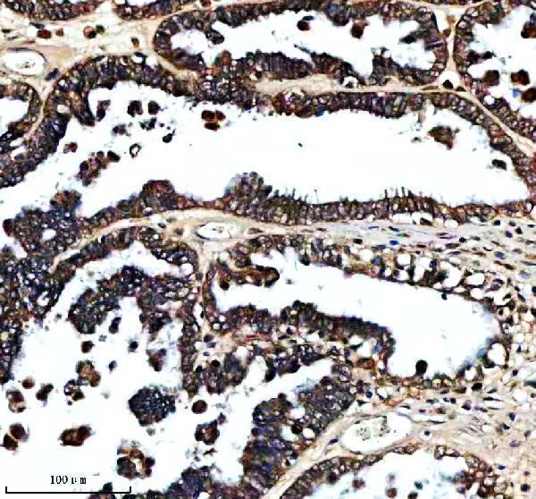
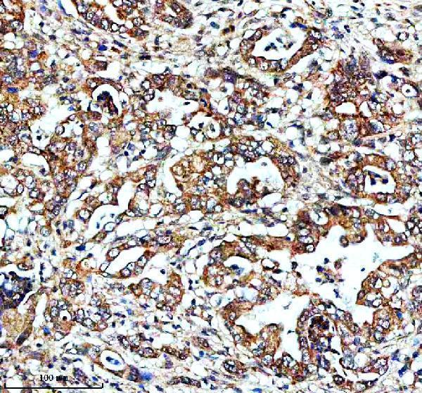
One anti-FXN antibody has real IHC data from human paraffin sections of ovarian and pancreatic cancer tissue (catalog A00842-1 IHC captions). No IF image is provided (catalog A00842-1).

Real IHC data IHC analysis of FXN using anti-FXN antibody (A00842-1). FXN was detected in a paraffin-embedded section of human ovarian cancer tissue. Heat mediated antigen retrieval was performed in EDTA buffer (pH 8.0, epitope retrieval solution). The tissue section was blocked with 10% goat serum. The tissue section was then incubated with 2 μg/ml rabbit anti-FXN Antibody (A00842-1) overnight at 4°C. Peroxidase Conjugated Goat Anti-rabbit IgG was used as secondary antibody and incubated for 30 minutes at 37°C. The tissue section was developed using HRP Conjugated Rabbit IgG Super Vision Assay Kit (Catalog # SV0002) with DAB as the chromogen.
Anti-FXN Antibody ®
Cat # A00842-1

A00842-1 will render with its human ovarian cancer paraffin-section IHC figure (catalog A00842-1 IHC caption). Its catalog also shows human pancreatic cancer paraffin-section IHC images and lists human reactivity (catalog A00842-1 IHC captions; catalog A00842-1 reactivity).

Which to pick: Choose A00842-1 for human tissue IHC: its paraffin-section captions report EDTA retrieval at pH 8.0 and primary antibody at 2 μg/ml, within the listed 2–5 μg/ml IHC range (catalog A00842-1 IHC captions; catalog A00842-1 dilution). The captions do not report a fixative, and the catalog provides no IF/ICC validation (catalog A00842-1 IHC captions; catalog A00842-1 applications). No cross-species option is listed; A00842-1 specifies human reactivity, and its clone is unreported (catalog A00842-1 reactivity; catalog A00842-1 clone).

Each figure is that product's own IHC / IF validation image from its datasheet.

References

  1. UniProt Consortium. UniProt entry Q16595 (FRDA_HUMAN, Frataxin, mitochondrial).
  2. Human Protein Atlas. FXN tissue IHC expression (reliability: Supported).
  3. Human Protein Atlas. FXN subcellular location (ICC-IF): Mainly localized to the mitochondria. In addition localized to the cytosol..
  4. Human Protein Atlas. FXN antibody validation summary (2 antibodies).
  5. Development of a secretable frataxin for enhanced efficacy in treating Friedreich's Ataxia. Molecular therapy. Advances 2026 — PMC13182795.
  6. In vivo overexpression of frataxin causes toxicity mediated by iron-sulfur cluster deficiency. Molecular therapy. Methods & clinical development 2022 — PMC8866050.
  7. AAV8 gene therapy reverses cardiac pathology and prevents early mortality in a mouse model of Friedreich's ataxia. Molecular therapy. Methods & clinical development 2024 — PMC10862410.
  8. Intrathecal delivery of frataxin mRNA encapsulated in lipid nanoparticles to dorsal root ganglia as a potential therapeutic for Friedreich's ataxia. Scientific reports 2016 — PMC4756688.
  9. PubMed PMID:8596916 — UniProt-cited evidence.
  10. PubMed PMID:15164053 — UniProt-cited evidence.
  11. PubMed PMID:15489334 — UniProt-cited evidence.