This website uses cookies to ensure you get the best experience on our website.


- Table of Contents
Plan chromogenic paraffin IHC for FXYD6 using kidney tubules or hepatocytes as high-staining controls (HPA tissue IHC). Start the IHC-validated antibody at 1:25 (datasheet: IHC-P) and assess the cytoplasmic pattern against a not-detected control such as adipocytes (HPA tissue IHC).

Expected localisation, validated protocols, controls and antibodies — the at-a-glance facts below, then the full design guide.
| Expected localisation | Cytoplasmic tissue staining (HPA tissue IHC); cell membrane location (UniProt) | |
| Staining pattern | Cytoplasmic staining in most tissues (HPA tissue IHC) | |
| Antigen retrieval | Citrate pH 6.0 HIER, 95–98 °C, 20 min (rule: cytoplasmic / membrane antigen) | |
| Positive control | Cerebellum+4 more · see all | |
| Negative control | Adipose tissue+4 more · see all |
| Fixation | Keep paraffin-section fixation consistent across samples. (standard IHC practice; not target-specific) | |
| Caveat | Endogenous liver peroxidase may raise DAB background (standard IHC practice; HPA tissue IHC: hepatocytes high) | |
| Regulation | Expression regulation unreported (UniProt) | |
| Isoform / epitope | 2 isoforms; check cytoplasmic C-terminal epitope coverage (UniProt; datasheet: C-term) |
The catalog antibody protocol is accompanied by three published chromogenic IHC protocols for FXYD6 (PMC3881923; PMC7874599; PMC11958163).
| Sample | Paraffin-embedded H. liver tissue; fixative not specified (datasheet A09541) |
| Fixation | Image fixative and duration unreported (datasheet A09541); verify before use. |
| Sectioning | 4–5 µm sections on charged slides (standard) |
| Deparaffinisation | Xylene, graded ethanol series to water (standard) |
| Antigen retrieval | Heat-induced epitope retrieval in citrate buffer, pH 6.0, 20 min at 95–98 °C (standard rule: cytoplasmic / membrane antigen) |
| Peroxidase block | 3% H2O2, 10 min, room temperature (standard) |
| Blocking | 10% normal serum of the secondary host, 30 min, room temperature (standard) |
| Primary antibody | Rabbit anti-FXYD6, 1:25 (datasheet A09541) |
| Primary incubation | Overnight at 4 °C (standard) |
| Detection | HRP-polymer secondary, DAB chromogen 5–10 min (standard) |
| Counterstain | Hematoxylin, blue, dehydrate and mount (standard) |
| Expected result | FXYD6-positive staining in cells in granular layer of cerebellum (HPA tissue IHC: High). HPA tissue profile: Cytoplasmic expression in most tissues. No signal in the no-primary control. |
FXYD6 is a cell-membrane protein with one transmembrane segment and an extracellular region spanning residues 19–35 (UniProt Q9H0Q3 topology). In paraffin IHC, expect chiefly cytoplasmic staining in cells of the cerebellar granular layer, kidney tubules, hepatocytes, and cerebral-cortex neuropil (HPA tissue IHC). HPA rates its tissue-IHC profile Enhanced for consistency with RNA expression; external verification remains pending (HPA tissue IHC).
| Strong staining in kidney tubule cells or hepatocytes, with a predominantly cytoplasmic appearance. | This matches two HPA High tissue-IHC observations and its broader cytoplasmic profile (HPA tissue IHC). Record the stained cell population and compartment; the cell-membrane assignment permits a peripheral component, but does not require a crisp membrane rim in every paraffin section (UniProt Q9H0Q3 subcellular location; HPA tissue IHC). |
| Predominantly nuclear staining on a paraffin IHC slide, especially when expected cytoplasmic staining is absent. | Treat this as discordant with the reported tissue-IHC profile and investigate an artefact before calling it specific (HPA tissue IHC; general IHC practice). HPA separately approves nucleoplasm and cytosol localisation in ICC-IF, so a nuclear signal alone cannot settle the interpretation across assays (HPA ICC-IF). |
| Strong staining of adipocytes or bone-marrow hematopoietic cells while a positive control stains as expected. | Both populations are listed as Not detected in HPA tissue IHC. Check morphology and detection controls for cross-reactivity or endogenous detection activity before assigning FXYD6 expression (HPA tissue IHC; general IHC practice). A discrepant result calls for review, not automatic exclusion of a biological difference. |
| Broad haze across cells, extracellular spaces, or the entire section, with little distinction between cell types. | This obscures the cell-specific pattern needed for interpretation. Review blocking, washes, primary-antibody concentration, and chromogen development; compare a no-primary control for detection background (general IHC practice). HPA's reported cytoplasmic expression in most tissues does not make uniform slide-wide haze a specific result (HPA tissue IHC). |
| Little or no staining in kidney tubules, hepatocytes, or cerebellar granular-layer cells. | These are High HPA tissue-IHC populations, so first examine control-tissue integrity, antibody conditions, retrieval, and detection performance (HPA tissue IHC; general IHC practice). A negative run cannot establish biological absence until the assay produces its expected positive-control signal (general IHC practice). |
| Compartment and topology | UniProt places FXYD6 at the cell membrane, with one transmembrane segment at residues 36–57; HPA describes tissue-IHC staining as cytoplasmic in most tissues (UniProt Q9H0Q3 topology; HPA tissue IHC). Interpret fine peripheral staining alongside the observed paraffin pattern rather than imposing a membrane-only scoring rule. |
| Choice of comparison tissue | Kidney tubule cells, hepatocytes, cerebellar granular-layer cells, cerebral-cortex neuropil, and seminal-vesicle glandular cells are High in HPA tissue IHC. Appendix, colon, and duodenal glandular cells are Medium; several listed populations are Not detected (HPA tissue IHC). Compare like cell types when assessing intensity. |
| Validation and interpretation limits | HPA reports an Enhanced tissue-IHC profile with external verification pending; antibody HPA041334 also has Enhanced IHC status (HPA tissue IHC; HPA antibodies). These support the reported pattern but do not establish the cause of every discordant stain or a target-specific fixation effect. |
| Processing and isoforms | UniProt lists a signal peptide at residues 1–18, a mature chain at 19–95, and 2 isoforms (UniProt Q9H0Q3). Antibody epitope and isoform coverage are unspecified here, so neither differential isoform staining nor processing-related signal loss can be predicted from this record. |
| IF/ICC Q: Should its localisation decide paraffin-IHC scoring? | A: HPA approves nucleoplasm and cytosol localisation in ICC-IF and lists Rh30 images (HPA ICC-IF). For this paraffin-IHC guide, score against the tissue-IHC cell and compartment pattern; the ICC-IF result is a separate assay observation (HPA tissue IHC; HPA ICC-IF). |
| Situation | Likely cause | Next action |
|---|---|---|
| A known-positive tissue is blank, including kidney tubules or hepatocytes. | The run may lack usable primary-antibody, retrieval, or detection signal; these cell types are High in HPA tissue IHC (HPA tissue IHC; general IHC practice). | Confirm tissue and section quality, then review the validated IHC antibody conditions, retrieval, reagent sequence, and a working positive-control slide (general IHC practice). Do not assign FXYD6-negative status from a failed positive control. |
| The section shows only nuclear staining in a proposed IHC-positive population. | This differs from HPA's chiefly cytoplasmic tissue-IHC profile, although nucleoplasm is an approved ICC-IF location (HPA tissue IHC; HPA ICC-IF). | Inspect morphology and compare the positive control, no-primary control, and antibody-validation information (general IHC practice). Report the assay-specific discrepancy rather than transferring an ICC-IF compartment call directly to paraffin IHC. |
| A nominally negative population stains strongly, such as adipocytes or bone-marrow hematopoietic cells. | HPA records these populations as Not detected; nonspecific binding or endogenous detection activity is a possible IHC explanation (HPA tissue IHC; general IHC practice). | Verify the cell identity, examine a no-primary control, and review blocking and detection steps (general IHC practice). Treat a persistent discrepancy as requiring independent confirmation before calling it FXYD6. |
| Diffuse brown haze makes cytoplasmic staining difficult to locate. | Background from binding, inadequate washing, or detection development can obscure cell boundaries (general IHC practice). | Compare the no-primary control; review antibody concentration, blocking, washes, and development time within the validated workflow (general IHC practice). Score only staining that remains distinct from background in identifiable cells. |
| A weak glandular signal appears in appendix, colon, or duodenum. | HPA rates these glandular populations Medium, whereas several suggested positive-control populations are High (HPA tissue IHC). | Compare cells of the same type and include a High HPA tissue-IHC control before calling the run insensitive (HPA tissue IHC; general IHC practice). Preserve the observed intensity in the record rather than forcing a binary call. |
| An IF/ICC image and a paraffin-IHC slide appear to disagree on compartment. | HPA approves nucleoplasm and cytosol in ICC-IF, while its tissue-IHC summary describes cytoplasmic expression in most tissues (HPA ICC-IF; HPA tissue IHC). | Document assay, cell type, and compartment separately; interpret this IHC slide against HPA tissue-IHC observations and its own controls (HPA tissue IHC; general IHC practice). Use the dedicated IF/ICC guide for IF assay decisions. |


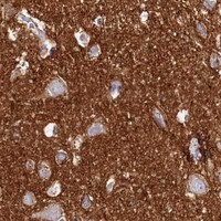

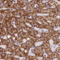


Comprehensive Human Protein Atlas IHC scoring per tissue (reliability: Enhanced — High consistency between antibody staining and RNA expression data. Pending external verification.). Rows are taken directly from the HPA tissue chart — click any row's HPA link to view the source.
| Tissue | Cell type | Level | Evidence | Source |
|---|---|---|---|---|
| Cerebellum | Cells in granular layer | High | Protein (IHC) | HPA → |
| Cerebral cortex | Neuropil | High | Protein (IHC) | HPA → |
| Kidney | Cells in tubules | High | Protein (IHC) | HPA → |
| Liver | Hepatocytes | High | Protein (IHC) | HPA → |
| Seminal vesicle | Glandular cells | High | Protein (IHC) | HPA → |
| Tissue | Cell type | Level | Evidence | Source |
|---|---|---|---|---|
| Adipose tissue | Adipocytes | Not detected | Protein (IHC) | HPA → |
| Adrenal gland | Glandular cells | Not detected | Protein (IHC) | HPA → |
| Bone marrow | Hematopoietic cells | Not detected | Protein (IHC) | HPA → |
| Bronchus | Respiratory epithelial cells | Not detected | Protein (IHC) | HPA → |
| Epididymis | Glandular cells | Not detected | Protein (IHC) | HPA → |
Troubleshoot FXYD6 staining in paraffin sections by checking retrieval, cellular distribution, controls, and scoring before interpreting chromogenic signal.
A09541 has paraffin-section IHC images from human liver and mouse kidney (A09541 image captions); its listed reactivity also includes rat (catalog: A09541). No IF image is supplied (catalog: A09541).
A09541 will render with its own IHC figure from a paraffin-embedded human liver section (A09541 image caption). Its second IHC caption shows paraffin-embedded mouse kidney; the application list includes IHC-P, and reactivity lists human, mouse and rat (A09541 image caption; catalog: A09541).
Which to pick: Choose A09541 for paraffin-section tissue IHC: it is a rabbit polyclonal antibody listed for IHC-P, with human liver and mouse kidney images (catalog: A09541; A09541 image captions). Its listed reactivity covers human, mouse and rat, although the supplied IHC captions show only human and mouse tissue (catalog: A09541; A09541 image captions). No IF/ICC application or image is supplied for A09541, so it is not an IF/ICC-validated choice here; the captions report paraffin embedding but do not report the fixative (catalog: A09541; A09541 image captions).