This website uses cookies to ensure you get the best experience on our website.


- Table of Contents
Plan FZD3 paraffin-section IHC around the observed general cytoplasmic tissue pattern (HPA tissue IHC) and its annotated cell membrane localisation (UniProt). Use the catalog antibody’s IHC protocol, and interpret staining with the reported low agreement between antibody staining and RNA expression in mind (HPA tissue IHC).

Expected localisation, validated protocols, controls and antibodies — the at-a-glance facts below, then the full design guide.
| Expected localisation | General cytoplasmic tissue staining (HPA tissue IHC); cell membrane localisation (UniProt) | |
| Staining pattern | General cytoplasmic staining across cell types (HPA tissue IHC) | |
| Antigen retrieval | Citrate pH 6 HIER, heat-mediated (datasheet A04680-1) | |
| Positive control | Adipose tissue+4 more · see all | |
| Negative control | None in HPA (detected in all 45 tissues); use no-primary + isotype controls |
| Fixation | Keep fixation consistent across sections. (standard IHC practice; not target-specific) | |
| Caveat | Staining has low agreement with RNA expression (HPA tissue IHC) | |
| Regulation | Abundance regulation is unspecified (UniProt) | |
| Isoform / epitope | 2 isoforms; map extracellular vs cytoplasmic epitopes (UniProt) |
The catalog antibody protocol uses citrate pH 6 retrieval (datasheet A04680-1). The published IHC protocols below report methods for human tissue and mouse hippocampal sections (PMC2453022; PMC7746375; PMC13617583).
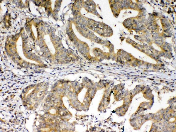
| Sample | Paraffin-embedded human colon cancer tissue; fixative not specified (datasheet A04680-1) |
| Fixation | Image fixative and duration unreported (datasheet A04680-1); verify before use. |
| Sectioning | 4–5 µm sections on charged slides (standard) |
| Deparaffinisation | Xylene, graded ethanol series to water (standard) |
| Antigen retrieval | Heat retrieval: Citrate pH 6, 20 min (datasheet A04680-1) |
| Peroxidase block | 3% H2O2, 10 min, room temperature (standard) |
| Blocking | 10% goat serum (datasheet A04680-1) |
| Primary antibody | Rabbit anti-FZD3, 1μg/ml (datasheet A04680-1) |
| Primary incubation | Overnight at 4 °C (datasheet A04680-1) |
| Detection | Streptavidin-biotin complex (SABC), DAB chromogen (datasheet A04680-1) |
| Counterstain | Hematoxylin, blue, dehydrate and mount (standard) |
| Expected result | FZD3-positive staining in adipocytes of adipose tissue (HPA tissue IHC: High). HPA tissue profile: General cytoplasmic expression. No signal in the no-primary control. |
FZD3 is a 7-pass cell-surface receptor, with an apical membrane location also reported (UniProt Q9NPG1 topology and subcellular location). In paraffin-section IHC, HPA instead describes a general cytoplasmic staining pattern, including high staining in adipocytes, glandular cells, hematopoietic cells, neurons, and cervical squamous epithelium (HPA tissue IHC). Treat that pattern as a reference, not proof of specificity: HPA rates the tissue IHC evidence Approved but reports low consistency with RNA expression (HPA tissue IHC).
| Cytoplasmic stain in adipocytes, colon or adrenal glandular cells, bone marrow hematopoietic cells, or caudate neurons. | This matches HPA's reported High staining in those cell populations and its general cytoplasmic profile (HPA tissue IHC). Compare staining within the identified cell population; HPA's low RNA–staining consistency limits how confidently intensity alone identifies FZD3 (HPA tissue IHC). |
| A cell-edge accent accompanies cytoplasmic staining in a cell population reported positive by HPA. | Membrane staining is biologically plausible for a cell-surface receptor with 7 transmembrane segments (UniProt Q9NPG1 topology and subcellular location). HPA's tissue IHC profile is generally cytoplasmic, so a sharp edge alone should not be made a required pass criterion (HPA tissue IHC). |
| Predominantly nuclear chromogen in a paraffin section. | This departs from HPA's general cytoplasmic tissue IHC profile and UniProt's membrane assignment (HPA tissue IHC; UniProt Q9NPG1 subcellular location). Investigate localisation and controls before calling it FZD3; HPA's nucleoplasmic finding comes from ICC-IF and does not validate nuclear IHC staining (HPA subcellular ICC-IF). |
| Strong staining in an unexpected cell population, or a similar deposit across unrelated cell types. | Cross-reactivity or endogenous detection activity are possibilities to check, not established diagnoses (general IHC practice). FZD3 is widely expressed, so a cell type absent from HPA's short positive list is not automatically negative (UniProt Q9NPG1 tissue specificity; HPA tissue IHC). |
| No stain in a section expected to contain HPA high-staining cells. | First check that the relevant cells are present and assess the run controls (general IHC practice). Absence in one section does not by itself overturn HPA's reported high staining, especially given the Approved rating and low RNA–staining consistency (HPA tissue IHC). |
| Tissue and cell population | HPA reports High staining in selected adipocytes, glandular cells, hematopoietic cells, caudate neurons, and cervical squamous epithelial cells, but Low staining in oral-mucosa squamous epithelial cells (HPA tissue IHC). Identify the cells before comparing sections (general IHC practice). |
| Topology and epitope location | FZD3 has extracellular, transmembrane, and cytoplasmic regions; its FZ domain spans residues 23–136 (UniProt Q9NPG1 topology and domains). The antibody epitope is unspecified here, so topology alone cannot predict which staining compartment that reagent will show. |
| Protein processing and variants | The signal peptide spans residues 1–22, and UniProt lists Long and Short isoforms (UniProt Q9NPG1 processing and isoforms). No epitope or isoform coverage is supplied, so neither feature supports an isoform-specific reading of the chromogen. |
| Evidence strength and antibody validation | HPA calls tissue IHC Approved while noting low consistency with RNA expression; CAB024995 is IHC Approved, with no IHC Enhanced designation supplied (HPA tissue IHC; HPA antibodies). Treat distribution as a comparison point, then interpret it with assay controls (general IHC practice). |
| IF/ICC: should nuclear signal be expected? | HPA reports mainly nucleoplasmic ICC-IF signal, with nuclear membrane and vesicle signal also approved (HPA subcellular ICC-IF). That is an IF observation; the paraffin IHC reference remains HPA's general cytoplasmic profile (HPA tissue IHC). |
| Situation | Likely cause | Next action |
|---|---|---|
| The expected high-staining cells are absent or hard to identify. | The chosen field or section may lack the relevant cell population (general IHC practice). | Inspect tissue architecture and select a field containing a documented HPA High population before judging the stain (HPA tissue IHC; general IHC practice). |
| Relevant cells are present, but chromogen is absent. | An assay step or reagent may have failed; this section alone does not identify which one (general IHC practice). | Review run controls, antibody dilution, retrieval, and detection setup as general IHC checks. No FZD3-specific retrieval condition or dilution is supplied (general IHC practice). |
| Most nuclei are strongly stained. | The pattern conflicts with the HPA tissue IHC profile; the separate ICC-IF nucleoplasmic observation cannot establish an IHC cause (HPA tissue IHC; HPA subcellular ICC-IF). | Compare the primary-omission control and cellular localisation, then reassess whether the signal is specific (general IHC practice). |
| Diffuse chromogen obscures cell boundaries. | Non-specific binding or excess detection background may reduce interpretability (general IHC practice). | Check blocking, antibody dilution, washes, and the primary-omission control; reassess staining only where cells remain identifiable (general IHC practice). |
| Chromogen appears broadly in unrelated cell types. | Endogenous detection activity or cross-reactivity is possible, but broad expression also limits tissue exclusion as a test (general IHC practice; UniProt Q9NPG1 tissue specificity). | Check the appropriate detection-background control and compare localisation with HPA's reported cytoplasmic pattern in identified cells (general IHC practice; HPA tissue IHC). |
| Only oral-mucosa squamous epithelium is weak or unstained. | HPA lists this cell population as Low, so a weak result there alone is a poor test of assay sensitivity (HPA tissue IHC). | Assess a section containing a documented HPA High population and its run controls before changing assay settings (HPA tissue IHC; general IHC practice). |


Comprehensive Human Protein Atlas IHC scoring per tissue (reliability: Approved — Low consistency between antibody staining and RNA expression data.). Rows are taken directly from the HPA tissue chart — click any row's HPA link to view the source.
| Tissue | Cell type | Level | Evidence | Source |
|---|---|---|---|---|
| Adipose tissue | Adipocytes | High | Protein (IHC) | HPA → |
| Adrenal gland | Glandular cells | High | Protein (IHC) | HPA → |
| Appendix | Glandular cells | High | Protein (IHC) | HPA → |
| Bone marrow | Hematopoietic cells | High | Protein (IHC) | HPA → |
| Caudate | Neuronal cells | High | Protein (IHC) | HPA → |
| Tissue | Cell type | Level | Evidence | Source |
|---|---|---|---|---|
| None in HPA: FZD3 is detected in all 45 scored tissues. Use a no-primary (secondary-only) and an isotype control instead. | ||||
Troubleshoot FZD3 chromogenic IHC by checking retrieval, controls, cellular localisation, and scoring before interpreting staining differences.
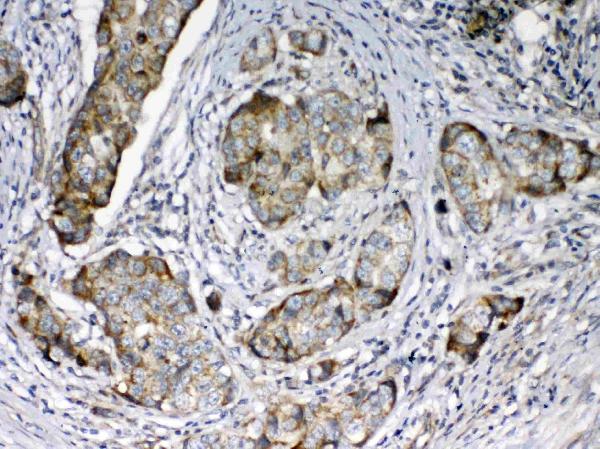
The IHC-validated anti-FZD3 antibody has paraffin-section images from human colon and mammary cancer and mouse and rat kidney (catalog IHC captions).
A04680-1 is listed for IHC in human, mouse and rat (catalog applications and reactivity). Its IHC images show paraffin sections of human colon and mammary cancer and mouse and rat kidney (catalog IHC captions).
Which to pick: For tissue IHC, choose A04680-1, a rabbit polyclonal antibody with paraffin-section images; the fixative is unreported (catalog dilution_raw; catalog IHC captions). For cross-species IHC, A04680-1 has images from human, mouse and rat tissue (catalog IHC captions). For IF/ICC, seek a separately validated reagent: A04680-1 has no listed IF/ICC application or IF image (catalog applications; catalog IF image alts).