This website uses cookies to ensure you get the best experience on our website.


- Table of Contents
Plan FZD6 paraffin IHC using high-staining colon glandular cells as a positive control (HPA tissue IHC). Compare the observed cytoplasmic pattern (HPA tissue IHC) with membrane localization (UniProt), and start within the catalog antibody’s 1:100–1:300 IHC range (datasheet: A04241-1).

Expected localisation, validated protocols, controls and antibodies — the at-a-glance facts below, then the full design guide.
| Expected localisation | Cytoplasmic tissue staining (HPA tissue IHC); cell membrane localization (UniProt) | |
| Staining pattern | Cytoplasmic signal in most tissues (HPA tissue IHC) | |
| Antigen retrieval | Citrate pH 6.0 HIER, 95–98 °C, 20 min (rule: cytoplasmic / membrane antigen) | |
| Positive control | Adrenal gland+4 more · see all | |
| Negative control | Lymph node |
| Fixation | Keep paraffin-section fixation consistent. (standard IHC practice; not target-specific) | |
| Caveat | Cytoplasmic IHC differs from membrane localization (HPA tissue IHC; UniProt) | |
| Regulation | No staining-intensity regulator specified (UniProt) | |
| Isoform / epitope | 2 isoforms; check epitope coverage and membrane side (UniProt) |
The catalog antibody’s IHC-P protocol is accompanied by four published FZD6 IHC methods (PMC5248601; PMC4578624; PMC8883113; PMC12311012).
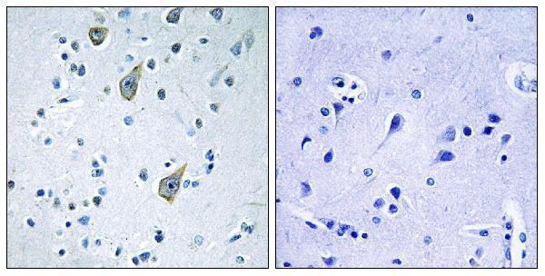
| Sample | Paraffin-embedded human brain tissue; fixative not specified (datasheet A04241-1) |
| Fixation | Image fixative and duration unreported (datasheet A04241-1); verify before use. |
| Sectioning | 4–5 µm sections on charged slides (standard) |
| Deparaffinisation | Xylene, graded ethanol series to water (standard) |
| Antigen retrieval | Heat-induced epitope retrieval in citrate buffer, pH 6.0, 20 min at 95–98 °C (standard rule: cytoplasmic / membrane antigen) |
| Peroxidase block | 3% H2O2, 10 min, room temperature (standard) |
| Blocking | 10% normal serum of the secondary host, 30 min, room temperature (standard) |
| Primary antibody | Rabbit anti-FZD6, 1:100-1:300 (datasheet A04241-1) |
| Primary incubation | Overnight at 4 °C (standard) |
| Detection | HRP-polymer secondary, DAB chromogen 5–10 min (standard) |
| Counterstain | Hematoxylin, blue, dehydrate and mount (standard) |
| Expected result | FZD6-positive staining in glandular cells of adrenal gland (HPA tissue IHC: High). HPA tissue profile: Cytoplasmic expression in most tissues. No signal in the no-primary control. |
FZD6 is a seven-pass membrane receptor with cell-surface, apical-membrane and intracellular-membrane localizations (UniProt O60353 topology and subcellular location). In paraffin-section IHC, HPA reports predominantly cytoplasmic staining across tissues, including high staining in several glandular and epithelial cell populations (HPA tissue IHC). Its IHC profile is Approved, with medium agreement between staining and RNA expression and pending external verification (HPA tissue IHC).
| Cytoplasmic staining in colon, duodenum or breast glandular cells, or bronchial respiratory epithelial cells. | These are HPA-reported high-staining cell populations (HPA tissue IHC). Record the stained cell type and distribution as well as intensity. Cytoplasmic staining is compatible with the reported IHC profile; membrane accentuation is biologically plausible from FZD6 localization, but is not required to match HPA tissue IHC (HPA tissue IHC; UniProt O60353 subcellular location). |
| Membrane-enriched signal in an appropriate epithelial cell population. | FZD6 has seven transmembrane segments and is annotated at the cell surface and apical membrane (UniProt O60353 topology and subcellular location). Interpret membrane staining alongside the tissue's cell identity and cytoplasmic IHC pattern. HPA's plasma-membrane localization is supported by ICC-IF, a separate application (HPA subcellular ICC-IF; HPA tissue IHC). |
| Predominantly nuclear or extracellular-deposit staining, without a coherent cellular pattern. | These compartments do not match the supplied FZD6 localization annotations or HPA tissue IHC summary (UniProt O60353 subcellular location; HPA tissue IHC). Treat the result as suspect and review controls and morphology before scoring it as FZD6. An isolated unusual compartment cannot establish a new localization from this evidence (general IHC interpretation practice). |
| Strong signal in an unexpected cell population, such as lymph-node non-germinal-center cells. | HPA reports FZD6 as not detected in that specific lymph-node population (HPA tissue IHC). If signal persists there, consider cross-reactivity or endogenous chromogen-generating activity and inspect the antibody-omission control (general chromogenic IHC practice). The HPA observation does not make every lymph-node cell a negative control (HPA tissue IHC). |
| Diffuse staining across cells and surrounding tissue, or no staining in a selected HPA high-staining population. | A uniform haze weakens cell-level interpretation and may reflect background from detection reagents or insufficient blocking or washing (general chromogenic IHC practice). Absence of signal in a suitable positive population warrants a run-level check before a biological negative call; HPA's Approved rating still has medium staining–RNA consistency and awaits external verification (HPA tissue IHC). |
| Tissue and cell selection | Use a named HPA high-staining population, such as colon glandular or bronchial respiratory epithelial cells, to judge whether a run can reveal the expected pattern (HPA tissue IHC). Salivary glandular cells, fibroblasts in soft tissue and adipocytes are listed as low, so a faint result in those cells is less informative (HPA tissue IHC). |
| Compartment and antibody epitope | FZD6 contains extracellular, transmembrane and cytoplasmic regions (UniProt O60353 topology). The supplied evidence gives no epitope for the IHC antibody, so it cannot predict which cellular face its stain represents or justify an FZD6-specific retrieval condition. Evaluate retrieval using the antibody's actual instructions and controls (general IHC practice). |
| Processing, glycosylation and isoforms | UniProt lists a cleaved signal peptide at residues 1–18, a mature chain at 19–706, glycosylation sites at 38 and 352, and two isoforms (UniProt O60353 processing, glycosylation and isoforms). Without an antibody epitope or isoform-specific IHC evidence, these features do not establish a distinct staining pattern or a reason to score weak tissue as negative. |
| Strength of tissue evidence | HPA labels the tissue IHC profile Approved and reports medium consistency with RNA plus pending external verification; its listed antibody, HPA017991, has IHC Approved status (HPA tissue IHC; HPA antibody validation). Use those data as a reference pattern, while requiring appropriate run controls for each experiment (general IHC practice). |
| IF/ICC interpretation — can it confirm localization? | On its separate guide page, IF/ICC can assess the HPA-supported plasma-membrane localization; primary-cilium and basal-body localizations are marked uncertain (HPA subcellular ICC-IF). These ICC-IF findings can inform interpretation but do not supply an IHC-P protocol or override the cytoplasmic tissue-IHC profile (HPA subcellular ICC-IF; HPA tissue IHC). |
| Situation | Likely cause | Next action |
|---|---|---|
| No signal in colon or duodenal glandular cells. | The selected cells are reported as high staining, so the run or section may have failed (HPA tissue IHC; general IHC practice). | Confirm cell identity and section quality; inspect a known-positive run control, primary-antibody application and detection steps before calling the tissue negative (general IHC practice). |
| Weak signal in salivary glandular cells, fibroblasts or adipocytes. | HPA lists these populations as low staining (HPA tissue IHC). | Check a named HPA high-staining cell population in the same run before changing conditions; record the low result by cell type and avoid treating it alone as assay failure (HPA tissue IHC; general IHC practice). |
| All compartments have diffuse chromogen. | Background from detection chemistry, endogenous activity or inadequate blocking or washing can obscure cellular staining (general chromogenic IHC practice). | Inspect an antibody-omission control, verify endogenous-activity blocking appropriate to the detection system, and review washing and counterstain before scoring FZD6 (general chromogenic IHC practice). |
| Unexpected nuclear staining dominates the section. | Nuclear localization is absent from the supplied FZD6 annotations and HPA tissue-IHC pattern (UniProt O60353 subcellular location; HPA tissue IHC). | Check control sections and morphology, then compare cytoplasmic staining in a named HPA high-staining population; do not count nuclear color alone as a confirmed positive (HPA tissue IHC; general IHC practice). |
| Strong staining appears in lymph-node non-germinal-center cells. | That population is reported as not detected; cross-reactivity or endogenous detection activity is possible (HPA tissue IHC; general chromogenic IHC practice). | Compare the same population with an antibody-omission control and inspect the positive control before interpreting the unexpected signal (general IHC practice). |
| Results differ between IHC and IF/ICC. | HPA describes cytoplasmic tissue-IHC expression but supported plasma-membrane localization by ICC-IF; the observations come from different applications (HPA tissue IHC; HPA subcellular ICC-IF). | Score each application against its own reported pattern and controls. For IF/ICC interpretation, use its separate guide; do not infer an IHC-P protocol or reject cytoplasmic IHC solely from ICC-IF images (HPA tissue IHC; HPA subcellular ICC-IF; general IHC practice). |


Comprehensive Human Protein Atlas IHC scoring per tissue (reliability: Approved — Medium consistency between antibody staining and RNA expression data. Pending external verification.). Rows are taken directly from the HPA tissue chart — click any row's HPA link to view the source.
| Tissue | Cell type | Level | Evidence | Source |
|---|---|---|---|---|
| Adrenal gland | Glandular cells | High | Protein (IHC) | HPA → |
| Appendix | Glandular cells | High | Protein (IHC) | HPA → |
| Breast | Glandular cells | High | Protein (IHC) | HPA → |
| Bronchus | Respiratory epithelial cells | High | Protein (IHC) | HPA → |
| Cervix | Squamous epithelial cells | High | Protein (IHC) | HPA → |
| Tissue | Cell type | Level | Evidence | Source |
|---|---|---|---|---|
| Lymph node | Non-germinal center cells | Not detected | Protein (IHC) | HPA → |
Troubleshoot FZD6 chromogenic IHC in paraffin sections by checking retrieval, staining compartment, controls and scoring before interpreting biological differences.
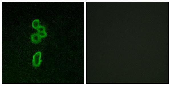
A04241-1 has IHC data from paraffin-embedded human brain tissue and IF data from MCF7 cells (catalog image captions). Its listed reactivity covers human and mouse (catalog: A04241-1).
A04241-1 is the only SKU with a card here; its IHC image shows paraffin-embedded human brain tissue with a peptide-blocked comparison (catalog IHC image caption). Its IF image shows MCF7 cells with a peptide-blocked comparison (catalog IF image caption).
Which to pick: For tissue IHC, choose A04241-1: IHC is listed at 1:100–1:300, and its own image documents paraffin-embedded human brain tissue; the fixative is unreported (catalog applications and dilution; catalog IHC image caption). For IF, A04241-1 has an MCF7 image, while A04241 lists IF and A04241T167 lists ICC; neither of the latter has an image in this payload (catalog applications and image captions). For human, mouse and rat IF or ICC work, consider A04241 or A04241T167 according to the listed application; their clonality is unreported, and neither lists IHC (catalog applications, reactivity and clone fields).