This website uses cookies to ensure you get the best experience on our website.


- Table of Contents
Real validated GADD45A Western blot protocols, expected-band and isoform facts, troubleshooting for weak or shifted signal, and recommended anti-GADD45A WB antibodies. Everything you need to plan the experiment before you commit precious samples.
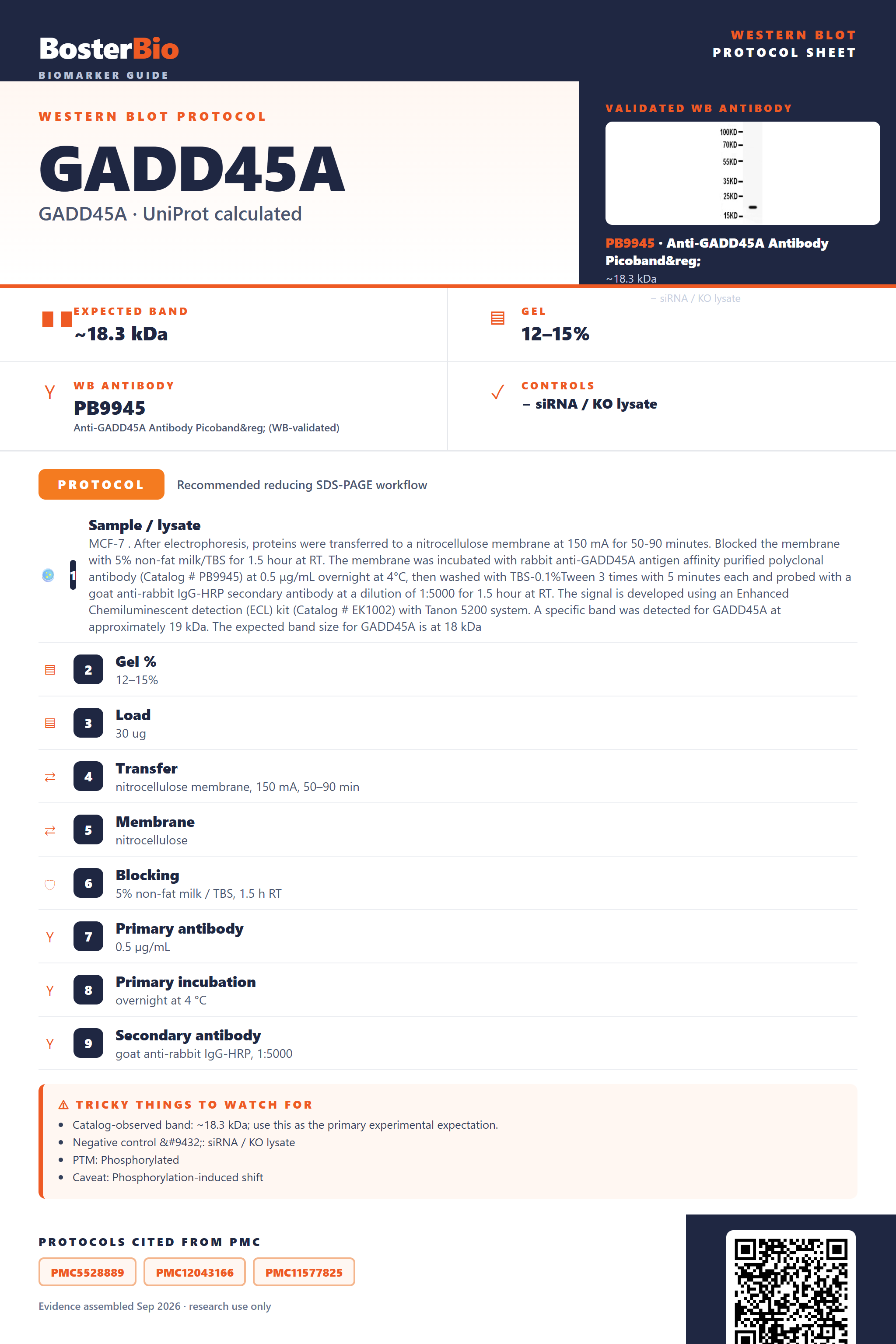
Expected bands, validated protocols, controls and antibodies — the at-a-glance facts below, then the full design guide.
| Expected band | ~18.3 kDa | |
| Observed band | ~19 kDa | |
| Gel | 12–15% | |
| Negative control | siRNA / KO lysate |
| PTM | Phosphorylated | |
| Caveat | Phosphorylation-induced shift | |
| Regulation | TNFα up | |
| Isoform | 2 isoform(s) |
Literature-validated Western blot parameters for GADD45A — gel percentage, transfer, blocking, antibody incubation and detection, extracted from published methods.
| Sample / lysate | MCF-7 . After electrophoresis, proteins were transferred to a nitrocellulose membrane at 150 mA for 50-90 minutes. Blocked the membrane with 5% non-fat milk/TBS for 1.5 hour at RT. The membrane was incubated with rabbit anti-GADD45A antigen affinity purified polyclonal antibody (Catalog # PB9945) at 0.5 μg/mL overnight at 4°C, then washed with TBS-0.1%Tween 3 times with 5 minutes each and probed with a goat anti-rabbit IgG-HRP secondary antibody at a dilution of 1:5000 for 1.5 hour at RT. The signal is developed using an Enhanced Chemiluminescent detection (ECL) kit (Catalog # EK1002) with Tanon 5200 system. A specific band was detected for GADD45A at approximately 19 kDa. The expected band size for GADD45A is at 18 kDa |
| Gel % | 12–15% |
| Load | 30 ug |
| Transfer | nitrocellulose membrane, 150 mA, 50–90 min |
| Membrane | nitrocellulose |
| Blocking | 5% non-fat milk / TBS, 1.5 h RT |
| Primary antibody | 0.5 µg/mL |
| Primary incubation | overnight at 4 °C |
| Secondary antibody | goat anti-rabbit IgG-HRP, 1:5000 |
| Wash | TBS-0.1% Tween, 3 × 5 min |
| Detection | ECL |
| Exposure / imaging | Tanon 5200 |
| Observed band | 19 kDa |
GADD45A has an 18.3 kDa predicted monomer backbone but runs at ~19 kDa on Western blots, likely reflecting its N-terminal phosphorylation and modest self-association tendency.
| Single band at ~19 kDa | Matches the empirical apparent mass for monomeric GADD45A, close to the 18.3 kDa predicted mass |
| Sharp, non-smeared band | GADD45A has no glycosylation sites, so no heterogeneous glycoform smearing is expected |
| Faint higher-mass band near double the monomer size | Reflects self-association into dimers or higher-order oligomers, which increases with protein concentration and can persist if reduction is incomplete |
| Doublet or two closely spaced bands | Consistent with expression of the two annotated splice isoforms (isoform 1 and isoform 2) |
| Band detected in unfractionated whole-cell lysate | GADD45A is nuclear rather than secreted, so it is retained intracellularly and readily detected without needing extracellular fractionation |
| Predicted monomer mass (18.3 kDa) | sets the baseline single-band position, closely matching the ~19 kDa empirically observed band |
| N-terminal phosphothreonine (Thr2) | can cause a slight upward mobility shift, contributing to migration a little above the unmodified predicted mass |
| Dimer/oligomer self-association | produces a faint higher apparent-mass band at roughly double the monomer size at higher protein concentrations or with incomplete reduction |
| Two annotated splice isoforms (isoform 1 and isoform 2) | can yield an additional band at a different apparent size depending on which isoform is expressed, without a defined per-isoform mass |
| Situation | Likely cause | Next action |
|---|---|---|
| No band in lysate | GADD45A is DNA-damage/growth-arrest inducible, so basal nuclear levels can be very low in untreated, unstressed cells | Treat cells with a genotoxic or growth-arrest stimulus as a positive control and enrich for nuclear protein before loading |
| Band higher than expected | GADD45A can self-associate into dimers and higher-order oligomers as concentration increases, and incomplete reduction can preserve these species on the gel | Increase reducing agent concentration and fully denature the sample before loading to resolve the monomer near 19 kDa |
| Multiple bands | Two annotated splice isoforms can be co-expressed at different apparent masses | Confirm isoform identity against known isoform sequence or epitope mapping rather than assuming nonspecific binding |
| Weak or no signal | As a stress-inducible protein, GADD45A expression is often low under normal growth conditions | Increase total protein loaded and/or induce expression with a DNA-damage or growth-arrest stimulus before harvest |
| Fragments below expected size | GADD45A has no annotated signal peptide or propeptide, so smaller bands are not a natural cleaved form and likely reflect proteolytic degradation during lysis | Use fresh lysate with protease inhibitors and keep samples cold throughout preparation |
| Broad smear instead of sharp band | GADD45A lacks glycosylation sites, so a diffuse smear is not expected from glycoform heterogeneity and more likely reflects overloading or aggregation | Reduce the amount of protein loaded per lane and avoid overheating the sample, which can promote aggregation |

Comprehensive Human Protein Atlas IHC scoring per tissue. Rows are taken directly from the HPA tissue chart — click any row's HPA link to view the source.
| Tissue | Cell type | Level | Evidence | Source |
|---|
| Tissue | Cell type | Level | Evidence | Source |
|---|
Deeper troubleshooting and optimisation questions for GADD45A, answered from its protein features.
BosterBio's GADD45A antibodies are among the best-performing WB antibodies on the market — well cited, thoroughly validated, and orthogonally cross-validated against negative tissues and complementary methods.
For GADD45A Western blotting we recommend Boster's anti-GADD45A antibody (PB9945), a best-performing, well-cited reagent that has been thoroughly validated, including a documented SDS-PAGE/WB image and orthogonal cross-validation against negative tissue and complementary detection methods for confident, reproducible results.
Which to pick: Only one Boster anti-GADD45A antibody is catalogued, PB9945, so it is the clear choice. It includes an actual validated Western blot image showing specific GADD45A detection, giving direct evidence of its performance before you order.