GAK / Cyclin-G-associated kinase · IHC design guide

Design Immunohistochemistry for GAK

Plan GAK chromogenic IHC in paraffin sections using the granular cytoplasmic pattern and high-staining glandular cells as references (HPA tissue IHC). The catalog antibody lists an IHC dilution range of 1:50–1:200 (datasheet).

Evidence assembled Oct 2026 · For research use; verify linked source records and product datasheet before use
Immunohistochemistry protocol sheet for GAK (IHC for GAK): expected localisation Granular cytoplasm in most tissues (HPA tissue IHC), antibody A04246-1, validated IHC image, and IHC protocol steps
Printable GAK IHC protocol sheet — expected localisation Granular cytoplasm in most tissues (HPA tissue IHC), antibody A04246-1, controls and protocol steps. Open the full GAK IHC guide →

GAK Immunohistochemistry Experimental Design Guide

Expected localisation, validated protocols, controls and antibodies — the at-a-glance facts below, then the full design guide.

Must know before staining
Expected localisation Granular cytoplasm in most tissues (HPA tissue IHC)
Staining pattern Glandular cells show granular cytoplasmic staining (HPA tissue IHC)
Antigen retrieval Citrate pH 6.0 HIER, 95–98 °C, 20 min (rule: cytoplasmic / membrane antigen)
Positive control ⓘ Appendix+4 more · see all
Negative control ⓘ Adipose tissue+4 more · see all
Important caveats
Reasons your staining may differ from the expected pattern.
Fixation Keep paraffin-section fixation consistent (standard IHC practice; not target-specific)
Caveat Some cell types lack staining despite the broad tissue pattern (HPA tissue IHC)
Regulation Slight expression peak in G1 (UniProt)
Isoform / epitope 2 isoforms; epitope coverage is unreported (UniProt; datasheet)
Section 1

Recommended GAK IHC & IF Protocols

The catalog antibody protocol is followed by a published IHC protocol for paraffin sections of mouse lung (PMC3192135).

Recommended immunohistochemistry (IHC-P) protocol parameters
SampleParaffin-embedded human kidney carcinoma tissue; fixative not specified (datasheet A04246-1)
FixationImage fixative and duration unreported (datasheet A04246-1); verify before use.
Sectioning4–5 µm sections on charged slides (standard)
DeparaffinisationXylene, graded ethanol series to water (standard)
Antigen retrievalHeat-induced epitope retrieval in citrate buffer, pH 6.0, 20 min at 95–98 °C (standard rule: cytoplasmic / membrane antigen)
Peroxidase block3% H2O2, 10 min, room temperature (standard)
Blocking10% normal serum of the secondary host, 30 min, room temperature (standard)
Primary antibodyRabbit anti-GAK, 1:50-1:200 (datasheet A04246-1)
Primary incubationOvernight at 4 °C (standard)
DetectionHRP-polymer secondary, DAB chromogen 5–10 min (standard)
CounterstainHematoxylin, blue, dehydrate and mount (standard)
Expected resultGAK-positive staining in glandular cells of appendix (HPA tissue IHC: High). HPA tissue profile: Cytoplasmic expression with a granular pattern in most tissues. No signal in the no-primary control.
💡Decision noteStart with citrate pH 6.0 HIER at 95–98 °C for 20 min (page retrieval rule). The published lung protocol uses autoclaved citrate (PMC3192135).
Section 2

What Is the Expected GAK Staining Pattern?

GAK should show granular cytoplasmic staining in most tissues, especially glandular cells of the appendix and gastrointestinal tract (HPA tissue IHC: Approved; medium consistency with RNA data). Perinuclear and trans-Golgi localization is consistent with UniProt O14976; GAK has no transmembrane segment (UniProt O14976 topology). Interpret staining by cell type and compartment together, since the HPA profile does not imply equal intensity in every cell.

What am I looking at on my slide?
Granular cytoplasmic staining in appendix or gastrointestinal glandular cells.This fits the reported pattern and high staining in these cells (HPA tissue IHC). Record the proportion and intensity of positive glandular cells; a clean surrounding background helps assess the pattern (general IHC practice).
Predominantly nuclear staining, or a continuous membrane rim replacing the granular cytoplasmic pattern.This conflicts with the reported tissue pattern (HPA tissue IHC) and the lack of a transmembrane segment (UniProt O14976 topology). Check controls and staining conditions before calling it GAK; the pattern alone cannot identify the artefact.
Strong signal confined to adipocytes, bronchial basal cells, or another listed HPA negative cell population.HPA reports GAK as not detected in those specific cell populations (HPA tissue IHC). Consider cross-reactivity or endogenous detection activity, and assess matched controls. A negative cell designation does not make its entire tissue negative.
Diffuse colour across cells and surrounding tissue, obscuring granules.A diffuse background does not match the granular cytoplasmic profile (HPA tissue IHC). It limits compartment scoring; review blocking, washes and detection controls (general IHC practice) before interpreting weak cellular colour.
No cellular signal in appendix or gastrointestinal glandular cells.These are reported high staining populations (HPA tissue IHC). An absent result warrants a check of the IHC assay and tissue quality; it does not, by itself, establish that the sample lacks GAK.
💡Expected GAK appearanceCall a positive result when glandular cells show clear, granular cytoplasmic staining at an intensity consistent with HPA's high category; isolated strong nuclear or uniform background colour is suspect (HPA tissue IHC; UniProt O14976 subcellular location).
How each factor affects the staining
Compartment and topology (UniProt O14976 subcellular location and topology).GAK is reported at the perinuclear region, trans-Golgi network, coated vesicles and focal adhesions, with no transmembrane segment. These annotations support a cytoplasmic granular interpretation; they do not require every structure to resolve in a chromogenic section.
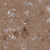
Tissue and cell context (HPA tissue IHC).HPA reports high staining in several glandular cell populations and neuropil, but low staining in hepatocytes, lung macrophages and other listed populations. Select a documented high staining cell population for an assay check; score each cell population separately.
Antibody validation and coverage (HPA antibodies; UniProt O14976 isoforms).The three listed antibodies have Approved IHC status; HPA027463 also has Supported ICC status (HPA antibodies). UniProt lists two GAK isoforms, but the payload gives no epitope or isoform coverage for an antibody. Do not infer which isoform a stain detects.
IF/ICC Q: What pattern can provide a localization comparison? (HPA subcellular ICC-IF).A: HPA reports mainly Golgi localization, with additional vesicles, in its ICC-IF summary (HPA subcellular ICC-IF). This supports a compartment comparison, while the chromogenic tissue interpretation remains grounded in tissue IHC (HPA tissue IHC).
Why is my staining missing, weak or wrong?
SituationLikely causeNext action
A documented high staining glandular population is blank (HPA tissue IHC).A failed stain or unsuitable section is possible; the absence alone cannot establish a biological negative (general IHC practice).Check the IHC-validated antibody's supplied IHC-P instructions, a known-positive section, detection reagents and tissue morphology. Record the controls before interpreting absence (general IHC practice).
Signal is widespread and granular detail is lost.Non-specific binding or detection background can obscure the reported granular cytoplasmic pattern (general IHC practice; HPA tissue IHC).Inspect a no-primary control, review blocking and washes, and adjust detection conditions according to the validated IHC procedure (general IHC practice). Reassess cell boundaries and background.
Only nuclei or continuous cell borders stain strongly.That distribution conflicts with HPA's cytoplasmic tissue profile; GAK lacks a transmembrane segment (HPA tissue IHC; UniProt O14976 topology).Compare a known-positive section and appropriate controls, then review antibody and detection conditions. Do not score the discordant compartment as confirmed GAK staining (general IHC practice).
A listed HPA negative cell population stains strongly.Cross-reactivity or endogenous detection activity is possible; HPA's negative call is specific to the named cell population (HPA tissue IHC).Check a no-primary control and inspect neighbouring cell types separately. Confirm the suspected signal with an independently validated IHC antibody if available (general IHC practice; HPA antibodies).
Signal is weaker than expected in a high staining population (HPA tissue IHC).Section or assay conditions may reduce visible signal (general IHC practice). No GAK-specific fixation or antigen-retrieval sensitivity is supplied.Compare a known-positive section processed in the same run; review the supplied IHC-P retrieval and detection instructions. Avoid assigning a GAK-specific fixation cause without evidence (general IHC practice).

Sample controls for GAK IHC & IF

🧪Run appendix first and require staining in its glandular cells (HPA: High in appendix glandular cells). Use adipose tissue adipocytes as the negative comparator (HPA: Not detected in adipocytes); the supplied HPA rows do not establish any cells within appendix as GAK-negative, so assess neighboring unstained cells against the control slide rather than assigning them negative status (HPA: appendix glandular-cell row).
Positive control tissue: Appendix (Glandular cells, HPA High)
Negative control tissue: Adipose tissue (HPA Not detected)
ICC-IF cell lines (HPA subcellular resource): HPA ICC-IF images show GAK in A-431, U-251MG, U2OS, with annotated localisation: Golgi apparatus (supported) (HPA subcellular).
Technical controls: Include a no-primary, secondary-only control and species-matched nonimmune rabbit IgG for the rabbit polyclonal catalog antibody; use GAK-knockout material as a biological negative if available (selected-SKU caption: rabbit pAb, goat anti-rabbit secondary, PBS in place of primary). Check appendix sections for endogenous peroxidase and, if using the caption’s avidin–biotin detection, endogenous biotin background (selected-SKU caption: avidin–peroxidase detection; standard IHC practice).
⚠️Feasibility: No target-specific fixation window, fixation effect, or retrieval dependency is reported in the supplied evidence; the selected-SKU paraffin-section caption does not state the fixative (selected-SKU caption: fixative unreported). The evidence does not establish whether frozen sections or IF would be easier for GAK, and it identifies no appendix-specific artefact (HPA: appendix tissue row; HPA subcellular: ICC-IF images available).

HPA tissue IHC evidence for GAK

Comprehensive Human Protein Atlas IHC scoring per tissue (reliability: Approved — Medium consistency between antibody staining and RNA expression data.). Rows are taken directly from the HPA tissue chart — click any row's HPA link to view the source.

Positive expression · recommended positive controls

TissueCell typeLevelEvidenceSource
Appendix Glandular cells High Protein (IHC) HPA →
Cerebral cortex Neuropil High Protein (IHC) HPA →
Colon Glandular cells High Protein (IHC) HPA →
Duodenum Glandular cells High Protein (IHC) HPA →
Gallbladder Glandular cells High Protein (IHC) HPA →

Undetected expression · recommended negative controls

TissueCell typeLevelEvidenceSource
Adipose tissue Adipocytes Not detected Protein (IHC) HPA →
Bronchus Basal cells Not detected Protein (IHC) HPA →
Fallopian tube Ciliated cells (cell body) Not detected Protein (IHC) HPA →
Nasopharynx Basal cells Not detected Protein (IHC) HPA →
Oral mucosa Squamous epithelial cells Not detected Protein (IHC) HPA →
Section 3

Advanced GAK IHC Tips

Troubleshoot GAK staining in paraffin sections by checking retrieval, compartment, controls and scoring; use IF as a separate validation route (HPA tissue IHC; HPA subcellular).

What retrieval should I try when GAK staining is weak in paraffin sections?
Start with citrate buffer at pH 6.0 for heat-induced antigen retrieval at 95–98 °C for 20 min (page retrieval setting). Let sections cool in buffer, then compare the same antibody dilution and detection conditions across a small retrieval series so that a stronger signal can be attributed to retrieval (standard IHC practice). If staining remains weak, test an EDTA-based alkaline retrieval condition on adjacent sections as a fallback, while watching for tissue damage and increased background (standard IHC practice). Judge improvement by granular cytoplasmic staining in appropriate cells, alongside a primary-omission control, rather than by total chromogen intensity alone (HPA tissue IHC; standard IHC practice).
Could fixation explain weak or uneven GAK staining?
GAK-specific sensitivity to fixation is unknown here: the A04246-1 image describes paraffin-embedded tissue but does not state its fixative (selected A04246-1 caption). Record fixative, fixation duration and processing history for each block, and compare adjacent sections at the same 1:50 primary dilution when assessing the pictured antibody (selected A04246-1 caption; standard IHC practice). If staining varies across a block, inspect morphology and section adhesion before changing retrieval or antibody concentration, because processing differences can alter accessible epitopes (standard IHC practice). Keep a matched primary-omission section and a consistently stained reference section in each run; neither HPA staining patterns nor GAK topology establishes a fixation-specific effect (standard IHC practice; HPA tissue IHC; UniProt O14976 topology).
Where should convincing GAK staining appear, and how should puncta be assessed?
Expect predominantly granular cytoplasmic staining in tissue sections, with possible perinuclear enrichment, rather than treating a uniformly nuclear signal as the expected pattern (HPA tissue IHC; UniProt O14976 subcellular). GAK is associated with the trans-Golgi network, clathrin-coated vesicles and focal adhesions, while HPA subcellular data chiefly support Golgi localisation with additional vesicles (UniProt O14976 subcellular; HPA subcellular). At 40× objective magnification, assess whether puncta sit within intact cell boundaries and recur in comparable cells on adjacent sections (standard IHC practice). Compare their distribution with the primary-omission control; a diffuse deposit over stroma or damaged edges does not establish vesicular GAK (standard IHC practice).
How can isoforms or epitope position change my interpretation of GAK staining?
GAK has 2 annotated isoforms, and the supplied record does not map the catalog antibody’s epitope to either one (UniProt O14976 isoforms; supplied antibody evidence). Request or verify the immunogen region before calling a negative section isoform-negative, and document whether that region is shared by both isoforms (standard IHC practice; UniProt O14976 isoforms). The protein contains kinase, tensin-type and C-terminal J domains, so epitope location matters when evaluating truncated constructs or domain-specific recognition (UniProt O14976 domains; standard IHC practice). Its annotated phosphorylation sites also make epitope accessibility a reasonable question to test experimentally, but they do not demonstrate a fixation or retrieval effect for this antibody (UniProt O14976 modified residues; supplied antibody evidence).
How should IF help resolve an ambiguous chromogenic GAK pattern?
Use IF as a separate validation experiment and multiplex GAK with a marker identifying the cell population scored in the IHC section; the supplied A04246-1 caption establishes only paraffin-section chromogenic staining (selected A04246-1 caption; standard IF practice). Select a fluorophore in a channel with low measured tissue autofluorescence, and image unstained and single-label controls before interpreting overlap (standard IF practice). Because GAK has no transmembrane segment and is reported in cytoplasmic, Golgi and vesicular compartments, optimise permeabilisation for access to the intracellular epitope rather than assuming surface staining (UniProt O14976 topology and subcellular; standard IF practice). Compare punctate signal with Golgi or vesicle markers and quantify overlap only after checking bleed-through and cell boundaries (HPA subcellular; standard IF practice).
What should I check when GAK IHC has widespread brown background?
First compare the stained section with a section processed without primary antibody; the A04246-1 caption reports a PBS-for-primary control with biotinylated secondary antibody and avidin-peroxidase detection (selected A04246-1 caption). If both sections stain, review the peroxidase block, endogenous biotin contribution, secondary-antibody binding and DAB development time before increasing primary-antibody dilution (selected A04246-1 caption; standard IHC practice). If only the test section stains diffusely, titrate around the reported 1:50 image dilution and compare matched sections under identical retrieval and development conditions (selected A04246-1 caption; standard IHC practice). Retain granular cytoplasmic staining in intact cells as the evaluation endpoint, rather than background intensity over extracellular material (HPA tissue IHC; standard IHC practice).
How should I score GAK staining across specimens? ⚠ ANSWER MARKED FOR VERIFICATION
Define the cell population and compartment before scoring, since the HPA tissue profile describes granular cytoplasmic GAK in most tissues (HPA tissue IHC). For a cellular region, report the percentage of positive cells and a prespecified 0–3 intensity category, then calculate an H-score from the percentages at each intensity (standard IHC scoring practice). If the question concerns puncta or sparse cells, report positive-cell density per mm² of viable tissue instead of treating empty area as negative cells (standard image-analysis practice). Normalise comparisons to the same tissue compartment, viable area, staining batch and detection settings, and retain control sections to identify batch drift (standard IHC practice).
How can I distinguish genuine GAK staining from tissue artefact?
A credible signal follows intact cell outlines and shows granular cytoplasmic or perinuclear distribution consistent with the tissue and subcellular evidence (HPA tissue IHC; UniProt O14976 subcellular). Check cell identity: HPA reports high staining in glandular cells of the appendix and colon, whereas its adipocyte entry is not detected; these observations are tissue-specific reference points (HPA tissue IHC). Treat edge-only staining, necrotic deposits and signal reproduced in the primary-omission section as potential artefacts, and examine endogenous peroxidase or biotin effects when using the captioned avidin-peroxidase workflow (selected A04246-1 caption; standard IHC practice). An isolated nuclear-only pattern needs independent validation before assignment to GAK (HPA tissue IHC; UniProt O14976 subcellular).
Boster reagents

Best GAK / Cyclin-G-associated kinase IHC Antibodies

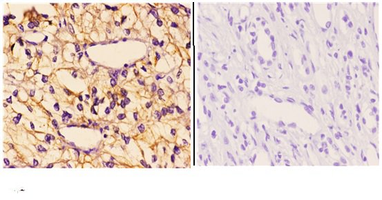
A04246-1 has real IHC data from a paraffin-embedded human kidney carcinoma section (IHC image caption). The catalog lists Human and Mouse reactivity (catalog: reactivity).

Real IHC data Immunohistochemistry (IHC) analyzes of GAK (Q129) pAb in paraffin-embedded human kidney carcinoma tissue at 1:50.showing Cytoplasm staining. Negative control (the right)Using PBS instead of primary antibody, secondary antibody is Goat Anti-Rabbit IgG-biotin followed by avidin-peroxidase.
Anti-GAK (Q129) Antibody
Cat # A04246-1

A04246-1 will render with an IHC figure showing cytoplasmic staining in paraffin-embedded human kidney carcinoma tissue at 1:50, alongside a no-primary control (IHC image caption). It lists IHC as its application and Human and Mouse as reactive species (catalog: applications and reactivity).

Which to pick: Choose A04246-1 for paraffin-section IHC: its own figure documents human tissue staining, and its listed IHC dilution is 1:50–1:200 (IHC image caption; datasheet: IHC dilution). The caption does not report the fixative (IHC image caption). For IF/ICC, no validated option is listed; for Mouse IHC, A04246-1 lists Mouse reactivity, but its figure documents only human tissue (catalog: applications and reactivity; IHC image caption).

Each figure is that product's own IHC / IF validation image from its datasheet.

References

  1. UniProt Consortium. UniProt entry O14976 (GAK_HUMAN, Cyclin-G-associated kinase).
  2. Human Protein Atlas. GAK tissue IHC expression (reliability: Approved).
  3. Human Protein Atlas. GAK subcellular location (ICC-IF): Mainly localized to the Golgi apparatus. In addition localized to vesicles..
  4. Human Protein Atlas. GAK antibody validation summary (3 antibodies).
  5. Neonatal lethality in knockout mice expressing the kinase-dead form of the gefitinib target GAK is caused by pulmonary dysfunction. PloS one 2011 — PMC3192135.
  6. Genkwanin mitigates renal fibrosis by reprogramming profibrotic signaling and restoring TFEB-mediated autophagy. Frontiers in pharmacology 2026 — PMC13518345.
  7. Exploration of Therapeutic Targets Using CDK4 Inhibitors for Head and Neck Mucosal Melanoma. Otolaryngology--head and neck surgery : official journal of American Academy of Otolaryngology-Head and Neck Surgery 2026 — PMC12948400.
  8. PubMed PMID:9299234 — UniProt-cited evidence.
  9. PubMed PMID:15489334 — UniProt-cited evidence.
  10. PubMed PMID:10625686 — UniProt-cited evidence.