This website uses cookies to ensure you get the best experience on our website.


- Table of Contents
Plan chromogenic GATA3 IHC in paraffin sections around the expected nuclear pattern (HPA tissue IHC). Compare kidney glomeruli, where staining is high, with adipocytes, where it is not detected, and assess unexpected signal in light of reported off-target staining (HPA tissue IHC).

Expected localisation, validated protocols, controls and antibodies — the at-a-glance facts below, then the full design guide.
| Expected localisation | Nuclear staining in selected tissues (HPA tissue IHC) | |
| Staining pattern | Nuclear signal in parathyroid glandular and skin basal cells (HPA tissue IHC) | |
| Antigen retrieval | EDTA pH 8.0 HIER, heat-mediated (datasheet A00593-1) | |
| Positive control | Kidney+4 more · see all | |
| Negative control | Adipose tissue+4 more · see all |
| Fixation | Keep fixation consistent across sections. (standard IHC practice; not target-specific) | |
| Caveat | Presumed off-target staining was observed (HPA tissue IHC) | |
| Regulation | Acts downstream of IL33 after injury (UniProt) | |
| Isoform / epitope | 2 isoforms; epitope coverage is unknown (UniProt) |
The catalog antibody’s EDTA retrieval protocol (datasheet A00593-1) is accompanied by published GATA3 IHC methods for lymphoma and genital tract tumors (PMC10969368; PMC11304466).
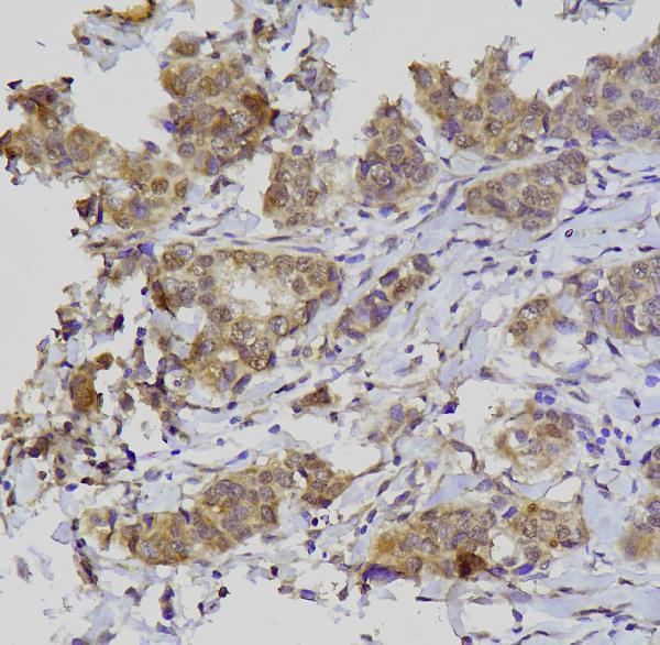
| Sample | Paraffin-embedded human mammary cancer tissue; fixative not specified (datasheet A00593-1) |
| Fixation | Image fixative and duration unreported (datasheet A00593-1); verify before use. |
| Sectioning | 4–5 µm sections on charged slides (standard) |
| Deparaffinisation | Xylene, graded ethanol series to water (standard) |
| Antigen retrieval | Heat retrieval: EDTA pH 8.0 (datasheet A00593-1); 20 min, 95–100 °C (standard) |
| Peroxidase block | 3% H2O2, 10 min, room temperature (standard) |
| Blocking | 10% goat serum (datasheet A00593-1) |
| Primary antibody | Rabbit anti-GATA3, 0.5-1μg/ml (datasheet A00593-1) |
| Primary incubation | Overnight at 4 °C (datasheet A00593-1) |
| Detection | Streptavidin-biotin complex (SABC), DAB chromogen (datasheet A00593-1) |
| Counterstain | Hematoxylin, blue, dehydrate and mount (standard) |
| Expected result | GATA3-positive staining in cells in glomeruli of kidney (HPA tissue IHC: High). HPA tissue profile: Nuclear expression in selected tissues, including parathyroid gland and skin. No signal in the no-primary control. |
GATA3 is a nuclear transcription factor with no transmembrane segment (UniProt P23771: subcellular location and topology). In paraffin-section IHC, expect staining in selected cell populations, including kidney glomerular cells, breast glandular cells and skin basal-layer cells (HPA: tissue IHC). HPA rates the tissue profile “Enhanced,” while reporting medium consistency with RNA data and presumed off-target binding that was disregarded (HPA: tissue IHC reliability).
| Distinct nuclear staining in the expected cells; kidney glomerular cells are strongly stained, while breast glandular cells show moderate staining (HPA: tissue IHC). | This supports the expected IHC pattern when the nuclear signal is confined to the relevant cells (UniProt P23771: nucleus; HPA: tissue IHC). Compare each cell population with its own HPA reference level; a moderate breast signal need not match the strong kidney signal (HPA: tissue IHC). |
| Predominantly cytoplasmic, membranous or extracellular color, with little convincing nuclear signal. | This is discordant with GATA3’s nuclear location and lack of a transmembrane segment (UniProt P23771: location and topology). Treat it as a possible staining artefact and inspect controls before interpreting it as target expression (general IHC practice). |
| Prominent staining in an unexpected cell population, such as adipocytes or adrenal glandular cells (HPA: both not detected in tissue IHC). | Consider cross-reactivity or endogenous detection activity (general IHC practice). HPA also reports presumed off-target binding in its tissue assessment; its “not detected” entries describe observed staining, not an absolute guarantee of absence (HPA: tissue IHC). |
| Weak color spread across nuclei, cytoplasm and surrounding tissue, without clear cell-specific nuclear contrast. | Diffuse background prevents a confident localization call (general IHC practice). The expected target location is nuclear, and HPA describes expression in selected tissues rather than uniform tissue-wide staining (UniProt P23771: nucleus; HPA: tissue IHC profile). |
| No nuclear staining in a kidney section’s glomerular cells (HPA: high tissue-IHC staining). | A missing signal in this known-positive population calls the assay result into question (HPA: tissue IHC; general IHC practice). Review slide quality, controls and detection conditions before calling another sample negative; HPA’s medium RNA–staining consistency also limits how broadly its tissue observations should be extrapolated (HPA: reliability). |
| Cell population and tissue | Expected intensity depends on the sampled cells: HPA reports high staining in kidney glomerular cells; medium in breast glandular cells, parathyroid glandular cells and skin basal-layer cells; and no detection in adipocytes (HPA: tissue IHC). Score the relevant cells, rather than averaging staining across the whole section (general IHC practice). |
| Antibody validation | The tissue-IHC profile has an Enhanced reliability label, with medium consistency against RNA data and presumed off-target binding disregarded (HPA: tissue IHC reliability). CAB016217 is IHC Enhanced, whereas HPA029730 has ICC Supported status with no IHC status listed; these are assay-specific validation entries (HPA: antibodies). |
| Nuclear location and topology | GATA3 is nuclear and has no transmembrane segment (UniProt P23771: location and topology). Nuclear contrast therefore carries the interpretation; a membrane outline alone does not match the supplied target-location evidence (UniProt P23771: location and topology). |
| Isoforms and processing | UniProt lists 2 isoforms, a single chain spanning residues 1–443, and no signal peptide or propeptide (UniProt P23771: isoforms and processing). No epitope map is supplied, so these annotations cannot establish whether a particular antibody detects both isoforms or predict a section-specific staining difference. |
| Endogenous detection activity | Endogenous enzyme activity can create chromogenic signal independently of primary-antibody binding (general IHC practice). Check the detection system and an appropriate negative control if unexpected color appears; this is a general assay concern, not a GATA3-specific effect established by HPA or UniProt. |
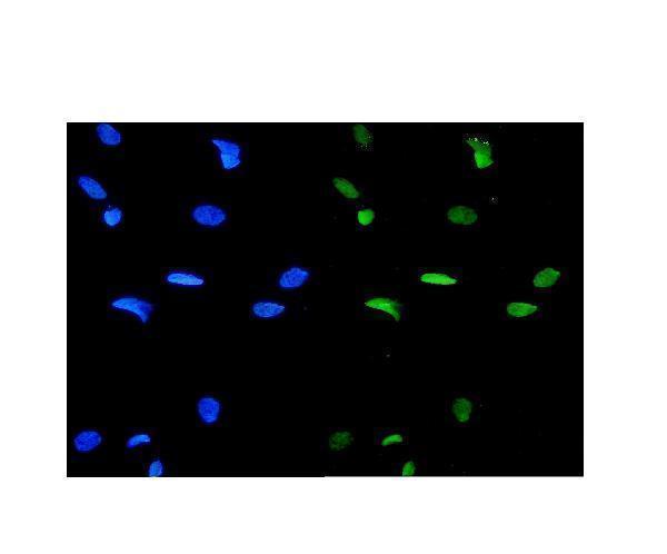
| IF/ICC expectation? | For a separate IF/ICC assay, the expected location is nucleoplasmic; HPA marks it supported and lists images from A-431, U-251MG and U2OS cells (HPA: subcellular ICC-IF). This localization answers the IF/ICC pattern question; the IHC tissue intensity entries do not specify an IF/ICC protocol (HPA: tissue IHC and subcellular ICC-IF). |
| Situation | Likely cause | Next action |
|---|---|---|
| Kidney glomerular cells lack visible nuclear stain (HPA: high tissue-IHC staining). | The staining run may have inadequate signal or an unsuitable control outcome (general IHC practice); the supplied sources do not identify a GATA3-specific fixation sensitivity. | Check section integrity, positive-control performance, primary-antibody application and chromogenic detection steps (general IHC practice). Repeat with a validated positive section before scoring the test section negative (HPA: kidney tissue IHC; general IHC practice). |
| Color is mainly cytoplasmic or membranous. | The compartment conflicts with the supplied nuclear localization and absent transmembrane segment (UniProt P23771: location and topology); nonspecific detection is possible (general IHC practice). | Inspect the nuclear counterstain and an appropriate negative control, then review blocking, washing and detection conditions (general IHC practice). Record only clearly localized nuclear signal as supporting the expected pattern (UniProt P23771: nucleus). |
| Adipocytes or adrenal glandular cells show conspicuous staining (HPA: not detected in tissue IHC). | Cross-reactivity or endogenous detection activity is plausible (general IHC practice). HPA’s reliability note reports presumed off-target binding that was disregarded (HPA: tissue IHC reliability). | Compare the suspect cells with a positive nuclear control and a detection negative control; assess whether color tracks cell type or processing background (general IHC practice). Treat the unexpected result cautiously against the HPA tissue observations. |
| The entire section has low-contrast brown haze. | Diffuse chromogen or nonspecific background can obscure nuclear localization (general IHC practice). | Review blocking, washes, primary-antibody concentration and chromogen development using the assay’s validated instructions (general IHC practice). Reassess only after nuclei can be distinguished from the surrounding tissue (UniProt P23771: nucleus; general IHC practice). |
| Breast or skin looks weaker than kidney. | The supplied reference levels differ: breast glandular and skin basal-layer cells are medium, while kidney glomerular cells are high (HPA: tissue IHC). | Score the appropriate cell population against its tissue-specific expectation (HPA: tissue IHC). Check positive controls if the signal is absent, but do not require all three tissues to reach the kidney reference intensity (HPA: tissue IHC). |
| An IF/ICC image appears diffuse despite an IHC-positive control. | HPA supports nucleoplasmic ICC-IF localization, while its tissue intensity observations come from IHC; performance in one assay does not itself establish the other assay’s staining quality (HPA: subcellular ICC-IF and tissue IHC). | For IF/ICC interpretation, look for nucleoplasmic contrast and evaluate that assay’s controls separately (HPA: subcellular ICC-IF; general immunofluorescence practice). Use the separate IF/ICC guide for method choices. |
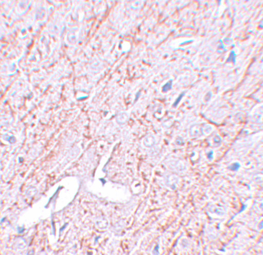
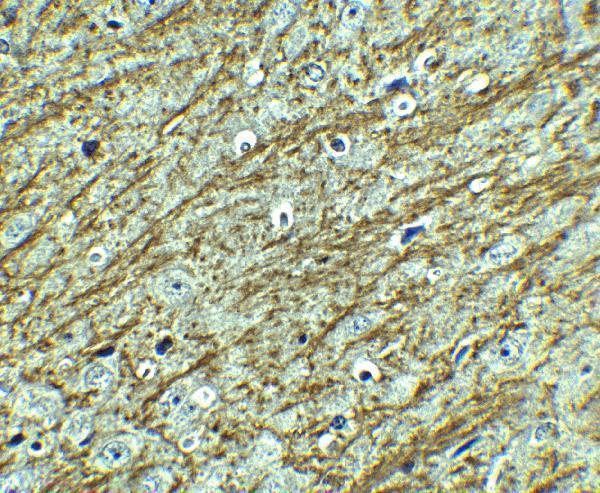
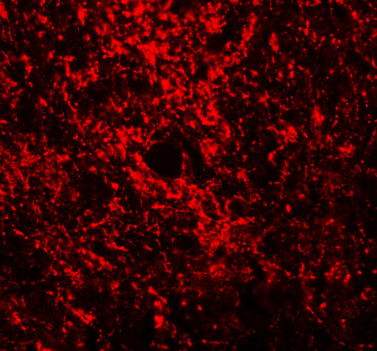
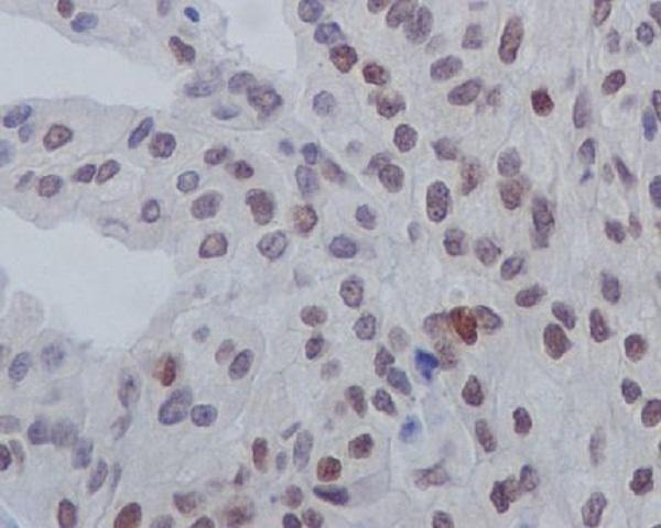


Comprehensive Human Protein Atlas IHC scoring per tissue (reliability: Enhanced — Medium consistency between antibody staining and RNA expression data. Presumed off target binding observed and disregarded.). Rows are taken directly from the HPA tissue chart — click any row's HPA link to view the source.
| Tissue | Cell type | Level | Evidence | Source |
|---|---|---|---|---|
| Kidney | Cells in glomeruli | High | Protein (IHC) | HPA → |
| Breast | Glandular cells | Medium | Protein (IHC) | HPA → |
| Epididymis | Glandular cells | Medium | Protein (IHC) | HPA → |
| Parathyroid gland | Glandular cells | Medium | Protein (IHC) | HPA → |
| Placenta | Syncytiotrophoblasts | Medium | Protein (IHC) | HPA → |
| Tissue | Cell type | Level | Evidence | Source |
|---|---|---|---|---|
| Adipose tissue | Adipocytes | Not detected | Protein (IHC) | HPA → |
| Adrenal gland | Glandular cells | Not detected | Protein (IHC) | HPA → |
| Appendix | Glandular cells | Not detected | Protein (IHC) | HPA → |
| Bone marrow | Hematopoietic cells | Not detected | Protein (IHC) | HPA → |
| Bronchus | Respiratory epithelial cells | Not detected | Protein (IHC) | HPA → |
Troubleshoot GATA3 staining in paraffin sections by checking nuclear localisation, tissue context and method controls before scoring chromogenic signal.
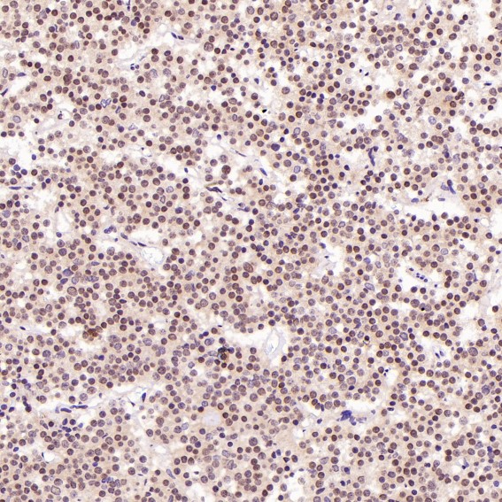
The catalog shows GATA3 IHC images from human, mouse, and rat tissues and IF images from human cells and mouse and rat brain (catalog image captions).
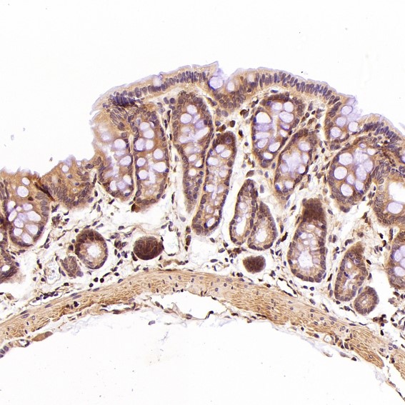
A00593-1 shows IHC in paraffin-embedded human mammary cancer tissue and IF in human cells (A00593-1 image captions); M00593 lists human reactivity and IHC/IF applications, although its displayed IHC image depicts rat intestine (M00593 reactivity, applications, and IHC caption). A00593 shows IHC and IF in mouse and rat brain (A00593 image captions); P00593 shows phospho-GATA3 (S308) IHC in paraffin-embedded human bladder cancer (P00593 IHC caption).
Which to pick: For human paraffin-section IHC, choose A00593-1: its image documents EDTA pH 8 retrieval and 1 μg/mL primary antibody, while the fixative is unreported (A00593-1 IHC caption). For human IF/ICC, A00593-1 has a human-cell IF image and an ICC application listing (A00593-1 IF caption and applications); for mouse or rat tissue IHC and IF, A00593 has images in both species and both applications listed (A00593 image captions and applications). Choose P00593 when staining phospho-GATA3 (S308) in human paraffin sections (P00593 title, reactivity, and IHC caption); treat M00593’s rat-intestine image cautiously because its catalog reactivity lists only human (M00593 IHC caption and reactivity).