This website uses cookies to ensure you get the best experience on our website.


- Table of Contents
Real validated GATA5 Western blot protocols, expected-band and isoform facts, troubleshooting for weak or shifted signal, and recommended anti-GATA5 WB antibodies. Everything you need to plan the experiment before you commit precious samples.
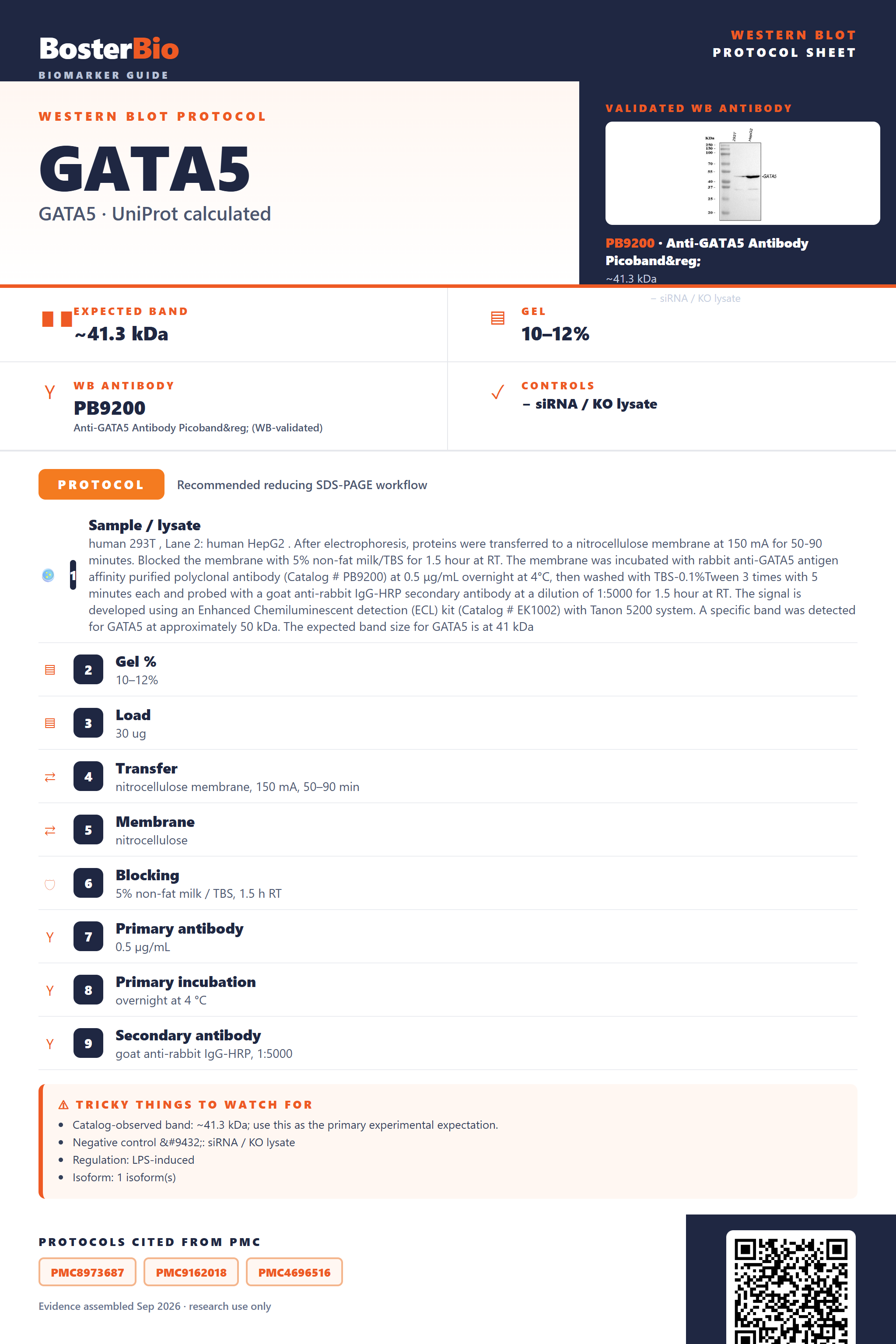
Expected bands, validated protocols, controls and antibodies — the at-a-glance facts below, then the full design guide.
| Expected band | ~41.3 kDa | |
| Observed band | ~50 kDa | |
| Gel | 10–12% | |
| Negative control | siRNA / KO lysate |
| PTM | — | |
| Caveat | — | |
| Regulation | LPS-induced | |
| Isoform | 1 isoform(s) |
Literature-validated Western blot parameters for GATA5 — gel percentage, transfer, blocking, antibody incubation and detection, extracted from published methods.
| Sample / lysate | human 293T , Lane 2: human HepG2 . After electrophoresis, proteins were transferred to a nitrocellulose membrane at 150 mA for 50-90 minutes. Blocked the membrane with 5% non-fat milk/TBS for 1.5 hour at RT. The membrane was incubated with rabbit anti-GATA5 antigen affinity purified polyclonal antibody (Catalog # PB9200) at 0.5 μg/mL overnight at 4°C, then washed with TBS-0.1%Tween 3 times with 5 minutes each and probed with a goat anti-rabbit IgG-HRP secondary antibody at a dilution of 1:5000 for 1.5 hour at RT. The signal is developed using an Enhanced Chemiluminescent detection (ECL) kit (Catalog # EK1002) with Tanon 5200 system. A specific band was detected for GATA5 at approximately 50 kDa. The expected band size for GATA5 is at 41 kDa |
| Gel % | 10–12% |
| Load | 30 ug |
| Transfer | nitrocellulose membrane, 150 mA, 50–90 min |
| Membrane | nitrocellulose |
| Blocking | 5% non-fat milk / TBS, 1.5 h RT |
| Primary antibody | 0.5 µg/mL |
| Primary incubation | overnight at 4 °C |
| Secondary antibody | goat anti-rabbit IgG-HRP, 1:5000 |
| Wash | TBS-0.1% Tween, 3 × 5 min |
| Detection | ECL |
| Exposure / imaging | Tanon 5200 |
| Observed band | 50 kDa |
GATA5 has a 41.3 kDa calculated backbone but typically runs near 50 kDa on Western blot, likely from anomalous SDS-PAGE migration of its zinc-finger DNA-binding domains.
| single band near 50 kDa | represents full-length GATA5 migrating slower than its 41.3 kDa calculated mass, consistent with the empirically observed band |
| no smear or ladder above the main band | GATA5 has no annotated glycosylation sites, so glycoform-driven smearing is not expected |
| no doublet near ~80-100 kDa | no inter-chain disulfide bonds are annotated, so a covalent SDS-resistant homodimer is not expected |
| single band without extra splice-variant bands | only one isoform is annotated, so additional bands are not expected from alternative splicing |
| stronger signal in nuclear-enriched preps than whole-cell lysate | GATA5 is annotated as a nuclear protein, so signal concentrates in the nuclear fraction |
| Predicted mass (UniProt) | calculated at 41.3 kDa from the 397-aa sequence, the baseline against which the observed band is compared |
| Zinc-finger/DNA-binding domain composition | basic, structured zinc-finger and DNA-binding regions can reduce SDS binding and slow migration, producing an apparent band around 50 kDa above the calculated mass |
| No annotated glycosylation sites | rules out glycosylation as a cause of the upward mass shift or of smearing |
| No annotated disulfide bonds or oligomer state | rules out covalent homodimer formation as a cause of a higher-molecular-weight band |
| Single annotated isoform | no alternative-splicing-driven band-size variation is expected |
| Situation | Likely cause | Next action |
|---|---|---|
| Band higher than expected | zinc-finger DNA-binding transcription factors like GATA5 often migrate anomalously slower on SDS-PAGE, giving an apparent ~50 kDa band versus the 41.3 kDa calculated mass | confirm identity with a knockdown or overexpression control and treat ~50 kDa as the expected native band rather than assuming degradation |
| No band in lysate | GATA5 is a low-abundance nuclear transcription factor, so whole-cell lysate without nuclear enrichment can dilute signal below detection | use a nuclear or nuclear-enriched extraction protocol and confirm GATA5 expression in the cell line before blotting |
| Multiple bands | only one isoform is annotated for GATA5, so extra bands are more likely nonspecific antibody binding or degradation than genuine splice variants | validate specificity with a knockdown/knockout or overexpression control and confirm which band matches the expected ~50 kDa size |
| Weak or no signal | low endogenous expression of this nuclear transcription factor in many cell types can yield weak signal | increase loading amount, use nuclear extraction, or test a cell line/tissue with known GATA5 expression |
| Fragments below expected size | degradation of the transcription factor during lysis can generate lower-molecular-weight fragments | add protease inhibitors during lysis, keep samples cold, and minimize freeze-thaw cycles |
| Broad smear instead of sharp band | GATA5 has no annotated glycosylation sites, so a broad smear is not expected from glycoform heterogeneity and more likely reflects overloading or poor transfer | reduce sample loading, optimize transfer conditions, and ensure complete denaturation before loading |

Comprehensive Human Protein Atlas IHC scoring per tissue. Rows are taken directly from the HPA tissue chart — click any row's HPA link to view the source.
| Tissue | Cell type | Level | Evidence | Source |
|---|
| Tissue | Cell type | Level | Evidence | Source |
|---|
Deeper troubleshooting and optimisation questions for GATA5, answered from its protein features.
BosterBio's GATA5 antibodies are among the best-performing WB antibodies on the market — well cited, thoroughly validated, and orthogonally cross-validated against negative tissues and complementary methods.
The featured anti-GATA5 antibody is a top-performing, extensively validated reagent for Western blot, with strong literature citation and orthogonal confirmation against negative tissue controls and complementary detection methods, giving confident, reproducible results for reliable GATA5 detection across experimental systems.
Which to pick: Only one Boster GATA5 antibody is catalogued, PB9200, so it is the default choice. It includes an actual Western blot validation image showing specific GATA5 detection under standard SDS-PAGE conditions, supporting its use for your experiment.