This website uses cookies to ensure you get the best experience on our website.


- Table of Contents
Plan GBF1 paraffin-section IHC using the catalog antibody’s 2–5 μg/ml range (datasheet A01817-1) and the granular cytoplasmic tissue pattern (HPA tissue IHC). Use cell-specific staining to choose controls; Golgi association is a molecular expectation (UniProt).

Expected localisation, validated protocols, controls and antibodies — the at-a-glance facts below, then the full design guide.
| Expected localisation | Granular cytoplasm in several tissues (HPA tissue IHC) | |
| Staining pattern | Granular cytoplasm in glandular and neuronal cells (HPA tissue IHC) | |
| Antigen retrieval | EDTA pH 8.0 HIER, heat-mediated (datasheet A01817-1) | |
| Positive control | Adrenal gland+4 more · see all | |
| Negative control | Adipose tissue+4 more · see all |
| Fixation | Matched tissue-IHC evidence does not establish the fixation claim. Validate the specimen-specific method before use. (selected-SKU IHC image A01817-1) | |
| Caveat | Tissue staining has only medium RNA concordance (HPA tissue IHC) | |
| Regulation | Ubiquitous expression (UniProt) | |
| Isoform / epitope | 4 isoforms; epitope coverage is unspecified (UniProt) |
Compare the catalog antibody’s IHC-P protocol (datasheet A01817-1) with the published human brain paraffin-section preparation (PMC13084705 methods).
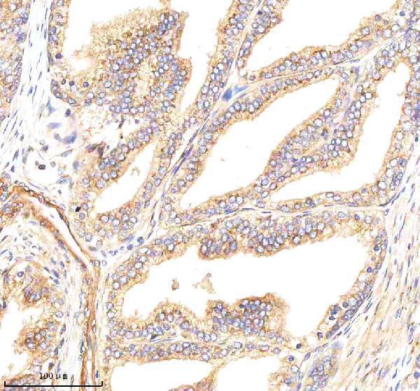
| Sample | Paraffin-embedded human prostate cancer tissue; fixative not specified (datasheet A01817-1) |
| Fixation | Image fixative and duration unreported (datasheet A01817-1); verify before use. |
| Sectioning | 4–5 µm sections on charged slides (standard) |
| Deparaffinisation | Xylene, graded ethanol series to water (standard) |
| Antigen retrieval | Heat retrieval: EDTA pH 8.0 (datasheet A01817-1); 20 min, 95–100 °C (standard) |
| Peroxidase block | 3% H2O2, 10 min, room temperature (standard) |
| Blocking | 10% goat serum (datasheet A01817-1) |
| Primary antibody | Rabbit anti-GBF1, 2-5 μg/ml (datasheet A01817-1) |
| Primary incubation | Overnight at 4 °C (datasheet A01817-1) |
| Detection | HRP-conjugated secondary, DAB chromogen (datasheet A01817-1) |
| Counterstain | Hematoxylin, blue, dehydrate and mount (standard) |
| Expected result | GBF1-positive staining in glandular cells of adrenal gland (HPA tissue IHC: Medium). HPA tissue profile: Granular cytoplasmic expression in several tissues. No signal in the no-primary control. |
GBF1 is a cytoplasmic protein associated with the Golgi, including the cis-Golgi network, and the ER–Golgi intermediate compartment; it has no transmembrane segment (UniProt Q92538). In paraffin-section IHC, expect granular cytoplasmic staining in several cell types, including glandular, respiratory epithelial, and neuronal cells reported at medium intensity (HPA tissue IHC). Treat this pattern as provisional because HPA rates tissue-IHC reliability Uncertain (HPA tissue IHC).
| Granular cytoplasmic staining in colon glandular cells, bronchial respiratory epithelium, or cerebellar Purkinje cells. | These are examples of medium staining reported by HPA (HPA tissue IHC). A compact perinuclear component is consistent with Golgi association (UniProt Q92538; HPA subcellular ICC-IF), but chromogenic IHC alone cannot identify the stained organelle. Score cell type, intensity, and distribution together. |
| Predominantly nuclear staining, or staining confined to cell surfaces or extracellular material. | That distribution does not match the reported granular cytoplasmic tissue pattern or supported Golgi location (HPA tissue IHC; HPA subcellular ICC-IF). Consider nonspecific binding, detection background, or a scoring error; morphology alone cannot determine which explanation applies. |
| Strong staining in adipocytes or duodenal glandular cells while the expected positive cells are weak. | HPA reports GBF1 as not detected in those cells, so disproportionate signal raises concern for cross-reactivity or detection background (HPA tissue IHC). It is not proof of a false positive: the tissue-IHC assessment is Uncertain, and absence of HPA staining does not establish biological absence (HPA tissue IHC). |
| Diffuse color covers most cells or tissue structures without a discernible granular cytoplasmic pattern. | The distribution is difficult to interpret as GBF1 because the reported tissue pattern is granular cytoplasmic (HPA tissue IHC). Review a no-primary control and the detection chemistry using general IHC practice; background, excessive detection signal, and poor contrast can obscure compartment and cell-type boundaries. |
| No signal in the chosen positive tissue, including the cells expected to stain. | First confirm that the section contains the relevant cell type: HPA reports medium staining in colon glandular cells, among other examples (HPA tissue IHC). An absent result can reflect assay performance or sampling; HPA's Uncertain tissue-IHC rating prevents treating one negative section as definitive evidence that GBF1 is absent (HPA tissue IHC). |
| Compartment and dynamics | GBF1 cycles rapidly on and off early Golgi membranes and is also listed in cytoplasm and the ER–Golgi intermediate compartment (UniProt Q92538). Expect a cytoplasmic pattern that may include Golgi-region enrichment, without requiring every positive cell to show an identical compact spot. |
| Tissue and cell selection | HPA reports medium staining in several glandular, respiratory epithelial, and neuronal populations, but not detected staining in selected other cells (HPA tissue IHC). Select and score a documented cell population rather than assigning one expected intensity to an entire tissue. |
| Validation limit | The tissue-IHC profile has Uncertain reliability; both listed HPA antibodies have Uncertain IHC assessments, while HPA037759 has Supported ICC assessment (HPA tissue IHC; HPA antibodies). These ratings support cautious interpretation and do not establish IHC specificity for a different catalog antibody. |
| Topology and processing | GBF1 has no transmembrane segment or signal peptide, and UniProt lists a single chain spanning residues 1–1860 (UniProt Q92538). A membrane outline or extracellular deposit is therefore not the expected primary pattern; these annotations do not establish how a particular epitope behaves in paraffin sections. |
| Isoforms and modified residues | UniProt lists four isoforms and multiple phosphorylated residues (UniProt Q92538). Their effect on staining cannot be predicted without the antibody epitope and validation data; do not interpret intensity differences as isoform expression or phosphorylation from this IHC pattern alone. |
| Situation | Likely cause | Next action |
|---|---|---|
| The expected positive cells show no chromogenic signal. | The relevant cells may be absent from the section, or the staining run may have failed; HPA's reported positives are cell-specific (HPA tissue IHC). | Check the section for an HPA-reported medium cell population, such as colon glandular cells, and compare a concurrently stained positive control (HPA tissue IHC; general IHC practice). Review the catalog antibody's IHC-P instructions for the actual retrieval, dilution, and detection settings. |
| Color is widespread and obscures cellular detail. | The reported GBF1 tissue pattern is granular cytoplasmic, so diffuse color may reflect assay background (HPA tissue IHC). | Compare with a no-primary control; assess blocking, washing, and detection development as general IHC practice. If an enzyme-based detection system is used, check its relevant endogenous activity control before attributing color to GBF1. |
| A nuclear or membrane-outline pattern dominates. | Those compartments conflict with HPA's granular cytoplasmic IHC profile and supported Golgi ICC-IF location (HPA tissue IHC; HPA subcellular ICC-IF). | Check counterstain and tissue morphology, then compare the suspect pattern with a no-primary control and an HPA-reported positive cell population (HPA tissue IHC; general IHC practice). Do not score compartment-mismatched color as a confirmed GBF1 positive. |
| Cells reported as not detected stain as strongly as the expected positives. | The discordance may reflect background or cross-reactivity, but HPA's tissue-IHC reliability is Uncertain (HPA tissue IHC). | Score each cell type separately and compare signal with a no-primary control and a documented medium population (HPA tissue IHC; general IHC practice). Treat disagreement as a reason to validate the catalog antibody in the specimen, not as proof that the HPA profile is universal. |
| Only a diffuse cytoplasmic haze remains after background checks. | GBF1 is listed in cytoplasm and cycles on and off Golgi membranes, although HPA describes granular cytoplasmic tissue staining (UniProt Q92538; HPA tissue IHC). | Assess whether reproducible granules or perinuclear enrichment can be resolved in the appropriate cells; compare the same compartments in controls (HPA tissue IHC; general IHC practice). If resolution is insufficient, record the result as cytoplasmic staining without claiming Golgi localization. |
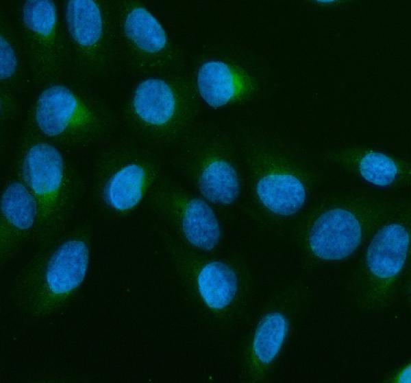
| Can IF/ICC resolve an ambiguous Golgi-region IHC pattern? | HPA reports a Supported Golgi location by ICC-IF, whereas its tissue-IHC profile is Uncertain (HPA subcellular ICC-IF; HPA tissue IHC). | Use the supported ICC-IF localization as a compartment cross-check, not as proof that paraffin-section IHC is specific (HPA subcellular ICC-IF; HPA tissue IHC). Consult the separate IF/ICC guide for that application; evaluate this slide with IHC controls and cell-specific tissue evidence. |

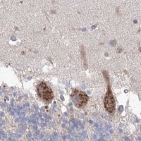


Comprehensive Human Protein Atlas IHC scoring per tissue (reliability: Uncertain — Medium consistency between antibody staining and RNA expression data.). Rows are taken directly from the HPA tissue chart — click any row's HPA link to view the source.
| Tissue | Cell type | Level | Evidence | Source |
|---|---|---|---|---|
| Adrenal gland | Glandular cells | Medium | Protein (IHC) | HPA → |
| Appendix | Glandular cells | Medium | Protein (IHC) | HPA → |
| Breast | Glandular cells | Medium | Protein (IHC) | HPA → |
| Bronchus | Respiratory epithelial cells | Medium | Protein (IHC) | HPA → |
| Caudate | Neuronal cells | Medium | Protein (IHC) | HPA → |
| Tissue | Cell type | Level | Evidence | Source |
|---|---|---|---|---|
| Adipose tissue | Adipocytes | Not detected | Protein (IHC) | HPA → |
| Bone marrow | Hematopoietic cells | Not detected | Protein (IHC) | HPA → |
| Duodenum | Glandular cells | Not detected | Protein (IHC) | HPA → |
| Hippocampus | Glial cells | Not detected | Protein (IHC) | HPA → |
| Liver | Cholangiocytes | Not detected | Protein (IHC) | HPA → |
Troubleshoot GBF1 staining in paraffin sections by checking retrieval, cytoplasmic localization, background controls, and scoring consistency (UniProt Q92538; HPA tissue IHC).
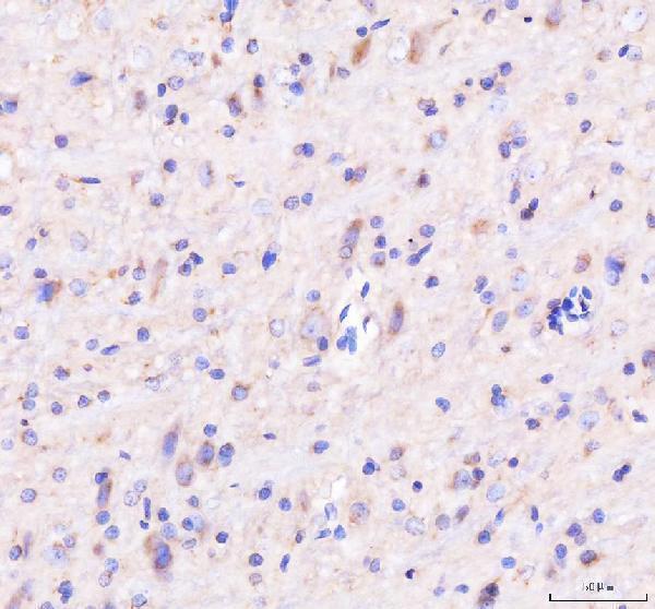
The IHC-validated antibody has paraffin-section images from human prostate and breast cancer tissue and rat and mouse brain (catalog IHC captions); IF data show U2OS cells (catalog IF caption).
A01817-1 is listed for IHC, ICC and IF in human, mouse and rat samples (catalog applications and reactivity). Its IHC captions show human prostate and breast cancer tissue and rat and mouse brain paraffin sections; its IF caption shows U2OS cells (catalog image captions).
Which to pick: Choose A01817-1 for paraffin-section IHC: its own caption documents EDTA retrieval at pH 8.0 and 2 μg/ml primary antibody in human prostate cancer tissue (A01817-1 IHC caption); the fixative is unreported (A01817-1 IHC caption). For IF/ICC, the same SKU has a U2OS cell image at 5 μg/ml (A01817-1 IF caption). For cross-species IHC, it lists human, mouse and rat reactivity and has paraffin-section images for all three; the host is rabbit and clonality is unspecified (catalog reactivity, host and IHC captions).